ABSTRACT
Multiple pregnancies in humans account for only 3% of pregnancies, 97-98% of which are twin pregnancies and the morbimortality is higher in the monochorionic twins when compared to dichorionic ones. The canine species is naturally multiparous, but the diagnosis of monochorionic twin pregnancy is not common. The objective of this report was to describe the ultrasonographic diagnosis of monochorionic twin pregnancies in two bitches [Pug (case 1) and Shih tzu (case 2)]. It was possible to verify the presence of one gestational vesicle containing two fetuses in each female by observing two heads or two bodies within the same placental site. These fetuses presented adequate viability and normal organogenesis. Their development was similar to the other fetuses. In case 1 they were stillborn and smaller than the other five live-born fetuses. The twins in case 2 were born alive, but they also appeared smaller when compared to the littermates. The gestational risks associated with this condition in pregnant bitches are still unknown, however, there are reports of fetal death in monochorionic pregnancies in this species. Therefore, ultrasonographic exam during pregnancy allows an early monochorionic diagnosis and monitoring the fetal viability could bring health benefits to both the female and the littermates.
Keywords:
gemelar pregnancy; monochorionic; canine fetus
RESUMO
As gestações múltiplas em humanos correspondem a apenas 3% das gestações, sendo 97-98% dessas gestações gemelares. Sabe-se que a morbimortalidade fetal é maior em gêmeos monocoriônicos do que nos dicoriônicos. A espécie canina é naturalmente multípara, mas o diagnóstico gestacional de gêmeos monocoriônicos não é comum. O objetivo deste relato é descrever o diagnóstico ultrassonográfico de gêmeos monocoriônicos em duas cadelas, sendo uma da raça Pug (caso 1) e outra da raça Shih-Tzu (caso 2). Foi possível verificar a presença de uma única vesícula gestacional contendo dois fetos em cada cadela, por meio da visibilização de duas cabeças ou de dois corpos dentro de uma mesma placenta. Esses fetos apresentavam viabilidade e organogênese adequadas e o grau de desenvolvimento era similar aos demais fetos da ninhada. No caso 1, os gêmeos nasceram mortos e de tamanho menor que os outros cinco fetos nascidos vivos. Os gêmeos do caso 2 nasceram vivos, mas também eram pequenos em relação aos irmãos da ninhada. Os riscos gestacionais associados a essa condição em cadelas ainda não são conhecidos, no entanto já existem relatos de morte fetal em gestação monocoriônica nessa espécie. Portanto, o exame ultrassonográfico durante a gestação permite um diagnóstico precoce da monocorionicidade, e a monitorização da viabilidade fetal pode trazer benefícios para a saúde da matriz, assim como para o restante da ninhada.
Palavras-chave:
gestação gemelar; monocoriônico; feto canino
INTRODUCTION
In the prenatal period, ultrasound is the best available tool to diagnose gestation, as well as to verify the viability and development of the litter since the embryonic period (Castro et al., 2011CASTRO, V.M.; MAMPRIM, M.J.; LOPES, M.D.; SARTOR, R. Acompanhamento da gestação em cadelas pelo exame ultrassonográfico: revisão de literatura. Vet. Zootec., v.18, p.9-18, 2011.). Early diagnosis occurs by visualization of gestational sacs at 10 to 20 days after luteinizing hormone (LH) peak in bitches. At that moment they are anechoic structures with a hyperechoic wall, which corresponds to the trophoblast. Between 23rd and 25th after the peak of LH, it is possible to identify the embryo, an elongated echogenic structure located within the gestational sac. A thin hyperechogenic layer internal to the uterus, corresponding to the developing placenta, surrounds it. The placenta becomes visible between 27 and 30 days after the LH peak and remains evident during organogenesis until the end of gestation (Mattoon and Nyland, 2015MATTOON, J.S.; NYLAND, T.G. Ovaries and uterus. In: MATTOON, J.S.; NYLAND, T.G. (Eds.). Small animal diagnostic ultrasound. 3.ed. St Louis: Elsevier Saunders, 2015. p.634-654.).
Multiple pregnancies in humans account for only 3% of pregnancies, 97-98% of which are twin pregnancies (Manso et al., 2011MANSO, P.; VAZ, A.; TABORDA, A.; SILVA, I.S. Corionicidade e complicações perinatais na gravidez gemelar: casuística de 10 anos. Acta Med. Port., v.24, p.695-698, 2011.). Twins are classified as dizygotic (when two eggs are fertilized by two spermatozoids) or monozygotic. In the second case, fertilization of a single ovum by one spermatozoid occurs with posterior division of the zygote. Depending on the moment of bipartition, the monozygotic twins can be dichorionic and diamniotics (complete and independent embryos, each one will have its own placenta and chorionic chamber), monochorionic and diamniotics (embryos share the same placenta and have a single chorion and separate amniotic chambers) or monochorionic and monoamniotics (single chorionic and amniotic chambers), different from the dizygotic ones that are always dichorionic and diamniotics (Silva et al., 2003SILVA, J.C.; CECATTI, J.G.; PIRES, H.M.B. et al. Assistência à gestação e parto gemelar. Rev. Ciênc. Med., v.12, p.173-183, 2003.).
According to the results obtained by Manso et al. (2011MANSO, P.; VAZ, A.; TABORDA, A.; SILVA, I.S. Corionicidade e complicações perinatais na gravidez gemelar: casuística de 10 anos. Acta Med. Port., v.24, p.695-698, 2011.), in women the morbimortality of multifetal pregnancy was superior to unifetal one, as well as it was higher in the monochorionic twins when compared to dichorionic ones. Therefore, an early ultrasound diagnosis, between the 11th and 13th week, is mandatory for adequate prenatal care to be instituted.
Bitches are naturally multiparous, so a meticulous ultrasonographic examination is necessary to access as many fetuses as possible. Despite the numerous litters, the diagnosis of monochorionic twin pregnancy in this species is not common (Joonè et al., 2016). The objective of this report was to describe the ultrasonographic diagnosis of monochorionic twin pregnancies in two bitches.
CASUISTRY
To confirm the pregnancy, two adult and healthy female dogs were submitted to image examination in the second trimester, using an ultrasound (Z6Vet, Mindray, China) with multifrequency 7.5MHz transducer, at a private veterinary clinic. One of the bitches was a 2-year-old Pug (case 1) and the other one was a Shih tzu (case 2).
During the ultrasonographic examination, an experienced operator confirmed gestation by the presence of fetal structures within the uterine silhouette. Uterus, placenta and amniotic fluid had no abnormalities. Adequate development and normal fetal heartbeats demonstrated gestational viability. It was possible to verify the presence of one gestational vesicle containing two fetuses in each female by observing two heads or two bodies within the same placental site, which indicated the presence of monochorionic twin pregnancy (Figures 1, 2). These fetuses presented adequate viability and normal organogenesis. Their development was similar to the other fetuses.
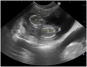
Ultrasonographic image of the skull of two distinct fetuses (1 and 2) sharing a single placental site (*) in a Pug bitch. The estimated gestational age was 38 days.
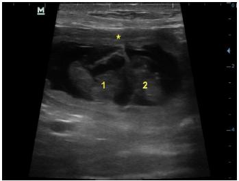
Ultrasonographic image of two concepts (1 and 2) sharing a single placental site (*) in a Shih tzu bitch. The estimated gestational age was 30 to 34 days.
Correlating ultrasonographic findings with the reproductive history, the gestational age in case 1 was approximately 38 days, when it was estimated the presence of six fetuses. After twenty days, a new evaluation was performed noting the absence of heartbeats in the twin fetuses. At night, they were stillborn, smaller than the other five live-born fetuses and they had their bodies covered by hair as expected for a full-term fetus. According to the owner, the twins presented no signs of maceration and they did not have any visible anomaly.
In case 2, gestational age was estimated from 30 to 34 days by using fetal echo biometry. It had an embryonic vesicle absorbed in left uterine horn. The heart rate of examined fetuses was within normal limits. The twins of this bitch were born alive, but they also appeared smaller when compared to the littermates. Both females had normal delivery.
DISCUSSION
The twins of cases 1 and 2 were visually smaller when compared to other canine neonates, similar to that observed in the report by Joonè et al. (2016), in which the monozygotic canine twins were born healthy, but significantly smaller than the other five of the litter.
Underdevelopment of twin fetuses, as well as cases of stillbirth, can be explained by the failure of a single placenta to supply the increased nutritional needs of two fetuses, which worsens in the third trimester of gestation when there is rapid growth of fetuses in the dog (Joonè et al., 2015). Fetal death of the pug bitch twins was detected exactly during this gestational period.
At approximately 58 days of gestation, ultrasonographic images of case 1 were compatible with fetal death for 1 or 2 days (the fetuses had no heart rate, the amniotic fluid was reduced and more echogenic). Similar to this case, Urhausen et al. (2017URHAUSEN, C.; WOLF, K.; BEINEKE, A. et al. Monochorial diamniotic dizygotic twins in a German Shepherd dog: a case report. Reprod. Domest. Anim., v.52, p.140-143, 2017.) evaluated the development of monochronic twins at 38, 41, and 48 days post ovulation (p.o.) using ultrasound, but at 52 days p.o. they were dead. After cesarean section, the twins were found to be monochorionic and diamniotic and one of them had an anasarca. In addition, DNA exams of neonatal tissues have shown that they developed from two different oocytes.
Considering that there are reports of dizygotic canine fetuses sharing the same placental site, karyotyping genetic analysis of blood and tissues is the only method capable to confirm monozygoze (Joonè et al., 2016). In the report described here, no test was performed to compare the DNA of the twins in either case. Therefore, it is not possible to affirm whether they were mono- or dizygotic and to determine whether the fetuses were mono- or diamniotic since the two bitches had normal deliveries and the owners had no experience to provide such information.
In the only report of genetically confirmed monozygotic twin dogs, the animals presented slight differences in pelage marks. For this reason, it is possible that these types of twins are not routinely identified in the litter, since dizygotic siblings in dogs may also be quite similar to each other (Joonè et al., 2016). At this point, ultrasound examination appears as a tool capable of reducing the number of undiagnosed twin pregnancies.
Twin pregnancies are probably not diagnosed because many bitches do not undergo any type of examination as prenatal follow-up. An example is the case of Joonè et al. (2016), in which the female had her first gestational veterinary care when presenting dystocia. Urhausen et al. (2017URHAUSEN, C.; WOLF, K.; BEINEKE, A. et al. Monochorial diamniotic dizygotic twins in a German Shepherd dog: a case report. Reprod. Domest. Anim., v.52, p.140-143, 2017.) described the only ultrasonographic diagnosis of monochorionic twin gestation in dogs. In the other cases of twins sharing the same placental site this fact was only observed at birth (Duke, 1946DUKE, K.L. Monozygotic twins in the dog. Anatom. Rec., v.94, p.35-41, 1946., Joonè et al., 2015, Joonè et al., 2016, Nottidge et al., 2007NOTTIDGE, H.O.; OMOBOWALE, O.; OLOPADE, J.O. et al. A case of craniothoracopagus (monocephalus thoracopagus tetrabrachius) in a dog. Anat. Histol. Embryol., v.36, p.179-181, 2007.).
The absence of adequate gestational monitoring and the late recognition of dystocia leads to higher rates of neonatal mortality and health risks for the female (Johnson, 2008JOHNSON, C.A. Pregnancy management in the bitch. Theriogenology, v.70, p.1412-1417, 2008.; Luz et al., 2015LUZ, M.R.; MÜNNICH, A.; VANNUCCHI, C.I. Novos enfoques na distocia em cadelas. Rev. Bras. Reprod. Anim., v.39, p.354-361, 2015.). The tests performed to differentiate monozygotic and dizygotic twins are only possible after birth (Joonè et al., 2016), therefore they do not contribute to the veterinarian management of the bitch. Thus, the present report shows ultrasonography as a relevant method from a practical point of view in the twinning diagnosis in the dog, since it was able to diagnose monochorionicity in the two described cases at an early stage.
In humans, morbimortality in monochorionic pregnancies is three to four times higher than in dichorionic pregnancies. This is related to an increased risk of spontaneous abortion or fetal death before the 24th week, spontaneous preterm delivery, below-normal birth weight and structural malformations. In addition, in the event of one fetus' death, the other twin has a higher risk of perinatal death and neurological damage due to twin-twin transfusion syndrome, which occurs only in monochorionicity events. Thus, the importance of an early diagnosis in addition to a cautious prenatal in cases of twinning pregnancy is already well established (Peralta and Barini, 2011PERALTA, C.F.A.; BARINI, R. Ultrassonografia obstétrica entre a 11ª e a 14ª semanas: além do rastreamento de anomalias cromossômicas. Rev. Bras. Ginecol. Obstet., v.33, p.49-57, 2011.).
Although the existence of these risks has not been defined in the dog, similar situations to what is reported in humans were described in the few cases described before and in the present report, as below average weight, malformations and fetal death (Joonè et al., 2016, Nottidge et al., 2007NOTTIDGE, H.O.; OMOBOWALE, O.; OLOPADE, J.O. et al. A case of craniothoracopagus (monocephalus thoracopagus tetrabrachius) in a dog. Anat. Histol. Embryol., v.36, p.179-181, 2007., Urhausen et al., 2017URHAUSEN, C.; WOLF, K.; BEINEKE, A. et al. Monochorial diamniotic dizygotic twins in a German Shepherd dog: a case report. Reprod. Domest. Anim., v.52, p.140-143, 2017.). Therefore, the ultrasound diagnosis is fundamental to guide the conduct of the veterinarian, such as suggesting a protocol of periodic exams during gestational follow-up, in order to guarantee delivery assistance and perform a cesarean section if necessary.
CONCLUSION
The presence of two fetuses sharing the same placental site on ultrasonography exam allows the diagnosis of monochorionic twins in canine species. The gestational risks associated with this condition in pregnant bitches are still unknown, however, there are reports of fetal death in monochorionic pregnancies in this species. Therefore, ultrasonographic exam during pregnancy allows an early monochorionicity diagnosis and monitoring the fetal viability could bring health benefits to both female and the littermates.
ACKNOWLEDGMENTS
The authors are thankful to the National Council for Technological and Scientific Development (CNPq) for conceding PQ-2 productivity grants (309199/2017-4).
REFERENCES
- CASTRO, V.M.; MAMPRIM, M.J.; LOPES, M.D.; SARTOR, R. Acompanhamento da gestação em cadelas pelo exame ultrassonográfico: revisão de literatura. Vet. Zootec., v.18, p.9-18, 2011.
- DUKE, K.L. Monozygotic twins in the dog. Anatom. Rec., v.94, p.35-41, 1946.
- JOHNSON, C.A. Pregnancy management in the bitch. Theriogenology, v.70, p.1412-1417, 2008.
- JOONE, C.J.; CRAMER, K.G.M.; NÖTHLING, J.O. Dizygotic monochorionic canine fetuses with blood chimaerism and suspected freemartinism. Reprod. Fertil. Dev., 2015. Available in: <http://dx.doi.org/10.1071/RD15174>. Accessed in: 19 Jan. 2019.
» http://dx.doi.org/10.1071/RD15174 - JOONE, C.J.; CRAMER, K.G.M.; NÖTHLING, J.O. The first case of genetically confirmed monozygotic twinning in the dog. Reprod. Domest. Anim., v.51, p.835-839, 2016.
- LUZ, M.R.; MÜNNICH, A.; VANNUCCHI, C.I. Novos enfoques na distocia em cadelas. Rev. Bras. Reprod. Anim., v.39, p.354-361, 2015.
- MANSO, P.; VAZ, A.; TABORDA, A.; SILVA, I.S. Corionicidade e complicações perinatais na gravidez gemelar: casuística de 10 anos. Acta Med. Port., v.24, p.695-698, 2011.
- MATTOON, J.S.; NYLAND, T.G. Ovaries and uterus. In: MATTOON, J.S.; NYLAND, T.G. (Eds.). Small animal diagnostic ultrasound. 3.ed. St Louis: Elsevier Saunders, 2015. p.634-654.
- NOTTIDGE, H.O.; OMOBOWALE, O.; OLOPADE, J.O. et al. A case of craniothoracopagus (monocephalus thoracopagus tetrabrachius) in a dog. Anat. Histol. Embryol., v.36, p.179-181, 2007.
- PERALTA, C.F.A.; BARINI, R. Ultrassonografia obstétrica entre a 11ª e a 14ª semanas: além do rastreamento de anomalias cromossômicas. Rev. Bras. Ginecol. Obstet., v.33, p.49-57, 2011.
- SILVA, J.C.; CECATTI, J.G.; PIRES, H.M.B. et al. Assistência à gestação e parto gemelar. Rev. Ciênc. Med., v.12, p.173-183, 2003.
- URHAUSEN, C.; WOLF, K.; BEINEKE, A. et al. Monochorial diamniotic dizygotic twins in a German Shepherd dog: a case report. Reprod. Domest. Anim., v.52, p.140-143, 2017.
Publication Dates
-
Publication in this collection
03 Apr 2020 -
Date of issue
Jan-Feb 2020
History
-
Received
08 Feb 2019 -
Accepted
25 July 2019