ABSTRACT
A captive adult male bush dog (Speothos venaticus) was referred to our Veterinary Medical Teaching Hospital from a local zoo due to a two-week history of progressive hind limb gait impairment and ataxia, non-responsive to clinical management. Computed tomography revealed decreased disc space at L3 - L4 level, with probable disc extrusion narrowing the right side of the spinal canal, compressing the spinal cord. We opted to surgically remove the disc material using both fenestration and right lateral pediculectomy (mini-hemilaminectomy) techniques. Twelve days after surgery there was mild residual proprioceptive ataxia. Gait was fully regained with remission of the neurological deficits around the 30th postoperative day. This is - to the best of our knowledge - the first successful report of a lumbar intervertebral disc extrusion in a bush dog (Speothos venaticus) surgically treated by pediculectomy and disc fenestration.
Keywords:
intervertebral disc; zoo animals; neurology; veterinary surgery
RESUMO
Um cachorro-vinagre (Speothos venaticus), macho, adulto, mantido em cativeiro, foi encaminhado ao Hospital Veterinário Universitário pelo zoológico local com histórico de duas semanas de comprometimento progressivo da marcha dos membros posteriores e ataxia, que não responderam ao tratamento clínico. Tomografia computadorizada revelou diminuição do espaço em disco no nível L3 - L4, com provável extrusão de disco estreitando o lado direito do canal vertebral, comprimindo a medula espinhal. Optamos por remover cirurgicamente esse material do disco usando técnicas de fenestração e pediculectomia lateral direita (mini-hemilaminectomia). Doze dias após a cirurgia, houve melhora na deambulação, com ataxia proprioceptiva residual leve. A marcha foi totalmente recuperada com remissão dos déficits neurológicos por volta do trigésimo dia de pós-operatório. Este é - até onde sabemos - o primeiro relato bem-sucedido de uma extrusão de disco intervertebral lombar em um cachorro-vinagre (Speothos venaticus) tratado cirurgicamente por pediculectomia e fenestração de disco.
Palavras-chave:
disco intervertebral; animais de zoológico; neurologia; cirurgia veterinária
INTRODUCTION
The bush dog (Speothos venaticus) is a small canid - rarely seen in nature - from Central and South America (DeMatteo and Loiselle, 2008). It is currently considered near threatened by the International Union for Conservation of Nature (Jorge et al., 2013JORGE, R.S.P.; BEISIEGEL, B.M.; LIMA, E.D. et al. Avaliação do risco de extinção do cachorro-vinagre Speothos venaticus (Lund, 1842) no Brasil. Biodiversity Bras., v.3, p.179-190, 2013.). It’s vulnerability is due to the toll of human progress and urban expansion over what was once their natural habitat, lack of protective laws added up to their susceptibility to fall ill from domestic dog (Canis lupus familiaris) diseases (Oliveira and Dalponte, 2008OLIVEIRA, T.G.; DALPONTE, J.C. Speothos venaticus (Lund, 1842). In: MACHADO, A.B.M.; DRUMMOND, G.M.; PAGLIA, A.P. (Eds.). Livro vermelho da fauna brasileira ameaçada de extinção, Brasília: MMA / Belo Horizonte: Fundação Biodiversitas, 2008. v.2, p.783-784.).
Intervertebral disc extrusion is the most common cause of spinal cord compression in domestic dogs (Braund, 1993BRAUND, K.G. Intervertebral disc disease. In: BOJRAB, M.J.; SMEAK, D.D.; BLOOMBERG, M.S. (Eds.). Disease mechanisms in small animal surgery. 2.ed. Philadelphia: Lea & Febiger, 1993. p.960-970.). It can be compromising by affecting pain and neurological deficits with eventually paralysis. Surgical treatment aims at removing the extruded disc material for spinal cord relieve. Several surgical techniques have been described in small animals (Sharp and Wheeler, 2005SHARP, N.; WHEELER, S. Thoracolumbar disc disease. In: SHARP, N.; WHEELER, S. (Eds.). Small animal spinal disorders. Diagnosis and surgery. 2.ed. Edinburgh: Elsevier, 2005. p.121-159.; Brisson, 2010BRISSON, B.A. Intervertebral disc disease in dogs. Vet. Clin. N. Am. Small Anim. Pract., v.40, p.829-858, 2010.). The literature covering spinal surgery in wild animals needs to be enhanced. Most of the common knowledge is translated from that of companion animals. Scarce reports mainly describe the outcomes of vertebral fractures caused by motor vehicle accidents (McEvoy, 2016).
Here we describe the clinical and surgical managements that allowed the patient to fully recover voluntary motor function within 30 days postoperatively.
CASE REPORT
A captive adult male bush dog (Speothos venaticus) was referred to our Veterinary Medical Teaching Hospital from a local zoo due to a two-week history of progressive hind limb gait impairment and ataxia, non-responsive to clinical management. At presentation, it was possible to observe abnormal wear and pressure sores at the toenails, but a more in depth neurological assessment was not feasible without proper pharmacologic restraint. Survey radiographs - taken under general anesthesia - revealed wedging of L3-L4 disc space (Figure 1). Further computed tomography scan revealed right lateral spinal cord compression due to hyperdense heterogeneous material (disc extrusion) at the same level (Figure 2). We performed surgical decompression using fenestration and lateral pediculectomy as treatment.
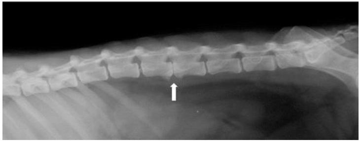
Lateral radiograph of the thoracolumbar spine of a bush dog (Speothos venaticus). Notice the L3-L4 collapsed disc space with ventral spondylosis deformans (white arrow) and opacification of the intervertebral foramen at the same level.
Cephalothin (30mg/kg) IV was administered 30 min prior to the surgical procedure. The patient was placed in a 23° left lateral recumbency, and its right thoracolumbar area was clipped and prepared for surgery. A dorsal paramedian skin incision (approximately 2.5cm parallel to the spinous process) was made from L1 to L5. Then the subcutaneous tissues, lumbodorsal fascia, and lateral epaxial muscles were incised and bluntly dissected.
We used Gelpi retractors and Freer elevators to assess the vertebrae and isolate the common neurovascular bundle. We fenestrated the intervertebral discs from L2 to L5 levels using a n° 11 scalpel blade. We then removed the adjacent pedicles from the L3-L4 vertebrae, avoiding damage to intervertebral foramens and articular processes. We assessed the vertebral canal by creating a window with pneumatic drill burr and removed the thin inner cortical layer using a 1mm Kerrison bone punch. We identified a large amount of extruded nucleus pulposus (Figure 3A) and gently removed (Figure 3B) it to alleviate the spinal cord with blunt instruments, making sure not to cause any harm and visually inspected medullar integrity to determine there was normal blood flow and no signs of necrotic tissue. After flushing with sterile saline, we approximated fascia and epaxial muscles using 2-0 absorbable monofilament suture in a simple continuous pattern. We closed the subcutaneous layer using 3-0 absorbable monofilament suture and closed the skin with surgical staples.
Computed tomography (CT) images of the lumbar spine of a bush dog. A. Sagital view in bone window setting showing calcified amorphous material wrapped in epidural fat occupying 25-50% of the vertebral canal (black arrow). B. Transverse 2-mm-thick CT slice of the L3-L4 space in bone window setting showing the ventrolateral position of the extruded material (black arrow). C. Transverse 2-mm-thick CT slice of the L3-L4 space in soft tissue window setting showing the ventrolateral position of the extruded material (black arrow).
Perioperative photograph of the mini-hemilaminectomy window created at the L3-L4 level of the bush dog. Note that no harm was caused to the articular processes (black asterisks). A. Extruded disc material (white arrow) compressing the spinal cord. B. Decompressed spinal cord (white asterisk) at normal anatomic position after material removal. It is possible to properly assess the floor of the vertebral canal by this surgical approach.
Immediate postoperative analgesia included methadone (0.3mg/kg BW, IM), meloxicam (0.2mg/kg BW, SC) and metamizole (25mg/kg BW, SC). The bush dog recovered uneventfully from the surgical procedure and remained hospitalized in a proper stall with restricted activity for the following weeks. Postoperative treatment consisted of: tramadol hydrochloride (4mg/kg BW, q8h, for 7 days), meloxicam (0.1mg/kg BW, q24h for 3 days), metamizole (25mg/ kg BW, q8h, for 7 days), enrofloxacin (5mg/kg BW, q12h, for 10 days), ketamine (0.25mg/kg BW, q12h, for 7 days) and gabapentin (10mg/kg BW, q12h for 30 days), all administered orally and it was kept wearing an e-collar.
Histopathological analysis of the extruded mass revealed chronically degenerated disc and calcified nucleus pulposus, confirming your hypothesis of a type I Hansen extrusion. Subjective neurological assessment was made daily by visual inspection of motor function and severity of ataxia. Ambulatory improvement was noted on the sixth postoperative day, when the patient regained walking by moving its pelvic limbs. Skin closure occurred uneventfully and the we removed the staples on the second week after surgery. Twelve days after surgery there was mild residual proprioceptive ataxia and we gradually increased the stall space to allow more room for exercise. Gait was fully regained with remission of the neurological deficits around the 30th postoperative day and we discharged the patient back to the zoo facilities.
DISCUSSION
This is, to the best of our knowledge, the first successful report of management of lumbar disc herniation with spinal cord compression in a bush dog (Speothos venaticus) by mini-hemilaminectomy (pediculectomy) and disc fenestration. Neurological impairment in wild animals, caused by traumatic injuries or degenerative diseases often makes survival impractical and may even lead to euthanasia (Cooper, 1996COOPER, J.E. Physical Injury. In: FAIRBROTHER, A.; LOCKE, L.; HOFF, G. (Eds.). Non infectious diseases of wildlife. 2.ed. London: Manson Publishing, 1996. p.157-172.).
Favorable outcomes of decompressive spinal surgery in wild animals are scarcely reported. Some authors have applied dorsal laminectomy in an alpaca (Barker et al., 2015BARKER, W.H.J.; WITTE, T.H.; DRIVER, C.J. et al. Dorsal laminectomy for treatment of cervical vertebral stenotic myelopathy in an alpaca. J. Am. Vet. Med. Assoc., v.246, p.1122-1128, 2015.) and in a two-toed amphiuma (Amphiuma means) (Waffa et al., 2012WAFFA, B.J.; MONTGERARD, A.C.; GRAFINGER, M.S. et al. Dorsal Laminectomy in a Two-Toed Amphiuma (Amphiuma Means). J. Zoo Wildl. Med., v.43, p.927-930, 2012.). Hemilaminectomy has been applied to a binturong (Arctictis binturong) (Spriggs et al., 2007SPRIGGS, M.; ARBLE, J.; MYERS, G. Intervertebral disc extrusion and spinal decompression in a binturong (Arctictis binturong). J. Zoo Wildl. Med., v.38, p.35-138, 2007.), a lion-tailed macaque (Macaca silenus) (Capitanio et al., 2008CAPITANIO, M.; ZIMMERMAN, D.M.; DOUGLASS, M. et al. Thoracolumbar hemilaminectomy and spinal cord decompression in a lion-tailed macaque (Macaca silenus). J. Zoo Wildl. Med., v.39, p.270-273, 2008.) and also tigers (Ketz-Riley et al., 2004; Flegel et al., 2008FLEGEL, T.; BÖTTCHER, P.; ALEF, M. et al. Continuous lumbar hemilaminectomy for intervertebral disc disease in an Amur tiger (Panthera tigris altaica). J. Zoo Wildl. Med., v.39, p.468-471, 2008.). But there is still a lot to be explored to enhance neurological knowledge concerning wild animals.
Intervertebral disc disease causing spinal cord compression, gait impairment and even paralysis quite commonly affects domestic dogs. There is typically chondroid degeneration of the annulus fibrosus with nucleus pulposus extrusion leading to compressive damage to the spinal cord (Brisson et al., 2011BRISSON, B.A.; HOLMBERG, D.L.; PARENT, J. et al. Comparison of the effect of single-site and multiple-site disk fenestration on the rate of recurrence of thoracolumbar intervertebral disk herniation in dogs. J. Am. Vet. Med. Assoc., v.238, p.1593-1600, 2011.). Despite interspecies differences, we translated surgical knowledge from the companion dog (Canis lupus familiaris) literature to treat this wild canid and reached a successful outcome. Clinical onset was similar to a type I Hansen disc extrusion and surgical decompression restored normal spinal cord hemodynamics and was crucial to clinical improvement (Brisson, 2010).
Mini-hemilaminectomy provided sufficient access to the ventral part of the vertebral canal in an efficient and less invasive approach than hemilaminectomy (Jeffery, 1988JEFFERY, N.D. Treatment of acute and chronic thoracolumbar disc disease by ‘mini hemilaminectomy’. J. Small Anim. Pract., v.29, p.611-616, 1988.; McCartney, 1997; Seim, 2002SEIM, H.B. Fundamentals of neurosurgery. In: FOSSUM, T.W. (Ed.). Small Animal Surgery. 2.ed. St. Louis, Missouri: Elsevier Health Sciences, 2002. p.1139-1158.; Brisson, 2010BRISSON, B.A. Intervertebral disc disease in dogs. Vet. Clin. N. Am. Small Anim. Pract., v.40, p.829-858, 2010.; Huska et al., 2014HUSKA, J.L.; GAITERO, L.; BRISSON, B.A. et al. Comparison of the access window created by hemilaminectomy and mini-hemilaminectomy in the thoracolumbar vertebral canal using computed tomography. Can. Vet. J., v.55, p.449-455, 2014.). This technique spares the articular processes and can be performed bilaterally without causing vertebral instability (since the cranial and/or caudal portions of the pedicles and most part of the dorsal lamina are left intact). Additionally, mini-hemilaminectomy can easily be converted to conventional hemilaminectomy if necessary (McCartney, 1997; Sharp and Wheeler, 2005SHARP, N.; WHEELER, S. Thoracolumbar disc disease. In: SHARP, N.; WHEELER, S. (Eds.). Small animal spinal disorders. Diagnosis and surgery. 2.ed. Edinburgh: Elsevier, 2005. p.121-159.). Dorsal laminectomy (Toombs and Waters, 2003TOOMBS, J.; WATERS, D. Intervertebral disc disease. In: SLATTER, D.H. (Ed.). Textbook of small animal surgery. 3.ed. Elsevier Health Sciences, 2003. p.1193-1208.) is also a viable (but more invasive) option to decompress the spinal cord and nerve roots, and may prolong the recovery time (Brisson, 2010).
The 23° lateral recumbency allowed a less invasive approach to the vertebral canal than hemilaminectomy (Roach et al., 2012ROACH, W.J.; THOMAS, M.; WEH, J.M. et al. Residual herniated disc material following hemilaminectomy in chondrodystrophic dogs with thoracolumbar intervertebral disc disease Vet. Comp. Orthop. Traumatol, v.25, p.109-115, 2012.) while providing proper access to the floor of the vertebral canal. It allowed effective removal of the extruded disc material (Huska et al., 2014HUSKA, J.L.; GAITERO, L.; BRISSON, B.A. et al. Comparison of the access window created by hemilaminectomy and mini-hemilaminectomy in the thoracolumbar vertebral canal using computed tomography. Can. Vet. J., v.55, p.449-455, 2014.; Marinho et al., 2014MARINHO, P.V.T.; ARIAS, M.V.B.; ZANI, C.C. et al. Hansen type II disc disease in dogs: pathophysiology, clinical surgical approach and controversies. Semina, v.35, p.1395-1413, 2014.). Partial lateral corpectomy could have been associated at this recumbency, had it been necessary (due to the two-week-history of clinical onset) (Flegel et al., 2011FLEGEL, T.; BOETTCHER, I.C., LUDEWIG, E. et al. Partial lateral corpectomy of the thoracolumbar spine in 51 dogs: assessment of slot morphometry and spinal cord decompression. Vet. Surg., v.40, p.14-21, 2011.; Vicente et al., 2013VICENTE, F.; BERNARD, F.; FITZPATRICK, D. et al. In vitro radiographic characteristics and biomechanical properties of the canine lumbar vertebral motion unit after lateral corpectomy, mini-hemilaminectomy and hemilaminectomy. Vet. Comp. Orthop. Traumatol, v.26, p.19-26, 2013.). The adjacent intervertebral discs were fenestrated to prevent further extrusions (Forterre et al., 2008FORTERRE, F.; KONAR, M.; SPRENG, D. et al. Influence of intervertebral disc fenestration at the herniation site in association with hemilaminectomy on recurrence in chondrodystrophic dogs with thoracolumbar disc disease: a prospective MRI study. Vet. Surg., v.37, p.399-405, 2008.; Brisson et al., 2011BRISSON, B.A.; HOLMBERG, D.L.; PARENT, J. et al. Comparison of the effect of single-site and multiple-site disk fenestration on the rate of recurrence of thoracolumbar intervertebral disk herniation in dogs. J. Am. Vet. Med. Assoc., v.238, p.1593-1600, 2011.; Aikawa et al., 2012AIKAWA, T.; FUJITA, H.; SHIBATA, M. et al. Recurrent thoracolumbar intervertebral disc extrusion after hemilaminectomy and concomitant prophylactic fenestration in 662 chondrodystrophic dogs. Vet. Surg. v.41, p.381-390, 2012.). This dorsolateral approach also benefits disc fenestration (Morelius et al., 2007MORELIUS, M.; BERGADANO, A.; SPRENG, D. et al. Influence of surgical approach on the efficacy of the intervertebral disk fenestration: a cadaveric study. J. Small Anim. Pract., v.48, p.87-92, 2007.; Tanaka et al., 2014TANAKA, N.; KITAGAWA, M.; ITO, M. et al. A modified lateral muscle-separation approach for mini-hemilaminectomy. Vet. Rec., v.173, p.296, 2014.).
Postoperative analgesia was also translated from the small animal literature. We opted to associate gabapentin since it is a promising postoperative analgesic tool, confirmed by recent studies (Ruel and Steagall, 2019RUEL, H.L.; STEAGALL, P.V. Adjuvant analgesics in acute pain management. Vet. Clin. N. Am. Small Anim. Pract., v.49, p.1127-1141, 2019.). We did not consider physical therapy in this case due to the animal’s aggressive behavior, even though it is known to be an important factor that improves recovery after neurological surgeries.
CONCLUSION
Our clinical and surgical conducts translated from domestic dogs to a bush-dog (Speothos venaticus) zoo animal were successful and provided an uneventful outcome, first of its kind to the best of our knowledge.
REFERENCES
- AIKAWA, T.; FUJITA, H.; SHIBATA, M. et al. Recurrent thoracolumbar intervertebral disc extrusion after hemilaminectomy and concomitant prophylactic fenestration in 662 chondrodystrophic dogs. Vet. Surg. v.41, p.381-390, 2012.
- BARKER, W.H.J.; WITTE, T.H.; DRIVER, C.J. et al. Dorsal laminectomy for treatment of cervical vertebral stenotic myelopathy in an alpaca. J. Am. Vet. Med. Assoc., v.246, p.1122-1128, 2015.
- BRAUND, K.G. Intervertebral disc disease. In: BOJRAB, M.J.; SMEAK, D.D.; BLOOMBERG, M.S. (Eds.). Disease mechanisms in small animal surgery. 2.ed. Philadelphia: Lea & Febiger, 1993. p.960-970.
- BRISSON, B.A. Intervertebral disc disease in dogs. Vet. Clin. N. Am. Small Anim. Pract., v.40, p.829-858, 2010.
- BRISSON, B.A.; HOLMBERG, D.L.; PARENT, J. et al. Comparison of the effect of single-site and multiple-site disk fenestration on the rate of recurrence of thoracolumbar intervertebral disk herniation in dogs. J. Am. Vet. Med. Assoc., v.238, p.1593-1600, 2011.
- CAPITANIO, M.; ZIMMERMAN, D.M.; DOUGLASS, M. et al. Thoracolumbar hemilaminectomy and spinal cord decompression in a lion-tailed macaque (Macaca silenus). J. Zoo Wildl. Med., v.39, p.270-273, 2008.
- COOPER, J.E. Physical Injury. In: FAIRBROTHER, A.; LOCKE, L.; HOFF, G. (Eds.). Non infectious diseases of wildlife. 2.ed. London: Manson Publishing, 1996. p.157-172.
- DEMATTEO, K.E.; LOISELLE, B.A. New data on the status and distribution of the bush dog (Speothos venaticus): Evaluating its quality of protection and directing research efforts. Bio Conserv., v.141, p.2494-2505, 2008.
- FLEGEL, T.; BOETTCHER, I.C., LUDEWIG, E. et al. Partial lateral corpectomy of the thoracolumbar spine in 51 dogs: assessment of slot morphometry and spinal cord decompression. Vet. Surg., v.40, p.14-21, 2011.
- FLEGEL, T.; BÖTTCHER, P.; ALEF, M. et al. Continuous lumbar hemilaminectomy for intervertebral disc disease in an Amur tiger (Panthera tigris altaica). J. Zoo Wildl. Med., v.39, p.468-471, 2008.
- FORTERRE, F.; KONAR, M.; SPRENG, D. et al. Influence of intervertebral disc fenestration at the herniation site in association with hemilaminectomy on recurrence in chondrodystrophic dogs with thoracolumbar disc disease: a prospective MRI study. Vet. Surg., v.37, p.399-405, 2008.
- HUSKA, J.L.; GAITERO, L.; BRISSON, B.A. et al. Comparison of the access window created by hemilaminectomy and mini-hemilaminectomy in the thoracolumbar vertebral canal using computed tomography. Can. Vet. J., v.55, p.449-455, 2014.
- JEFFERY, N.D. Treatment of acute and chronic thoracolumbar disc disease by ‘mini hemilaminectomy’. J. Small Anim. Pract., v.29, p.611-616, 1988.
- JORGE, R.S.P.; BEISIEGEL, B.M.; LIMA, E.D. et al. Avaliação do risco de extinção do cachorro-vinagre Speothos venaticus (Lund, 1842) no Brasil. Biodiversity Bras., v.3, p.179-190, 2013.
- KETZ-RILEY, C.J.; GALLOWAY, D.S.; HOOVER, J.P. et al. Paresis secondary to an extradural hematoma in a Sumatra tiger (Panthera tigris sumatrae). J. Zoo Wildl. Med., v.35, p.208-215, 2004.
- MARINHO, P.V.T.; ARIAS, M.V.B.; ZANI, C.C. et al. Hansen type II disc disease in dogs: pathophysiology, clinical surgical approach and controversies. Semina, v.35, p.1395-1413, 2014.
- MCCARTNEY, W. Partial pediculectomy for the treatment of thoracolumbar disc disease. Vet. Comp. Orthop. Traumatol., v.10, p.63-67, 1997.
- MCEVOY, F.J. Spine - conditions not related to intervertebral disc disease. In: KIRBERGER, R.M.; MCEVOY, F.J. (Eds.). BSAVA manual of canine and feline musculoskeletal imaging. 2.ed. Gloucester: BSAVA, 2016. 408p.
- MORELIUS, M.; BERGADANO, A.; SPRENG, D. et al. Influence of surgical approach on the efficacy of the intervertebral disk fenestration: a cadaveric study. J. Small Anim. Pract., v.48, p.87-92, 2007.
- OLIVEIRA, T.G.; DALPONTE, J.C. Speothos venaticus (Lund, 1842). In: MACHADO, A.B.M.; DRUMMOND, G.M.; PAGLIA, A.P. (Eds.). Livro vermelho da fauna brasileira ameaçada de extinção, Brasília: MMA / Belo Horizonte: Fundação Biodiversitas, 2008. v.2, p.783-784.
- ROACH, W.J.; THOMAS, M.; WEH, J.M. et al. Residual herniated disc material following hemilaminectomy in chondrodystrophic dogs with thoracolumbar intervertebral disc disease Vet. Comp. Orthop. Traumatol, v.25, p.109-115, 2012.
- RUEL, H.L.; STEAGALL, P.V. Adjuvant analgesics in acute pain management. Vet. Clin. N. Am. Small Anim. Pract., v.49, p.1127-1141, 2019.
- SEIM, H.B. Fundamentals of neurosurgery. In: FOSSUM, T.W. (Ed.). Small Animal Surgery. 2.ed. St. Louis, Missouri: Elsevier Health Sciences, 2002. p.1139-1158.
- SHARP, N.; WHEELER, S. Thoracolumbar disc disease. In: SHARP, N.; WHEELER, S. (Eds.). Small animal spinal disorders. Diagnosis and surgery. 2.ed. Edinburgh: Elsevier, 2005. p.121-159.
- SPRIGGS, M.; ARBLE, J.; MYERS, G. Intervertebral disc extrusion and spinal decompression in a binturong (Arctictis binturong). J. Zoo Wildl. Med., v.38, p.35-138, 2007.
- TANAKA, N.; KITAGAWA, M.; ITO, M. et al. A modified lateral muscle-separation approach for mini-hemilaminectomy. Vet. Rec., v.173, p.296, 2014.
- TOOMBS, J.; WATERS, D. Intervertebral disc disease. In: SLATTER, D.H. (Ed.). Textbook of small animal surgery. 3.ed. Elsevier Health Sciences, 2003. p.1193-1208.
- VICENTE, F.; BERNARD, F.; FITZPATRICK, D. et al. In vitro radiographic characteristics and biomechanical properties of the canine lumbar vertebral motion unit after lateral corpectomy, mini-hemilaminectomy and hemilaminectomy. Vet. Comp. Orthop. Traumatol, v.26, p.19-26, 2013.
- WAFFA, B.J.; MONTGERARD, A.C.; GRAFINGER, M.S. et al. Dorsal Laminectomy in a Two-Toed Amphiuma (Amphiuma Means). J. Zoo Wildl. Med., v.43, p.927-930, 2012.
Publication Dates
-
Publication in this collection
14 Aug 2020 -
Date of issue
Jul-Aug 2020
History
-
Received
11 July 2019 -
Accepted
30 Mar 2020