Abstract
OBJECTIVE: To determine the contribution of percutaneous biopsy with core cutting needle in the diagnosis of mediastinal tumors. METHOD: Retrospective review of 22 patients with mediastinal lesions who were submitted to percutaneous core cutting needle biopsy, oriented, but not guided by computer assisted tomography of the thorax, between 1999 and 2002. RESULTS: Percutaneous biopsy with core cutting needle provided adequate material in 18/22 cases, with a total positive sample rate of 82%. In 4/22 cases, the material was insufficient to define the diagnosis (18%). Percutaneous core cutting needle biopsy established a specific histologic diagnosis in 82% of the patients: 8/22 (36%) lymphoma; 5/22 (28%) thymoma; 2/22 (11%) thymic carcinoma; 1/22 (6%) metastatic adenocarcinoma; 1/22 (6%) neuroectodermic primitive tumor; and 1/22 (6%) plasmocytoma. All the patients were submitted to a thoracic X-ray after the biopsy. No complications were found in these patients. CONCLUSION: Percutaneous core cutting needle biopsy oriented, but not guided by computer assisted tomography of the thorax, is an easy and safe procedure which can provide a precise diagnosis in most mediastinal tumors, and can prevent the exploratory thoracic surgery in inoperable or chemotherapy-treated cases.
Biopsy; Mediastinal lesions; Mediastinal neoplasms
ORIGINAL ARTICLE
Transthoracic biopsy with core cutting needle for the diagnosis of mediastinal tumors* * Work performed in the Department of Thoracic Surgery of the Hospital do Câncer Rio de Janeiro
Mauro ZamboniI; Deborah C. LannesI; Walter RorizII (te sbct); Aureliano CavalcantiII (te sbct); Emanuel B. TorquatoII (te sbct); Samuel Z. de BiasiIII; Edson ToscanoIV (te sbct)
IPneumologist
IIThorax surgeon. Specialist degree from the Brazilian Society of Thoracic Surgery.
IIIThoracic surgeon.
IVPneumologist. Thoracic surgeon. Specialist degree from the Brazilian Society of Thoracic Surgery.
Correspondence Correspondence to Praça da Cruz Vermelha, 23 20230-130 Rio de Janeiro, RJ Tel. (21) 2537-5562 E-mail: zamboni@iis.com.br
ABSTRACT
OBJECTIVE: To determine the contribution of percutaneous biopsy with core cutting needle in the diagnosis of mediastinal tumors.
METHOD: Retrospective review of 22 patients with mediastinal lesions who were submitted to percutaneous core cutting needle biopsy, oriented, but not guided by computer assisted tomography of the thorax, between 1999 and 2002.
RESULTS: Percutaneous biopsy with core cutting needle provided adequate material in 18/22 cases, with a total positive sample rate of 82%. In 4/22 cases, the material was insufficient to define the diagnosis (18%). Percutaneous core cutting needle biopsy established a specific histologic diagnosis in 82% of the patients: 8/22 (36%) lymphoma; 5/22 (28%) thymoma; 2/22 (11%) thymic carcinoma; 1/22 (6%) metastatic adenocarcinoma; 1/22 (6%) neuroectodermic primitive tumor; and 1/22 (6%) plasmocytoma. All the patients were submitted to a thoracic X-ray after the biopsy. No complications were found in these patients.
CONCLUSION: Percutaneous core cutting needle biopsy oriented, but not guided by computer assisted tomography of the thorax, is an easy and safe procedure which can provide a precise diagnosis in most mediastinal tumors, and can prevent the exploratory thoracic surgery in inoperable or chemotherapy-treated cases.
Key words: Biopsy, needle. Mediastinal lesions. Mediastinal neoplasms.
Introduction
Transthoracic biopsy with cutting needle is an accepted technique in the diagnostic evaluation of mediastinal mass. This method avoids more invasive diagnostic procedures, such as mediastinoscopy, thoracoscopy or exploratory thoracotomy (1-3). Most of the times, the biopsy is performed with a thin needle, able to collect sufficient material for the cytopathologic and microbiologic exam, but unable to collect material for the hystopathologic study (4). Biopsy with core cutting needle provides a tissue fragment in which, in addition to the hystopathologic study, it is possible to perform others, such as electronic microscopy, immunohystochemistry, and analysis of surface tumoral markers, which increase the diagnostic specificity, being extremely useful for the diagnosis of mediastinal masses (5-7).
In the present study, we report our experience with percutaneous biopsy with cutting needle as an initial diagnostic procedure in patients with mediastinal tumors.
Objective
The purpose of this study was to evaluate the contribution of transthoracic biopsy with core cutting needle for the diagnosis of mediastinal masses.
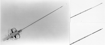
In all our cases, we have used the cutting needle Tru Core (med Tech Tru-Core, Gainsville, FL, USA) (Figure 1) as an initial invasive diagnostic procedure for mediastinal mass patients.
Methods
We carried out a retrospective analysis of transthoracic puncture with cutting needle, oriented, but not guided by thoracic computerized tomography, in 22 patients in the period from 1999 to 2002.
A procedure is guided by computerized tomography when is performed together with the biopsy, guiding and orienting the needle position. The transthoracic biopsy with cutting needle is oriented by the computerized tomography when we use an already performed thoracic computerized tomography to guide the positioning of the needle.
The group of patients was composed of 15 women and 7 men, with ages varying from 8 to 79 years, and the median age was 40 years.
The lesions were radiologically presented as solid and homogenous in all cases. The lesions diameters varied from 3 to 10 cm and in 77% of the cases, the tumors diameter was larger than 4 cm. All lesions were located at less than 2 cm under the skin. The tumor was in the anterior mediastinum in 18 patients (91%) and in the posterior mediastinum in four (9%).
All biopsies were performed in the ambulatory procedure room of the Thoracic Surgery Department, without sedation and under skin anesthesia and needle course with 5 mL of lidocaine at 2% with no vasoconstrictor, and in the ambulatory.
Transthoracic biopsies were carried out with an 18-gauge needle, as previously described. This needle was designed to remove tissue from the lesion for a histopathological study by means of a mechanism that triggers a small cutting canulla, capable of removing adequate fragments for the histopathological exam inside a 1.7 cm stylet (8).
An average of two biopsies were performed in each patient, and additional material was collected whenever necessary. After the biopsy, the patient was submitted to a thoracic X-ray, and no cases of pneumothorax, nor cases of intra-thoracic bleeding, nor of blood in the sputum were observed.
The biopsy results were classified as sufficient and insufficient, based on the presence or absence of neoplastic cells in the samples collected by transthoracic biopsy with cutting needle. The material was considered insufficient when it did not represent the pathological lesion necrotic material, blood and was analyzed separately.
Results
Twenty two patients underwent transthoracic biopsy of mediastinal masses with cutting needle during the study period.
The diagnosis was defined by this procedure in 18 (82%) of the patients: eight (44%) lymphomas; five (28%) thymomas; two (11%) thymus carcinomas; one (6%) metastatic adenocarcinoma; one (6%) primitive neuroectodermic tumor; and one (6%) plasmocytoma.
In the four patients (18%) whose material was insufficient for the diagnosis, this was achieved by surgical biopsy: two (50%) lymphomas; one (25%) thymus carcinoma and one (25%), dysgerminoma.
Discussion
Mediastinal tumors are traditionally diagnosed by mediatinoscopy, thoracoscopy or exploratory thoracotomy. These procedures require general anesthesia and hospital admission. The purpose of our study was to evaluate the clinical usefulness of transthoracic biopsy with cutting needle to diagnose these tumors, and to determine whether this could be an initial diagnostic procedure in such cases.
Most of the tumors were located in the anterior mediastinum. The most common pathologies were lymphoproliferative neoplasias, epithelial neoplasias, and thymomas. These findings are similar to those reported in the literature (3-7,9).
To know the nature of the mediastinal tumor is essential for the correct therapeutic planning. For instance, thymomas are primarily treated with surgery, whereas lymphomas are treated with chemotherapy associated or not to radiotherapy. Patients with metastatic disease need this definition, for subsequent treatment depends on it.
Transthoracic biopsy with cutting needle showed high specificity in our group of patients, being positive in 82% of them.
Pneumothorax is the most common complication of the cutting needle transthoracic biopsy reported in the literature; however, we did not have a single case in our group of patients (8,10). The absence of pneumothorax in our patients can probably be explained by the superficial location of the lesions all located at less than 2 cm of the thoracic wall. Other complications reported in the literature, such as hemoptysis (1.6%) and pain (3.2%), were not observed in our group (11,12). Many mediastinal diseases are treated clinically, such as lymphomas, or are clearly not resectable, such as metastatic neoplasias; therefore, the hystopathological diagnosis is obviously essential for these patients.
Based on our experience, we believe that transthoracic biopsy with cutting needle is a safe procedure and easy to perform, defining the diagnostic in most mediastinum malignant tumors. It can often replace extensive surgical procedures, such as thoracoscopy or thoracotomy, which require general anesthesia and hospital admission, and are associated with higher morbidity and high cost.
Conclusion
Because this procedure is easy to perform and safe, with the least complications and high diagnostic yield, we recommend the transthoracic biopsy with cutting needle as an initial diagnostic method for the evaluation of patients with mediastinal tumors, especially those which are not resectable at the thoracic computerized tomography.
Received for publication on 1/14/03
Approved after revision on 3/6/03.
- 1. Adler OB, Rosenberg A, Peleg H. Fine needle aspiration biopsy of mediastinal masses: evaluation of 136 experiences. Am J Radiol 1983; 140:893-6.
- 2. Weisbrod GL, Lyons DJ, Tao LC, Chamberlain DW. Percutaneous fine needle apiration biopsy of mediastinal lesions. Am J Radiol 1984;143: 525-9.
- 3. Herman SJ, Holub RV, Weisbrod GL, Chamberlain DW. Anterior mediastinal masses: utility of thoracic needle biopsy. Radiology 1991; 180:167-70.
- 4. Quinn SF, Demlow T, Dunkley B. Temno biopsy needle: evaluation of efficacy and safety in 165 biopsy procedures. AJR Am J Roentgenol 1992;158:641-3.
- 5. Bocking A, Klose KC, Kyll HJ, Hauptmann S. Cytologic versus histologic evaluation of needle biopsy of the lung, hilum, and mediastinum. Acta Cytol 1995;35:463-71.
- 6. Ben Yehuda D, Polliack A, Dkon E, Sherman Y, Bebenshart P, Lotan H, et al. Image-guided core needle biopsy in malignant lymphoma. Experience with 100 patients that suggests the technique is reliable. J Clin Oncol 1996;14:2431-4.
- 7. Klein JS, Salomon G, Stewart EA. Transthoracic needle biopsy with coaxially placed 20-gauge automated cutting needle: results in 122 patients. Klein Radiol 1996;198:715-20.
- 8. Stevens GM, Jackman RJ. Outpatient needle biopsy of the lungs: its safety and utility. Radiology 1984;151:301-3.
- 9. Morrisey B, Adams H, Gibbs AR, Crane MD. Percutaneous needle biopsy of the mediastinum. Thorax 1993;48:632-7.
- 10. Conces DS, Schwenk GR, Doering PR, Glant MD. Thoracic needle biopsy. Chest 1987;91:813-6.
- 11. Lalli AF, McCormack LJ, Zelch M, Reich NE, Belovich D. Aspiration biopsy of chest lesions. Radiology 1978;127:35-40.
- 12. Crosby JH, Hager B, Hoeg K. Transthoracic fine needle aspiration. Cancer 1985;56:3504-7.
Publication Dates
-
Publication in this collection
24 Sept 2003 -
Date of issue
June 2003
History
-
Accepted
06 Mar 2003 -
Received
14 Jan 2003