Abstracts
OBJECTIVE:
Preoperative estimation of the length and diameter of the semitendinosus (ST) and gracilis (G) tendons can assist surgeons and allow them to have the opportunity to choose alternative grafts. The aim of this study was to investigate whether anthropometric measurements such as height, weight and body mass index (BMI) or the patient's age and sex have any correlation with the thickness and the length of ST and G tendons.
METHODS:
Data were gathered from 64 patients who underwent the surgical procedure of anterior cruciate ligament reconstruction using the tendons of the ST and G muscles as grafts, between June 2012 and August 2013. Variables such as age, sex, weight, height, body mass index (BMI) and length and diameter of the tendons of the ST and G muscles were analyzed.
RESULTS:
There was a positive correlation between the height and total diameter of the quadruple graft (r = 0.254; p = 0.043), total length of the ST tendon (r = 0.450; p < 0.01), diameter of the double ST (r = 0.270; p = 0.031), triple ST (r = 0.347; p = 0.005), length of G tendon (r = 0.249; p = 0.047) and diameter of the double-G (r = 0.258; p = 0.039). However, age (r = -0.015; p = 0.908), weight (r = 0.165; p = 0.193) and body mass index (r = 0.012; p = 0.926) showed no correlation.
CONCLUSION:
Our results show that age, weight and BMI did not correlate with the diameter and length of the graft, while the height had a positive correlation with the total length of the flexor tendons and the diameter of the graft from the flexors (ST and G).
Anterior cruciate ligament; Anthropometry; Transplantation autologous; Tendons
OBJETIVO:
A estimativa pré-operatória do comprimento e do diâmetro dos tendões semitendinoso (ST) e grácil (G) pode auxiliar e permitir que os cirurgiões tenham a oportunidade de escolher opções de enxerto. O objetivo deste estudo foi pesquisar se existe correlação entre as medidas antropométricas, como altura, peso, índice de massa corpórea (IMC), idade e sexo do paciente, com a espessura e o comprimento dos tendões ST e G.
MÉTODOS:
Entre junho de 2012 e agosto de 2013, foram coletados os dados de 64 pacientes que se submeteram ao procedimento cirúrgico de reconstrução do ligamento cruzado anterior em que se usaram como enxerto os tendões dos músculos ST e G. Foram analisadas variáveis como idade, sexo, peso, altura e índice de massa corporal (IMC), comprimento e diâmetro dos tendões dos músculos ST e G.
RESULTADOS:
Houve uma correlação positiva entre altura e diâmetro total do enxerto de quádruplo (r = 0,254 p = 0,043), comprimento total do tendão ST (r = 0,450, p < 0,01), diâmetro do ST duplo (r = 0,270 p = 0,031), ST triplo (r = 0,347 p = 0,005), comprimento do tendão G (r = 0,249 p = 0,047) e diâmetro do G duplo (r = 0,258 p = 0,039). No entanto, idade (r = -0,015 p = 0,908), peso (r = 0,165 p = 0,193) e índice de massa corporal (r = 0,012 p = 0,926) não apresentaram correlação.
CONCLUSÃO:
Nossos resultados mostram que idade, peso e IMC não se correlacionaram com o diâmetro e comprimento do enxerto, enquanto a altura tinha uma correlação positiva com o comprimento total dos tendões flexores e com o diâmetro do enxerto dos flexores (ST e G).
Ligamento cruzado anterior; Antropometria; Transplante autólogo; Tendões
Introduction
The growing trend toward practicing sports, together with greater complexity of sports movements has increasingly been causing knee joint injuries to appear. The anterior cruciate ligament (ACL) is among most commonly affected ligaments, and surgical treatment is chosen for most patients who present pain and instability.11. Dunn WR, Lyman S, Lincoln AE, Amoroso PJ, Wickiewicz T, Marx RG. The effect of anterior cruciate ligament reconstruction on the risk of knee reinjury. Am J Sports Med. 2004;32(8):1906-14. The autologous grafts most frequently used in ACL reconstructions are the patellar, gracilis (G), semitendinosus (ST) and quadriceps tendons. Each technique has its adherents and indications, and selecting the graft depends on many factors, including the surgeon's preference and the patient's age and level of activity. The surgical technique using the tendons of the ST and G muscles as grafts presents results that are similar to those from the patellar tendon technique and enable smoother and less painful rehabilitation. The main disadvantages of this technique include the individual variability in length and thickness of the graft from the tendons and the potential complications during graft harvesting.22. Prodromos CC, Han YS, Keller BL, Bolyard RJ. Stability results of hamstring anterior cruciate ligament reconstruction at 2- to 8 -year follow- up. Arthroscopy. 2005;21(2):138-46., 33. Biau DJ, Tournoux C, Katsahian S, Schranz PJ, Nizard RS. Bone-patellar tendon-bone autografts versus hamstring autografts for reconstruction of anterior cruciate ligament: meta- analysis. BMJ. 2006;332(7548):995-1001.,44. Aglietti P, Giron F, Buzzi R, Biddau F, Sasso F. Anterior cruciate ligament reconstruction: bone-patellar tendon-bone compared with double semitendinosus and gracilis tendon grafts. A prospective, randomized clinical trial. J Bone Joint Surg Am. 2004;86-A(10):2143-55.and55. Fu FH, Bennett CH, Lattermann C, Ma CB. Current trends in anterior cruciate ligament reconstruction. Part 1: biology and biomechanics of reconstruction. Am J Sports Med. 1999;27(6):821-30. After the tendons have been removed from the semitendinosus and gracilis muscles, and before the graft has been constructed, surgeons are faced with many possibilities for the final configuration of the graft. These include using the semitendinosus tendon in double, triple or quadruple form alone, or in double form in association with the tendon of the gracilis muscle, also in double form (thus making a quadruple graft), or even other combinations. Preoperative estimation of the length and diameter of the ST and G tendons may help and allow surgeons to have the opportunity to choose another graft option if the tendon that might be harvested does not meet the needs of a given patient. However, no method capable of precisely determining the length and diameter of ST and G tendons before ACL reconstruction surgery is performed has yet been described in the literature.
The objective of this study was to investigate whether anthropometric measurements such as the patient's height, weight, body mass index (BMI), age and sex would have any correlation with the thickness and length of the hamstring tendons (ST and G).
Materials and methods
This sample comprised patients who attended consultations with one of the three surgeons in our team who are accredited by the Brazilian Knee Surgery Society (SBCJ). For all of these patients, ACL reconstruction surgery was indicted. Out of 200 ACL reconstruction operations performed between June 2012 and August 2013, data were gathered from 64 patients who underwent ACL reconstruction surgery in which the graft used was the tendons of the ST and G muscles. The operations were performed in three private-sector hospitals in Porto Alegre, by three surgeons who were specialists in knee surgery and members of the Brazilian Knee Surgery Society (SBCJ). Individuals who had undergone previous surgery on the same knee that was operated on this occasion, those with other associated ligament injuries, those in whom the gracilis tendon was not used in constructing the graft and those who did not agree to participate in the study or did not sign the informed consent statement were excluded from the sample. The study was conducted among patients who were evaluated through anamnesis, physical examination and magnetic resonance imaging, and for whom there was an indication for surgical reconstruction of the ACL in which the ST and G tendons were used to construct the graft. This investigation did not change the routine of steps to be followed during the surgical procedure indicated. The data-gathering did not add risk to the procedure. This study was approved by our university and by the research ethics committees of the institutions involved.
The following variables were analyzed: age, sex, weight, height, body mass index (BMI), length of the tendon of the semitendinosus muscle, length of the tendon of the gracilis muscle, diameter of the tendon of the semitendinosus muscle when folded in the middle (double) and in three parts (triple), diameter of the tendon of the gracilis muscle when folded in the middle (double) and the diameter of the tendons of the semitendinosus and gracilis muscles when folded in the middle and grouped (quadruple).
Firstly, demographic data (age and sex) and anthropometric information (weight and height) reported at the time of the surgery were obtained and noted in the medical file. Soon afterwards, during the surgical procedure, measurements on the tendons of the semitendinosus and gracilis muscles that were used for constructing the grafts were made.
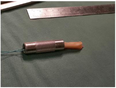
The technique for obtaining the grafts during the ACL reconstruction surgery consisted of an oblique anteromedial incision in the proximal tibia, at the level of the insertion of the ST and G muscles. Following this, the tendon of the ST muscle was dissected and was then deinserted against the bone and removed using a graft extractor. The same procedure was repeated in relation to the tendon of the gracilis muscle. After the tendons had been prepared through removal of the muscle layer, the length (cm) and diameter (MM) of the grafts were obtained using a ruler and measuring cylinder that had previously been sterilized. The diameters of the semitendinosus and gracilis grafts were measured using a set of cylindrical tubes ranging in dimension from 6 to 12 mm, in steps of 0.5 mm between cylinders. Both of the tendons were measured. In the case of the tendon derived from the semitendinosus muscle, its total length and diameter were measured twice. Firstly, it was folded in the middle (double) and then it was folded into three parts (triple). The total length and diameter of the graft derived from the gracilis muscle were measured with the tendon folded in the middle (double). Lastly, the length and diameter of the two tendons folded in the middle and grouped together (quadruple) were measured. The measurements were made without suturing thread, which might have hindered obtaining the real dimensions of the tendons (Fig. 1 and Fig. 2).
The data gathered were analyzed through descriptive and analytical statistics. IN the descriptive phase, means and standard deviations were used. In the analytical phase, the variables were compared in relation to the different lengths and diameters of the tendons of the semitendinosus and gracilis muscles. The t test for independent samples was used to identify differences in the clinical and intraoperative variables. Pearson's correlation coefficient (r) and simple linear regression were used to determine the relationship between the result variables (diameter of the quadruple graft and the lengths of the semitendinosus and gracilis tendons) and the predictive variables (age, sex, height, weight and BMI). Differences were considered to be significant when P values were less than 0.05. The analysis was done using the SPSS software, version 17.0.
Results
Among the 64 individuals analyzed between June 2012 and August 2013, 60 (94%) were male and four (6%) were female. The patients' mean age was 31.78 ± 8.26 years (range: 15-48 years); among the females, it was 32 ± 13.49 years, and among the males, it was 31.76 ± 7.97 years. The mean height was 1.77 ± 0.08 cm (range: 1.52-1.96 cm); mean weight 82.43 ± 12.89 kg (range: 56-115 kg); and mean BMI 26.14 ± 3.74 (range: 21.60-37.55). The mean tendon lengths measured during the operation were 28.75 ± 2.91 cm (range: 23-36 cm) for the semitendinosus and 25.28 ± 3.81 cm (range: 14-34 cm) for the gracilis. The means tendon thicknesses (diameters) were 6.24 ± 0.75 mm (range: 4.5-8 mm) for the double semitendinosus; 7.32 ± 0.76 mm (range: 6-9 mm) for the triple semitendinosus; and 6.24 ± 0.75 mm (range: 4.5-8 mm) for the double gracilis. The mean total diameter of the quadruple tendon graft (ST and G) was 8.03 ± 0.72 mm (range: 7-10 mm). During the study period, we did not obtain any quadruple flexor grafts smaller than 7 mm or larger than 10 mm (Table 1).
In total, 75% of the patients had quadruple flexor grafts of diameter 7-8 mm, while 25% of the grafts were larger than 8 mm. The frequencies of different diameters of quadruple flexor grafts were as follows: 7 mm (22%), 7.5 mm (3%), 8 mm (50%), 9 mm (23.5%) and 10 mm (1.5%) (Fig. 3).
There were positive correlations between height and the total diameter of the quadruple graft from the flexor tendons (r = 0.254; p = 0.043); total length of the semitendinosus tendon (r = 0.450; p < 0.01); diameter of the double semitendinosus (r = 0.270; p = 0.031); diameter of the triple semitendinosus (r = 0.347; p = 0.005); total length of the gracilis tendon (r = 0.249; p = 0.047); and diameter of the double gracilis (r = 0.258; p = 0.039). However, age (r = -0.015; p = 0.908), weight (r = 0.165; p = 0.193) and body mass index (r = 0.012; p = 0.926) did not present any correlation with the total diameter of the quadruple graft or with the lengths of the semitendinosus and gracilis tendons (Table 2).
Discussion
Use of flexor tendon grafts has become an increasingly popular choice among orthopedic surgeons for ACL reconstructions.66. Gianotti SM, Marshall SW, Hume PA, Bunt L. Incidence of anterior cruciate ligament injury and other knee ligament injuries: a national population-based study. J Sci Med Sport. 2009;12(6):622-7. Anatomical studies have demonstrated that the mean diameter of the normal ACL is approximately 11 mm, with a range from 6 to 12 mm. Although the parameters for acceptable graft size that are necessary in order to achieve a satisfactory result after ACL reconstruction have not been clearly defined, a diameter of at least 7 mm has traditionally been recommended.77. Hamada M, Shino K, Horibe S, Mitsuoka T, Toritsuka Y, Nakamura N. Changes in cross-sectional area of hamstring anterior cruciate ligament grafts as a function of time following transplantation. Arthroscopy. 2005;21(8): 917-22.,88. Grood ES, Walz-Hasselfeld KA, Holden JP, Noyes FR, Levy MS, Butler DL, et al. The correlation between anterior-posterior translation and cross-sectional area of anterior cruciate ligament reconstructions. J Orthop Res. 1992;10(6):878-85.and99. Scott WN, Insall JN. Injuries of the knee. In: Rockwood DP Jr, Green DP, Bucholz RW, editors. Rockwood and Green's fractures in adults. Philadelphia: Lippincott Williams & Wilkins; 1996. p. 1799-816. However, in a recent study, greater incidence of ACL reconstruction failure was shown with grafts smaller than 8 mm.1010. Magnussen RA, Lawrence JT, West RL, Toth AP, Taylor DC, Garrett WE. Graft size and patient age are predictors of early revision after anterior cruciate ligament reconstruction with hamstring autograft. Arthroscopy. 2012;28(4):526-31. Studies on animal models that evaluated the influence of graft diameter on graft strength and resistance have demonstrated that in grafts smaller than 8 mm, failure due to tearing of the tendon material occurs, while in grafts larger than 10 mm, failure occurs at the fixation screws, due to traction.1111. Teli M, Chiodini F, Sottocasa R, Villa T. Influence of the diameters of tendon graft and bone tunnel in hamstring ACL reconstruction. A bovine model. Chir Organi Mov. 2005;90(3):281-5. Thus, a capacity to predict which patients present greater risk of having hamstring tendons of insufficient diameter, before the operation, may be beneficial for surgical planning, thereby avoiding disagreeable intercurrences during the operation.77. Hamada M, Shino K, Horibe S, Mitsuoka T, Toritsuka Y, Nakamura N. Changes in cross-sectional area of hamstring anterior cruciate ligament grafts as a function of time following transplantation. Arthroscopy. 2005;21(8): 917-22., 88. Grood ES, Walz-Hasselfeld KA, Holden JP, Noyes FR, Levy MS, Butler DL, et al. The correlation between anterior-posterior translation and cross-sectional area of anterior cruciate ligament reconstructions. J Orthop Res. 1992;10(6):878-85.,99. Scott WN, Insall JN. Injuries of the knee. In: Rockwood DP Jr, Green DP, Bucholz RW, editors. Rockwood and Green's fractures in adults. Philadelphia: Lippincott Williams & Wilkins; 1996. p. 1799-816.and1010. Magnussen RA, Lawrence JT, West RL, Toth AP, Taylor DC, Garrett WE. Graft size and patient age are predictors of early revision after anterior cruciate ligament reconstruction with hamstring autograft. Arthroscopy. 2012;28(4):526-31.
In our patients, we used a variety of graft combinations, such as triple ST alone, quadruple ST alone, double ST with double G, triple ST with double G and triple ST with triple G. The aim was always to seek grafts of diameter greater than 8 mm, of adequate length.
The capacity to predict the diameter and length of the graft may be useful for certain surgical techniques and may provide options for the surgeon. Through a biomechanical study, Hamner et al.1212. Hamner DL, Brown CH, Steiner ME, Hecker AT, Hayes WC. Hamstring tendon grafts for reconstruction of the anterior cruciate ligament: biomechanical evaluation of the multiple strands and tensioning techniques. J Bone Joint Surg Am. 1999;81(4):549-57. demonstrated that the resistance, stiffness and biomechanical properties of the graft tendon are affected by its diameter. However, the results from the few studies in the literature correlating patients' anthropometric data with the length and diameter of flexor grafts continue to be contradictory.
In our study, we investigated the predictive value of simple anthropometric measurements such as height, weight, BMI and age in relation to the lengths of the semitendinosus and gracilis flexor tendons and the diameter of the quadruple graft from the flexors. Our results showed that age, weight and BMI did not correlate with the length or diameter of the graft, while height had a positive correlation with the total length of the grafts from the semitendinosus (r = 0.450; p < 0.01) and gracilis (r = 0.249; p = 0.047), and also with the diameter of the quadruple graft from the flexors (r = 0.254; p = 0.043). A deeper analysis using step-by-step regression revealed that height was the clinical parameter of greatest importance for predicting the thickness of the graft.
Tuman et al.1313. Tuman JM, Diduch DR, Rubino LJ, Baumfeld JA, Nguyen HS, Hart JM. Predictors for hamstring graft diameter in anterior cruciate ligament reconstruction. Am J Sports Med. 2007;35(11):1945-9. analyzed 106 patients who underwent ACL reconstruction and reported that among the anthropometric data analyzed in their study, such as height, weight, sex and age, height was the best predictor for the diameter of grafts from the hamstrings in both sexes, and particularly among women. From their analysis, they concluded that women presented graft diameters that were significantly smaller than those of men, and that in patients whose height was less than 147 cm, it was likely that the graft would be insufficient, with a diameter of less than 7 mm.
Ma et al.1414. Ma CB, Keifa E, Dunn W, Fu FH, Harner CD. Can pre- operative measures predict quadruple hamstring graft diameter? Knee. 2010;17(1):81-3. retrospectively analyzed preoperative anthropometric data from 536 patients. Their results showed that height and sex were significant predictors for the diameter of flexor tendon grafts. Male patients had grafts that were significantly larger than those of females. It was also demonstrated that height was a specific indicator solely for the men, without any significant results for the women.
In a study on 119 patients who underwent ACL reconstruction, Schwartzberg et al.1515. Schwartzberg R, Burkhart B, Lariviere C. Prediction of hamstring tendon autograft diameter and length for anterior cruciate ligament reconstruction. Am J Orthop (Belle Mead NJ). 2008;37(3):157-9. reported that there was a strong correlation between leg length and the length of the flexor tendons used as grafts (r = 0.73; p < 0.001). In the same study, weight (r = 0.51; p < 0.001) and leg length (r = 0.42; p < 0.001) only showed moderate correlations with graft diameter. All the other correlations were weak in relation to height. Weight measurements and age did not show any relationship with the length and diameter of the flexor tendons, and these findings were concordant with the results reported by Schwartzberg et al.1515. Schwartzberg R, Burkhart B, Lariviere C. Prediction of hamstring tendon autograft diameter and length for anterior cruciate ligament reconstruction. Am J Orthop (Belle Mead NJ). 2008;37(3):157-9.
In analyses by Treme et al.1616. Treme G, Diduch DR, Billante MJ, Miller MD, Hart JM. Hamstring graft size prediction: a prospective clinical evaluation. Am J Sports Med. 2008;36(11):2204-9. on the length and diameter of the semitendinosus and gracilis tendons of 50 patients who underwent ACL reconstruction, a strong correlation was found between the length of the graft and the patient's height and lower-limb length. The diameter of the graft was correlated with the patient's weight and thigh circumference.
Although our study did not confirm all the positive correlations of other studies,1212. Hamner DL, Brown CH, Steiner ME, Hecker AT, Hayes WC. Hamstring tendon grafts for reconstruction of the anterior cruciate ligament: biomechanical evaluation of the multiple strands and tensioning techniques. J Bone Joint Surg Am. 1999;81(4):549-57., 1313. Tuman JM, Diduch DR, Rubino LJ, Baumfeld JA, Nguyen HS, Hart JM. Predictors for hamstring graft diameter in anterior cruciate ligament reconstruction. Am J Sports Med. 2007;35(11):1945-9., 1414. Ma CB, Keifa E, Dunn W, Fu FH, Harner CD. Can pre- operative measures predict quadruple hamstring graft diameter? Knee. 2010;17(1):81-3., 1515. Schwartzberg R, Burkhart B, Lariviere C. Prediction of hamstring tendon autograft diameter and length for anterior cruciate ligament reconstruction. Am J Orthop (Belle Mead NJ). 2008;37(3):157-9.,1616. Treme G, Diduch DR, Billante MJ, Miller MD, Hart JM. Hamstring graft size prediction: a prospective clinical evaluation. Am J Sports Med. 2008;36(11):2204-9.and1717. Pichler W, Tesch NP, Schwantzer G, Fronhöfer G, Boldin C, Hausleitner L, et al. Differences in length and cross-section of semitendinosus and gracilis tendons and their effect on anterior cruciate ligament reconstruction: a cadaver study. J Bone Joint Surg Br. 2008;90(4):516-9. our results favored the notion that individual anthropometric variables such as height may be predictors for the length and diameter of the flexor tendons (ST and G) that are used in ACL reconstruction. Our results also showed that height had a positive correlation with the total length of the semitendinosus and gracilis tendons and with the diameters of double ST, triple ST, double G and quadruple grafts, and they were also concordant with the findings of Tuman et al.1313. Tuman JM, Diduch DR, Rubino LJ, Baumfeld JA, Nguyen HS, Hart JM. Predictors for hamstring graft diameter in anterior cruciate ligament reconstruction. Am J Sports Med. 2007;35(11):1945-9. Moreover, our results demonstrated that height was the most important variable, especially among women, whereas Schwatzberg et al.1515. Schwartzberg R, Burkhart B, Lariviere C. Prediction of hamstring tendon autograft diameter and length for anterior cruciate ligament reconstruction. Am J Orthop (Belle Mead NJ). 2008;37(3):157-9. found that height had a weak correlation with thigh diameter and that weight and lower-limb diameter had a moderate correlation with graft diameter.
There is limited evidence in the literature correlating the patient's fitness and physical activity level with the length and diameter of the flexor tendons. Treme et al.1616. Treme G, Diduch DR, Billante MJ, Miller MD, Hart JM. Hamstring graft size prediction: a prospective clinical evaluation. Am J Sports Med. 2008;36(11):2204-9. reported that they had not found any correlation between the diameter of the flexor grafts and the patient's sports and activity levels. Similar results were recorded by Pichler et al.1717. Pichler W, Tesch NP, Schwantzer G, Fronhöfer G, Boldin C, Hausleitner L, et al. Differences in length and cross-section of semitendinosus and gracilis tendons and their effect on anterior cruciate ligament reconstruction: a cadaver study. J Bone Joint Surg Br. 2008;90(4):516-9. in their study on cadavers.
Other studies in the literature have used radiological imaging examinations such as computed tomography and magnetic resonance imaging (MRI) in an attempt to predict the length and diameter of flexor grafts before the operation. Although some studies have shown good correlations between measurements of cross-sectional area and tendon thickness, others have not shown any correlation.1818. Wernecke G, Harris IA, Houang MT, Seeto BG, Chen DB, MacDessi SJ. Using magnetic resonance imaging to predict adequate graft diameters for autologous hamstring double-bundle anterior cruciate ligament reconstruction. Arthroscopy. 2011;27(8):1055-9.,1919. Yasumoto M, Deie M, Sunagawa T, Adachi N, Kobayashi K, Ochi M. Predictive value of preoperative 3-dimensional computer tomography measurement of semitendinosus tendon harvested for anterior cruciate ligament reconstruction. Arthroscopy. 2006;22(3):259-64.and2020. Hamada M, Shino K, Mitsuoka T, Abe N, Horibe S. Cross-sectional area measurement of the semitendinosus tendon for anterior cruciate ligament reconstruction. Arthroscopy. 1998;14(7):696-701. Nevertheless, the lack of standardization in relation to the level at which measurements are made and regarding the trustworthiness and precision of the examinations gives rise to limitations that make the results obtained questionable. Yasumoto et al.1919. Yasumoto M, Deie M, Sunagawa T, Adachi N, Kobayashi K, Ochi M. Predictive value of preoperative 3-dimensional computer tomography measurement of semitendinosus tendon harvested for anterior cruciate ligament reconstruction. Arthroscopy. 2006;22(3):259-64. analyzed the preoperative length of the semitendinosus tendon using 3D computed tomography and were able to predict the length of the tendon. However, they did not find any relationship between the images and the diameters. The study by Hamada et al.2020. Hamada M, Shino K, Mitsuoka T, Abe N, Horibe S. Cross-sectional area measurement of the semitendinosus tendon for anterior cruciate ligament reconstruction. Arthroscopy. 1998;14(7):696-701. showed that MRI was impractical for evaluating the sizes of the tendons, given that a long sweep time would be needed and the cost is high.
Conclusion
The capacity to precisely predict the length and diameter of the hamstring tendons (ST and G) used in ACL reconstructions continues to be an important factor in decision-making and in choosing the best surgical technique and the appropriate graft. Our results showed that age, weight and BMI did not correlate with the diameter and length of the graft, while height showed a positive correlation with the total length of the flexor tendons and with the diameter of the graft from the flexors (ST and G).
Acknowledgements
Special thanks to the physicians Luiz Antonio Silveira Simões Pires and Guilherme Isler Pereira, who contributed equally to data-gathering and conducting this study.
References
-
1Dunn WR, Lyman S, Lincoln AE, Amoroso PJ, Wickiewicz T, Marx RG. The effect of anterior cruciate ligament reconstruction on the risk of knee reinjury. Am J Sports Med. 2004;32(8):1906-14.
-
2Prodromos CC, Han YS, Keller BL, Bolyard RJ. Stability results of hamstring anterior cruciate ligament reconstruction at 2- to 8 -year follow- up. Arthroscopy. 2005;21(2):138-46.
-
3Biau DJ, Tournoux C, Katsahian S, Schranz PJ, Nizard RS. Bone-patellar tendon-bone autografts versus hamstring autografts for reconstruction of anterior cruciate ligament: meta- analysis. BMJ. 2006;332(7548):995-1001.
-
4Aglietti P, Giron F, Buzzi R, Biddau F, Sasso F. Anterior cruciate ligament reconstruction: bone-patellar tendon-bone compared with double semitendinosus and gracilis tendon grafts. A prospective, randomized clinical trial. J Bone Joint Surg Am. 2004;86-A(10):2143-55.
-
5Fu FH, Bennett CH, Lattermann C, Ma CB. Current trends in anterior cruciate ligament reconstruction. Part 1: biology and biomechanics of reconstruction. Am J Sports Med. 1999;27(6):821-30.
-
6Gianotti SM, Marshall SW, Hume PA, Bunt L. Incidence of anterior cruciate ligament injury and other knee ligament injuries: a national population-based study. J Sci Med Sport. 2009;12(6):622-7.
-
7Hamada M, Shino K, Horibe S, Mitsuoka T, Toritsuka Y, Nakamura N. Changes in cross-sectional area of hamstring anterior cruciate ligament grafts as a function of time following transplantation. Arthroscopy. 2005;21(8): 917-22.
-
8Grood ES, Walz-Hasselfeld KA, Holden JP, Noyes FR, Levy MS, Butler DL, et al. The correlation between anterior-posterior translation and cross-sectional area of anterior cruciate ligament reconstructions. J Orthop Res. 1992;10(6):878-85.
-
9Scott WN, Insall JN. Injuries of the knee. In: Rockwood DP Jr, Green DP, Bucholz RW, editors. Rockwood and Green's fractures in adults. Philadelphia: Lippincott Williams & Wilkins; 1996. p. 1799-816.
-
10Magnussen RA, Lawrence JT, West RL, Toth AP, Taylor DC, Garrett WE. Graft size and patient age are predictors of early revision after anterior cruciate ligament reconstruction with hamstring autograft. Arthroscopy. 2012;28(4):526-31.
-
11Teli M, Chiodini F, Sottocasa R, Villa T. Influence of the diameters of tendon graft and bone tunnel in hamstring ACL reconstruction. A bovine model. Chir Organi Mov. 2005;90(3):281-5.
-
12Hamner DL, Brown CH, Steiner ME, Hecker AT, Hayes WC. Hamstring tendon grafts for reconstruction of the anterior cruciate ligament: biomechanical evaluation of the multiple strands and tensioning techniques. J Bone Joint Surg Am. 1999;81(4):549-57.
-
13Tuman JM, Diduch DR, Rubino LJ, Baumfeld JA, Nguyen HS, Hart JM. Predictors for hamstring graft diameter in anterior cruciate ligament reconstruction. Am J Sports Med. 2007;35(11):1945-9.
-
14Ma CB, Keifa E, Dunn W, Fu FH, Harner CD. Can pre- operative measures predict quadruple hamstring graft diameter? Knee. 2010;17(1):81-3.
-
15Schwartzberg R, Burkhart B, Lariviere C. Prediction of hamstring tendon autograft diameter and length for anterior cruciate ligament reconstruction. Am J Orthop (Belle Mead NJ). 2008;37(3):157-9.
-
16Treme G, Diduch DR, Billante MJ, Miller MD, Hart JM. Hamstring graft size prediction: a prospective clinical evaluation. Am J Sports Med. 2008;36(11):2204-9.
-
17Pichler W, Tesch NP, Schwantzer G, Fronhöfer G, Boldin C, Hausleitner L, et al. Differences in length and cross-section of semitendinosus and gracilis tendons and their effect on anterior cruciate ligament reconstruction: a cadaver study. J Bone Joint Surg Br. 2008;90(4):516-9.
-
18Wernecke G, Harris IA, Houang MT, Seeto BG, Chen DB, MacDessi SJ. Using magnetic resonance imaging to predict adequate graft diameters for autologous hamstring double-bundle anterior cruciate ligament reconstruction. Arthroscopy. 2011;27(8):1055-9.
-
19Yasumoto M, Deie M, Sunagawa T, Adachi N, Kobayashi K, Ochi M. Predictive value of preoperative 3-dimensional computer tomography measurement of semitendinosus tendon harvested for anterior cruciate ligament reconstruction. Arthroscopy. 2006;22(3):259-64.
-
20Hamada M, Shino K, Mitsuoka T, Abe N, Horibe S. Cross-sectional area measurement of the semitendinosus tendon for anterior cruciate ligament reconstruction. Arthroscopy. 1998;14(7):696-701.
-
☆
Work performed at Hospital São Lucas, Pontifícia Universidade Católica do Rio Grande do Sul (PUC-RS), Hospital Mãe de Deus and Hospital Divina Providência, Porto Alegre, RS, Brazil.
Publication Dates
-
Publication in this collection
Mar-Apr 2016
History
-
Received
01 Sept 2014 -
Accepted
05 May 2015