Abstract
Sulfated polysaccharides derived from seaweed have shown great potential for use in the development of new drugs. In this study, we observed that a low-molecular-weight sulfated polysaccharide from Caulerpa racemosa, termed CrSP, could interact with secretory phospholipase A2 (sPLA2) isolated from Crotalus durissus terrificus venom. When native sPLA2 (14 kDa) was incubated with CrSP, they formed a molecular complex (sPLA2:CrSP) with a molecular mass of 32 kDa, approximately. Size exclusion chromatography experiments suggested that CrSP formed a stable complex with sPLA2. We belived that sPLA2 and SPCr are involved an ionic interaction between negatively charged CrSP and the positively charged basic amino acid residues of sPLA2, because this interaction induced significant changes in sPLA2 enzymatic and pharmacological activities. CrSP caused a significant increase in sPLA2 enzymatic and bactericidal activity and increased its edematogenic effect. A pharmacological assay showed that the myotoxic activity of sPLA2:CrSP is unrelated to its enzymatic activity and that sPLA2:CrSP may have a practical application as a natural antibacterial agent for use in humans and commercially raised animals.
Crotalus durissus terrificus; edematogenic effect; myotoxic activity; seaweed; secretory; phospholipase A2; sulfated polysaccharides
Sulfated polysaccharide extracted of the green algae Caulerpa racemosa increase the enzymatic activity and paw edema induced by sPLA2 from Crotalus durissus terrificus venom
Camila L. PiresI; Selma D. RodriguesI; Daniel BristotI; Henrique Hessel GaetaI; Daniela de Oliveira ToyamaII; Wladimir Ronald Lobo FariasIII; Marcos Hikari ToyamaI
ILaboratório de Biologia Molecular e Peptídeos, Universidade Estadual Paulista, Campus do Litoral Paulista, Brazil,
IICentro de Ciências Biológicas e da Saúde, Universidade Presbiteriana Mackenzie, Brazil,
IIIDepartamento de Engenharia de Pesca, Centro de Ciências Agrárias, Universidade Federal do Ceará, Brazil
Correspondence Correspondence: Camila Lehnhardt Pires Laboratório de Biologia Molecular e Pepitideos UNESP, Campus do Litoral Paulista Praça Infante Dom Henrique s/nº, Parque Bitaru 11330-900 São Vicente-SP, Brazil lehnhardtpires@clp.unesp.br Tel. + 55 13 8809 7200
ABSTRACT
Sulfated polysaccharides derived from seaweed have shown great potential for use in the development of new drugs. In this study, we observed that a low-molecular-weight sulfated polysaccharide from Caulerpa racemosa, termed CrSP, could interact with secretory phospholipase A2 (sPLA2) isolated from Crotalus durissus terrificus venom. When native sPLA2 (14 kDa) was incubated with CrSP, they formed a molecular complex (sPLA2:CrSP) with a molecular mass of 32 kDa, approximately. Size exclusion chromatography experiments suggested that CrSP formed a stable complex with sPLA2. We belived that sPLA2 and SPCr are involved an ionic interaction between negatively charged CrSP and the positively charged basic amino acid residues of sPLA2, because this interaction induced significant changes in sPLA2 enzymatic and pharmacological activities. CrSP caused a significant increase in sPLA2 enzymatic and bactericidal activity and increased its edematogenic effect. A pharmacological assay showed that the myotoxic activity of sPLA2:CrSP is unrelated to its enzymatic activity and that sPLA2:CrSP may have a practical application as a natural antibacterial agent for use in humans and commercially raised animals.
Keywords:Crotalus durissus terrificus; edematogenic effect; myotoxic activity; seaweed; secretory; phospholipase A2; sulfated polysaccharides
Introduction
The natural products of seaweed and other marine organisms represent one of the new frontiers in the exploration for bioactive compounds. The sulfated polysaccharides (SP) found in marine seaweed are known to have many physiological and biological activities, including anticoagulant, anti-viral, anti-tumor, anti-inflammatory and antioxidant effects (Silva et al., 2011). Marine algae contain a high concentration of SP, which are heterogeneous and complex macromolecules that are important for algal physiology; these molecules perform ionic, mechanical and osmotic functions and are components of the extracellular matrix (Pomin & Mourão, 2008). Due to their relative abundance in algae, these compounds have been investigated for their therapeutic potential (Rodrigues et al., 2012). Caulerpa racemosa (Cr), a large, edible green alga, is widely distributed in tropical and subtropical areas of Brazil and other countries (Ji et al., 2008). C. racemosa contains SP with anticoagulant and antiviral activity, and recently it has been shown that SP fractions from C. racemosa have significant antitumor activity (Ji et al., 2008). However, additional biological activities have not been described for the SP that have been isolated from green algae of the genus Caulerpa.
There is evidence that heparin that has been chemically treated can interact with phospholipase A2 (PLA2). Dicciaani and colleagues (1991) showed that heparin binding to the N-terminal region of pancreatic PLA2 inhibits the interaction of the enzyme with a micellar substrate. Besides, the standard pharmaceutical dose of heparin might inhibit human class II secretory PLA2 (sPLA2) and regulate its biological effects (Dua & Cho, 1994). Heparin is also capable of modulating the pharmacological activity of venom-derived sPLA2 (Kini, 2005), and this modulation appears to involve conformational changes in the secondary structure of sPLA2 (Lin et al., 2000). Heparin is a complex polysaccharide that can be extracted from numerous animal sources, including pig intestines and cattle lungs. Extracted heparin contains several contaminants, including a heterogeneous group of oversulfated glycosaminoglycans (OSGAG), which may mediate multiple pathophysiologic responses (Ramacciotti et al., 2011). In addition, OSGAG-contaminated heparin can produce anaphylaxis through contact system activation and can also increase the risk of HIT and HIT-associated anaphylactoic reactions (Warkentin & Greinacher, 2009). Thus, the therapeutic use of heparin is limited due to its side effects, such as anaphylaxis and the risk of hemorrhage (Mourão & Pereira, 1999; Nader et al., 2001).
Algae-derived sulfated polysaccharides have been substituted for heparin for use as anticoagulants, although they are chemically distinct from heparin. Group II sPLA2 enzymes have been found at inflammatory sites in animal models, as well as in synovial fluid from patients with rheumatoid arthritis and other human inflammatory diseases, and a correlation has been observed between serum sPLA2 levels and disease state (Ramacciotti et al., 2011; Warkentin & Greinacher, 2009). Moreover, exogenous administration of PLA2, such as snake venom sPLA2, induces or exacerbates the inflammatory response in animals (Cirino, 1998; Fuentes et al., 2002). Structural analyses have revealed that snake venom sPLA2 has a similar molecular profile compared with human secretory PLA2, as well as a conserved catalytic site (Gil et al., 1997). The control and modulation of the structure and function of secretory phospholipase A2 are crucial for managing the pathophysiology of inflammatory disease. Due to the role of PLA2 in the inflammatory process, there is interest in identifying PLA2 inhibitors for therapeutic use. Sulfated polysaccharides from seaweeds, which have been used to control inflammation, are one such potential inhibitor. Our focus on SP from green algae distinguishes our work from that on the better-studied red and brown seaweeds. Because venom-derived sPLA2 has been characterized as a proinflammatory agent and is very similar to the human sPLA2, we investigated the effect of a highly purified sulfated polysaccharide fraction isolated from the green algae Caulerpa racemosa on the edematogenic activity of sPLA2 from Crotalus durissus terrificus venom. Ours is the first study to evaluate this and other biological and pharmacological activities induced by the interaction of this SP with the sPLA2 protein (Câmara et al., 2003; Lee et al., 1999).
Materials and Methods
Venom, animal use, and general reagents
Venom from Crotalus durissus terrificus was kindly donated by the Institute of Butantan (São Paulo, Brazil). Solvents, chemicals and reagents used in protein purification and characterization were of HPLC grade or higher and were acquired from Sigma-Aldrich Chemicals, Merck (USA), and Bio-Rad (USA). The female Swiss mice (22 g) used in the pharmacological assays were obtained from the Multidisciplinary Center of Biological Investigations (CEMIB-UNICAMP). All animal experiments were approved by the State University of Campinas Ethics Committee (São Paulo, Brazil) and the number is 2898-1. The seaweeds used to extraction was collected at Fortaleza city and the exsiccate is located in the Phycology Herbarium from Labomar - Ocean Sciense Institute at Ceará Federal University and the number is 2385.
Purification of sPLA2 from Crotalus durissus terrificus venom
Whole venom was fractionated as previously described by Oliveira and colleagues (2003). Dried venom (10 mg) was dissolved in Tris-HCL buffer (1 M, pH 7.0) and clarified by centrifugation (4,500 g, 1 min). The supernatant was injected onto a molecular exclusion HPLC column (Superdex 75, 1 × 60 cm, Pharmacia), and the chromatographic run was performed with a 0.2 mL/min flow rate for fraction elution. Absorbance was monitored at λ 280 nm. The separated crotoxin-like fraction was immediately lyophilized. The crotoxin-like fraction was then subjected to reversed-phase chromatography using a µ-Bondapak C18 column (0.39 × 30 cm) with a 1 mL/min flow rate for fraction elution. The chromatography was monitored at λ 280 nm. sPLA2 was eluted using a non-linear gradient with buffer A (0.1% trifluoroacetic acid in Milli-Q water) and buffer B (66% acetonitrile in buffer A).
Purification of sulfated polysaccharides from the green seaweed Caulerpa racemosa (CrSP)
For the purification and isolation of sulfated polysaccharides from Caulerpa racemosa, we followed the method and protocol described by Farias and colleagues (2000) and modified by Lehnhardt Pires and colleagues (2013). Five grams of dry seaweed was triturated and hydrated in 250 mL of 0.1 M sodium acetate at pH 5.0, 5 mM cysteine and 5 mM EDTA. Immediately afterward, 17 mL of crude papain solution (30 mg/mL) was added, and the mixture was incubated in a water bath at 60 °C for 24 h. The material was then filtered and centrifuged (14,000 g, 30 min, 4 °C). After this step, 16 mL cetylpyridinium chloride (CPC) was added to a final concentration of 10%, and the solution was incubated at room temperature for 24 h to allow the precipitation of polysaccharides. The SP were washed with 500 mL CPC, dissolved in 174 mL of 2 M NaCl:ethanol (100:15, v:v) in a water bath at 60 °C, and precipitated again by the addition of 305 mL absolute ethanol and incubation for 24 h at 4°C. The material was centrifuged again and washed successively with 500 mL absolute ethanol and 300 mL 80% ethanol. The SPs were dried in an oven at 60 °C for 24 h to obtain the SP-rich fraction. The crude extracts were purified on a DEAE-cellulose resin equilibrated in 0.1 M sodium acetate at pH 5.0, 5 mM cysteine, and 5 mM EDTA. The polysaccharides adsorbed on the ion exchange resin were eluted with 1.5 M NaCl using gel equilibration buffer. The quantification of the SP was performed using the metachromatic reaction using a spectrophotometer at 525 nm.
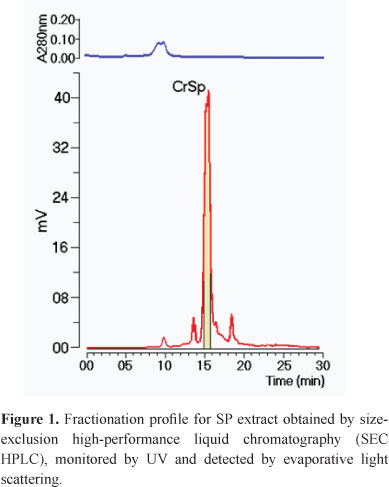
After ion exchange chromatography, we performed a final purification step on an SEC HPLC. We used high-performance liquid chromatography equipment from Jasco (pump Model PU-2080; a Chronav system controller; a Rheodyne injector equipped with a 20 µL loop; an FP-2020 fluorescence detector; an MD-2015/2018 diode arrangement detector; and an ELC-2041 evaporative light scattering detector). Samples from the first chromatographic step were dissolved in a Tris - HCl (1M, pH 7.8), and the resulting solution was clarified by centrifugation at 4,500 x g for 5 min. The resulting supernatant was applied to the TSKgel G3000SWXL silica base SEC column (0.78 x 30 cm), which was previously equilibrated with the sample dilution buffer. The elution was performed at a flow rate of 1 mL/min. SEC coupled with "on-line" laser light scattering (LS), and ultraviolet (UV) detection provides an elegant approach to determining the molecular weights of proteins and their complexes in solution. We used a protein marker blue dextran (BD, 2000 kDa), β-amylases (BA, 225 kDa), bovine serum albumin (BSA, Sigma, 66 kDa) that form a natural dimmer of 120 kDa, Ovalbumin (OVA, 43 kDa), Carbonic anhydrase (CA, 29 kDa), Ribonuclease (RA, 14 kda). This columns TSK gel can be used for characterization of protein and other molecules such as polysaccharides, carbohydrates and oligosscharides. The estimation of the molecular mass of SP was done using the BD, BA, BSA, OVA, CA, RA. All samples were subjected analysis under same chromatographic condition of buffer, flow rate and columns. For monitoring the protein elution, we used λ 280nm and the monitoring the sulfated polysaccharides was used a fluorescence detection. The elution was performed at a flow rate of 1 mL/min, and highly purified SP were detected using the ELC-2041 evaporative light scattering detector (Jasco).
Incubation of sPLA2 with CrSP
The incubation of CrSP with sPLA2 was conducted as described by Toyama (2010). CrSP was dissolved in saline solution. Purified sPLA2 (1.0 mg, 80 nmol/mL) was dissolved in 1 mL of water. After complete homogenization, 500 µL of CrSP solution (4 mg/mL, 160 nmol) was added and incubated for 60 min in a water bath at 37 °C. Samples (200 µL) were loaded onto silica-based GFC columns (TSKgel G3000SW) to separate the modified sPLA2 (sPLA2:CrSp) from native sPLA2 and CrSP. Samples were eluted using a continuous gradient of buffer (Tris-HCl 1 M, pH 7.8) at a constant flow rate of 1.0 mL/min. The chromatographic run was monitored at λ 280 nm.
Measurement of sPLA2 activity
sPLA2 activity was measured, following protocols described by Rigden (2003) and modified by Toyama (2003), in 96-well plates, using 4-nitro-3-octanoyloxybenzoic acid (4N3OBA, BIOMOL, USA) as a substrate. Enzyme activity, expressed as the initial velocity of the reaction (Vo), was calculated based on the increase in absorbance after 20 min. All assays were performed with absorbance at λ 425 nm using a SpectraMax 340 multiwell plate reader (Molecular Devices, Sunnyvale, CA). After the addition of native sPLA2, sulfated polysaccharides, or sPLA2 pre-incubated with sulfated polysaccharides (20 µg), the reaction mixture was incubated for up to 40 min at 37 °C, and absorbance was read at 10 min intervals.
Mouse paw edema assay
The paw edema assay was performed using the protocol described in Cotrim (2011). Female Swiss mice (22 g) were anesthetized with halothane by inhalation. Posterior paw edema was induced by a single subplantar injection of one of the following: 0.9% sterile saline (control group), native sPLA2, CrSP and sPLA2 pre-incubated with CrSP. We inject a total volume of 25 µL in all treatments. However, to sPLA2 the 1:1 proportion was maintained by addition 12.5 µL saline solution at PLA2 solution. The same procedure was followed by a treatment which received only CrSP. Paw volume was measured before the injection and at selected time points thereafter (15, 30, 60, 120, 180, and 360 min) using a hydro-plethysmometer (model 7150, Ugo Basile, Italy). All samples were dissolved in a 0.9% sterile saline solution. The results were expressed as an increase in paw volume (mL) calculated by subtracting the initial volume. The area under the curve was calculated using the trapezoidal rule, and the results were expressed as total edema volume (mL per paw).
Myotoxic activity
The presence of creatine kinase (CK) was assayed using the CK-NAc kit (Laborlab). Native sPLA2, sPLA2:CrSp, 0.9% sterile saline (control group) and CrSP (15 µg/µL in 50 µL) was injected into the left gastrocnemius muscle of female Swiss mice (22 g, n=5). After 3 h, the mice were anesthetized, and blood was collected from the abdominal vena cava into tubes containing heparin as an anticoagulant. The plasma was stored at 4 °C for a maximum of 12 h before assaying. The amount of CK was then determined using 4 µL of plasma, which was incubated for 3 min at 37 °C with 1.0 mL of the reagent according to the kit protocol. Activity was expressed in U/L.
Antibacterial activity
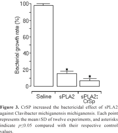
The antibacterial activity of sPLA2, CrSP and sPLA2:CrSP was assayed as described by Cotrim (2011). Clavibacter michiganensis michiganensis cells were harvested from fresh agar plates and suspended in sterile distilled water (A600 nm = 3 ×10⁸ CFU/mL). Aliquots of bacterial suspension were diluted to 103 colony-forming units/mL (CFU/mL) and incubated with sPLA2, CrSP and sPLA2:CrSP samples (75 µg/mL) for 60 min at 28 ºC. Survival was assayed on nutrient agar (Difco) plates (n=5). For antibacterial assays, electron microscopic assessments of morphologic alterations were performed in the presence of 0.9% sterile saline (negative control), sPLA2, and sPLA2 pre-incubated with sulfated polysaccharides.
Statistical analysis
The results are expressed as the means±SD. The data were analyzed using Student's t test. The level of significance was set at p<0.05.
Results and Discussion
In this study, we present the purification of a low-molecular-weight sulfated polysaccharide that is soluble in aqueous solution, termed CrSP, which has a molecular mass of approximately 15 kDa. CrSP was obtained after two chromatographic steps using an ion exchange (DEAE-cellulose resin column) followed by a molecular size exclusion column (HPLC SEC) (Figure 1). Sulfated polysaccharides of high molecular weight from Caulerpa racemosa have previously been isolated and characterized by other groups. Using DEAE-ion exchange column chromatography followed by molecular exclusion chromatography on Sepharose 4B columns, Ji and colleagues (2008) identified four major groups of sulfated polysaccharides with molecular masses between 100 and 1395 kDa from samples of Caulerpa racemosa collected on the coast of China. Chattopadhyay and colleagues (2007) isolated another group of sulfated polysaccharides with an apparent molecular mass of 80 kDa from Caulerpa racemosa collected on the coast of India. This work also used two chromatographic steps: ion exchange and size exclusion chromatography on S-300 Sephacryl. Work by Rodrigues and colleagues (2011) suggests that sulfated polysaccharides from Caulerpa racemosa can exhibit a large molecular mass range, from 10 to 216 kDa; the SP obtained from the DEAE-cellulose column were high-molecular-weight macromolecules or macromolecular aggregates. This finding agrees with Melo (2002). Thus, the CrSP found in this study is the first low-molecular-weight sulfated polysaccharide isolated from Caulerpa racemosa and may be present as part of a larger aggregate.
The curve for enzymatic activity of sPLA2 showed that CrSP induced an increase in phospholipase A2 activity. The concentration of CrSP that induced a 50% increase in activity was estimated to be 0.25 mg/mL (Figure 2a). In Figure 2b, we show that sPLA2 pre-incubated with CrSP possessed increased catalytic activity and capacity, and had a modified saturation point for catalytic sites. Most SP isolated from algae are capable of inhibiting the enzymes and factors involved in the coagulation cascade (Pereira et al., 2005; Jiao et al., 2011). These results show that CrSP was able to enhance the enzymatic activity of sPLA2. sPLA2 is known to possess antibacterial activity against Gram-positive and Gram-negative bacteria. Its high concentration in inflammatory fluids and human tears is consistent with the notion that this activity is a major function of this protein (Buckland & Wilton, 2000). In addition, Group IIA sPLA2 is the most potent among mammalian secreted PLA2 against Gram-positive bacteria, but additional antibacterial compounds, for example, the bactericidal/permeability-increasing protein, are needed to kill Gram-negative bacteria. The mechanisms of sPLA2 binding to the bacterial surface and its bactericidal activities are based on the positive charge of the PLA2 protein and its phospholipolytic enzymatic activity, respectively (Nevalainen et al., 2008). The results in Figure 3 show that the antibacterial activity of sPLA2 pre-incubated with CrSP was twofold higher than that of native sPLA2. Thus, the increased antibacterial activity of sPLA2 was due to the enhanced enzymatic activity induced by CrSP.

The phospholipase A2 extraction and purification was performed by two chromatography steps, using SEC and ion exchange, respectively. However, we used SEC coupled with "on-line" detectors as this column provides an elegant approach for determining the molecular weights of proteins and their complexes in solution. Because light scattering provides the weight-average molecular weight (MW) of all species in solution, SEC serves a critical size-fractionation function (Folta-Stogniew & Williams, 1999). Chromatographic analysis of CrSP, sPLA2 and sPLA2:CrSP was performed under the same chromatographic conditions (Figure 4). The sPLA2 and sPLA2:CrSP chromatographic runs was monitored at λ 280 nm, and the CrSP chromatographic run was monitored using relative fluorescence. The analysis of the chromatographic profile of CrSP showed the presence of one main peak that eluted at 15.25 min, whereas native sPLA2 eluted at 16.2 min. These results suggest that CrSP has an approximate molecular mass of 15 kDa, but the experiments were conducted with molecular marks. The molecular mass of sPLA2 was determined to be approximately 14 kDa, and the molecular mass of sPLA2:CrSP was estimated to be 32 kDa.
There is evidence that sulfated polysaccharides are capable of binding proteins through several different mechanisms. As highly acidic macromolecules, they can bind non-specifically to any basic region on a protein surface at low ionic strength, and such interactions are not likely to be physiologically significant (Mulloy, 2005). The molecular interaction of algae-derived sulfated polysaccharides with sPLA2 is not well studied, but it is known that sPLA2 has several basic amino acid residues at the N-terminus, at the C-terminus and on the outside of the α-helical structure of sPLA2 (Oliveira et al., 2002). Our chromatography results showed that there is an interaction between sPLA2 and CrSP as the mass from the incubed proteins increased. This finds may indicated a possible interaction in the surface of sPLA2 basic region with a highly acidic group in CrSP.
Our study of mouse paw edema shows that CrSP:sPLA2 increased sPLA2-induced edema (Figure 5). The native sPLA2 induced a swelling of 0.26±0.03 mL in the first 15 min of the experiment, and, the sPLA2:CrSP sample was able to induce edema up to 0.317±0.02 mL (Figure 5a). CrSP did not significantly change the maximum edema induced by native sPLA2. These initial results suggest that once the initial edema was induced by sPLA2:CrSP, only sPLA2 appears to have interacted with the target cells. The edema induced by sPLA2 depends not only on the enzymatic activity of sPLA2 but also on regions of sPLA2 near the calcium-binding loop (Oliveira et al., 2008; Toyama et al., 2011). It has been established that the N-terminal region of PLA2 enzymes is essential for enzymatic activity and is involved in cell membrane phospholipid recognition (Ali et al., 1999). Because CrSP was able to increase the enzymatic activity of sPLA2, CrSP could also be involved in modulating the interaction of sPLA2 with target cells. The increased enzymatic activity of sPLA2 induced by CrSP could be involved in the extended paw edema observed over the time period of 60 to 360 minutes (Figure 5b).

The effect of myotoxic Lys49 proteins is exerted locally at the site of injection, in contrast to the systemic action of other types of myotoxins, such as PLA2 or PLA2 complexes from elapids and some viperids. The myotoxic effect induced by snake venom-derived sPLA2 involves the presence of highly basic amino acid residues that cause muscle cell destabilization and myonecrosis (Rigden et al., 2003; dos Santos et al., 2008). The results observed from the SEC showed that sPLA2 is able to establish a heterodimeric complex with CrSP, which is stabilized by the interaction between the acid groups of CrSP and the basic residues of sPLA2. The results shown in Figure 5b confirm the results shown in Figure 4: impairment of basic residues by CrSP binding prevents the induction of a myotoxic effect. Despite of myotoxic inhibition by complex sPLA2:CrSP be independent of the enzymatic activity of PLA2, this shows another type of association between these two components, which still favors interesting pharmacological effects attributed to sulfated polysaccharide from green algae Caulerpa racemosa.
Ophidian accidents represent a great public health problem in developing countries. Snake bites are often dangerous accidents that require immediate medical treatment to neutralize the venon toxic effects (Andrade et al., 2013). There are reports in the literature which show that the efficiency of antivenom is limited to the time it takes to make the administration of the antibody solution. However, in Brazil, hospital care and effective antivenom administration happen after first hour after snake bit (Offerman et al., 2002; Gutiérrez et al., 2009). A feared complication of South America and North America viper snake bites is extended myonecrosis destruction, for example, large amount of muscle tissue, which can lead to amputation of the bitten limb and permanent disability. In the case of myonecrosis induced by Bothrops sp., the sPLA2 is the main factor involved in pathology. Although currently available antiserum can neutralize the toxic effect of snake venom, the venom myotoxic effects is not completely neutralized (Nuchpraryoon & Garner, 2000; Marsh & Williams, 2007). Thus the sulfated polysaccharide of this alga do not decrease the edema frame, the drug was capable of virtually abolish myonecrosis, suggesting its potential therapeutic use as an adjuvant in antiserum, and the purpose of being able to increase the neutralizing capacity the antiserum. The conventional serum therapy has some problems involving the reaction of the immune system of patients injected with antibodies, such as hypersensitivity, especially in children. As the polysaccharide tested in this work within our tests showed no capability either edematogenic effect or myonecrosis, this may indicate a potential therapeutic use against the action of phospholipase A2 activity with myotoxins.
Conclusion
Our study showed that the sulfated polysaccharide extracted from the green macroalgae Caulerpa racemosa led the increase of the edematogenic effect, but inhibited the myotoxic activity. We believe that there is an interaction between sPLA2 and CrSP as the mass from the incubed proteins increased and this interaction may be related with some active site responsible for myotoxic activity, but activated the PLA2 site responsible for edematogenic effect which potentiated inflammatory effect. Despite of myotoxic activity be independent of the enzymatic activity of PLA2, this region was inhibited showing the association with this compounds which have important pharmacological effects attributed to sulfated polysaccharide from Caulerpa racemosa.
Acknowledgments
The authors thank the Research Foundation of the State of São Paulo (FAPESP), Process number 2011/06704-4 and Process nº 2011/14241-4, for financial support.
Authors' contributions
CLP, DB, and HHG performed the biochemical and pharmacological experiments. WRLF collected the Caulerpa racemosa and isolated the sulfated polysaccharide. SDR, DOT and MHT coordinated and designed the experiments. CLP and MHT wrote the manuscript. All authors read and approved the final manuscript.
Received 5 Feb 2013
Accepted 26 Jun 2013
- Ali SA, Alam JM, Stoeva S, Schütz J, Abbasi A, Zaidi ZH, Voelter W 1999. Sea snake Hydrophis cyanocinctus venom. I. Purification, characterization and N-terminal sequence of two phospholipases A2. Toxicon 37: 1505-1520.
- Andrade FG, Eto SF, Ferraro ACNS, Marioto DTG, Vieira NJ, Cheirubim AP, Ramos S P, Venâncio EJ 2013. The production and characterization of anti-bothropic and anti-crotalic IgY antibodies in laying hens: A long term experiment. Toxicon 66: 18-24.
- Buckland AG, Wilton DC 2000. The antibacterial properties of secreted phospholipases A(2). Biochim Biophys Acta 1488: 71-82.
- Câmara P, Esquisatto L, Camargo E, Ribela M, Toyama M, Marangoni S, De Nucci G, Antunes E 2003. Inflammatory edema induced by phospholipases A2 isolated from Crotalus durissus sp. in the rat dorsal skin: A role for mast cells and sensory c-fibers. Toxicon 41: 823-829.
- Chattopadhyay K, Adhikari U, Lerouge P, Ray B 2007. Polysaccharides from Caulerpa racemosa: purification and structural features. Carbohyd Polym 68: 407-415.
- Cirino G 1998. Multiple controls in inflammation. Extracellular and intracellular phospholipase A2, inducible and constitutive cyclooxygenase, and inducible nitric oxide synthase. Biochem Pharmacol 55: 105-111.
- Cotrim CA, de Oliveira SC, Diz Filho EB, Fonseca FV, Baldissera LJr, Antunes E, Ximenes RM, Monteiro HS, Rabello MM, Hernandes MZ, de Oliveira Toyama D, Toyama MH 2011. Quercetin as an inhibitor of snake venom secretory phospholipase A2. Chem Biol Interact 189: 9-16.
- Dicciaani MB, Lilly-Stauderman M, McLean LR, Balasubramaniam A, Harmony JA 1991. Heparin prevents the binding of phospholipase A2 to phospholipid micelles: importance of the amino-terminus. Biochemistry 30: 9090-9097.
- dos Santos ML, Fagundes FH, Teixeira BR, Toyama MH, Aparicio R 2008. Purification and preliminary crystallographic analysis of a new Lys49-PLA2 from B. jararacussu. Int J Mol Sci 9: 736-750.
- Dua R, Cho W 1994. Inhibition of human secretory class II phospholipase A2 by heparin. Eur J Biochem 221: 481-490.
- Farias WRL, Valente AP, Pereira MS, Mourao PAS 2000. Structure and anticoagulant activity of sulfated galactans. Isolation of a unique sulfated galactan from the red algae Botryocladia occidentalis and comparison of its anticoagulant action with that of sulfated galactans from invertebrates. J Biol Chem 275: 29299-29307.
- Folta-Stogniew E, Williams KR 1999. Determination of molecular masses of proteins in solution: implementation of an HPLC size exclusion chromatography and laser light scattering service in a core laboratory. J Biomol Tech 10: 51-63.
- Fuentes L, Hernández M, Nieto M, Sánchez Crespo M 2002. Biological effects of group II secreted phosholipase A (2). FEBS Lett 531: 7-11.
- Gil B, Sanz M, Terencio M, Gunasegaran R, Payá M, Alcaraz M 1997. Morelloflavone, a novel biflavonoid inhibitor of human secretory phospholipase A2 with anti-inflammatory activity. Biochem Pharmacol 53: 733-740.
- Gutiérrez J M, Escalante T, Rucavado A 2009. Experimental pathophysiology of systemic alterations induced by Bothrops asper snake venom. Toxicon 54: 976-987.
- Ji H, Shao H, Zhang C, Hong P, Xiong H 2008. Separation of the polysaccharides in Caulerpa racemosa and their chemical composition and antitumor activity. J Appl Polym Sci 110: 1435-1440.
- Jiao G, Yu G, Zhang J, Ewart HS 2011. Chemical structures and bioactivities of sulfated polysaccharides from marine algae. Mar Drugs 9: 196-223.
- Kini RM 2005. Structure-function relationships and mechanism of anticoagulant phospholipase A2 enzymes from snake venoms. Toxicon 45: 1147-1156.
- Lee W, Toyama M, Soares, A, Giglio J, Marangoni S, Polikarpov I 1999. Crystallization and preliminary X-ray diffraction studies of piratoxin III, a d-49 phospholipase A2 from the venom of Bothrops pirajai. Acta Crystallogr D 55: 1229-1230.
- Lehnhardt Pires C, Rodrigues SD, Bristot D, Gaeta HH, Toyama DO, Farias WRL, Toyama MH 2013. Evaluation of macroalgae sulfated polysaccharides on the Leishmania (L.) amazonensis promastigote. Mar Drugs 11: 934-943.
- Lin YH, Huang WN, Lee SC, Wu WG 2000. Heparin reduces the alpha-helical content of cobra basic phospholipase A(2) and promotes its complex formation. Int J Biol Macromol 27: 171-176.
- Marsh N, Williams V 2005. Practical applications of snake venom toxins in haemostasis. Toxicon 45: 1171-1181.
- Melo MRS, Feitosa JPA, Freitas ALP, Paula RCM 2002. Isolation and characterization of soluble sulfated polysaccharide from the red seaweed Gracilaria cornea. Carbohyd Polym 49: 491-498.
- Mourão PAS, Pereira MS 1999. Searching for alternatives to heparin: sulfated fucans from marine invertebrates. Trends Cardiovas Med 9: 225-232.
- Mulloy B 2005. The specificity of interactions between proteins and sulfated polysaccharides. An Acad Bras Cien 77: 651-664.
- Nader HB, Pinhal MAS, Baú EC, Castro RAB, Medeiros GF, Chavante SF, Leite EL, Trindade ES, Shinjo SK, Rocha HAO, Tersariol ILS, Mendes A, Dietrich CP 2001. Development of new heparin-like compounds and other antithrombotic drugs and their interaction with vascular endothelial cells. Braz J Med Biol Res 34: 699-709.
- Nevalainen TJ, Graham GG, Scott KF 2008. Antibacterial actions of secreted phospholipases A2. Review. Biochim Biophys Acta 1781: 1-9.
- Nuchpraryoon I, Garner P 2000. Interventions for preventing reactions to snake antivenom. Cochrane Database Syst Rev 2: 2153.
- Offerman SR, Bush SP, Moynihan JA, Clark RF 2002. Crotaline fab antivenom for the treatment of children with rattlesnake envenomation. Pediatrics 110: 968-971.
- Oliveira DG, Toyama MH, Novello JC, Beriam LO, Marangoni S 2002. Structural and functional characterization of basic PLA2 isolated from Crotalus durissus terrificus venom. J Protein Chem 21: 161-168.
- Oliveira D, Toyama M, Martins A, Havt A, Nobre A, Marangoni S, Câmara P, Antunes E, de Nucci G, Beliam L, Fonteles M, Monteiro H 2003. Structural and biological characterization of a crotapotin isoform isolated from Crotalus durissus cascavella venom. Toxicon 42: 53-62.
- Oliveira SC, Fonseca FV, Antunes E, Camargo EA, Morganti RP, Aparício R, Toyama DO, Beriam LO, Nunes EV, Cavada BS, Nagano CS, Sampaio AH, Nascimento KS, Toyama MH 2008. Modulation of the pharmacological effects of enzymatically-active PLA2 by BTL-2, an isolectin isolated from the Bryothamnion triquetrum red alga. BMC Biochem 6: 16.
- Pereira MG, Benevides NM, Melo MR, Valente AP, Melo FR, Mourão PA 2005. Structure and anticoagulant activity of a sulfated galactan from the red alga, Gelidium crinale. Is there a specific structural requirement for the anticoagulant action? Carbohydr Res 340: 2015-2023.
- Pomin VH, Mourão PAS 2008. Structure, biology, evolution, and medical importance of sulfated fucans and galactans. Glycobiology 18: 1016-1027.
- Ramacciotti E, Clark M, Sadeghi N, Hoppensteadt D, Thethi I, Gomes M, Fareed J 2011. Review: contaminants in heparin: review of the literature, molecular profiling, and clinical implications. Clin Appl Thromb Hemost 17: 126-135.
- Rigden D, Hwa L, Marangoni S, Toyama M, Polikarpov I 2003. The structure of the D49 phospholipase A2 piratoxin III from Bothrops pirajai reveals unprecedented structural displacement of the calcium-binding loop: Possible relationship to cooperative substrate binding. Acta Crystallogr D 59: 255-262.
- Rodrigues JA, Vanderlei ES, Silva LM, Araújo IW, Queiroz IN, Paula GA, Abreu TM, Ribeiro NA, Bezerra MM, Chaves HV, Lima V, Jorge RJ, Monteiro HS, Leite EL, Benevides NM 2012. Antinociceptive and anti-inflammatory activities of a sulfated polysaccharide isolated from the green seaweed Caulerpa cupressoides. Pharmacol Rep 64: 282-292.
- Rodrigues JAG, Queiroz INL, Quinderé ALG, Vairo BC, Mourão PAS, Benevides NMB 2011. An antithrombin-dependent sulfated polysaccharide isolated from the green alga Caulerpa cupressoides has in vivo anti- and prothrombotic effects. Cienc Rural 41: 634-639.
- Silva RO, dos Santos GM, Nicolau LA, Lucetti LT, Santana AP, Chaves LS, Barros FC, Freitas AL, Souza MH, Medeiros JV 2011. Sulfated-polysaccharide fraction from red algae Gracilaria caudata protects mice gut against ethanol-induced damage. Mar Drugs 9: 2188-2200.
- Toyama M, de Oliveira D, Beriam L, Novello J, Rodrigues-Simioni L, Marangoni S 2003. Structural, enzymatic and biological properties of new PLA(2) isoform from Crotalus durissus terrificus venom. Toxicon 41: 1033-1038.
- Toyama MH, Toyama DO, Torres VM, Pontes GC, Farias WR, Melo FR, Oliveira SC, Fagundes FH, Diz Filho EB, Cavada BS 2010. Effects of low molecular weight sulfated galactan fragments from Botryocladia occidentalis on the pharmacological and enzymatic activity of sPLA2 from Crotalus durissus cascavella. Protein J 29: 567-571.
- Toyama DO, Diz-Filho EB, Cavada BS, da Rocha BA, de Oliveira SC, Cotrim CA, Soares VC, Delatorre P, Marangoni S, Toyama MH 2011. Umbelliferone induces changes in the structure and pharmacological activities of Bn IV, a phospholipase A(2) isoform isolated from Bothrops neuwiedi. Toxicon 57: 851-860.
- Warkentin TE, Greinacher A 2009. Heparin-induced anaphylactic and anaphylactoid reactions: two distinct but overlapping syndromes. Expert Opin Drug Saf 8: 129-144.
 Correspondence:
 Publication Dates
-
Publication in this collection
02 Aug 2013 -
Date of issue
Aug 2013
History
-
Received
05 Feb 2013 -
Accepted
26 June 2013