Abstracts
The aim of the present paper is to compare and correlate the take of nerve segments in a severely crushed nerve. Forty adult Wistar rats had their right sciatic nerve by a "Péan-Murphy" forceps for 40 minutes. In Group 1 (n=20), a segmentar serection in the crushed sciatic nerve was made. A sural nerve segment from the opposite hindpaw was placed in the gap. In Group 2 (n=20), a lontudinal insision in the epineurium of the lesioned sciatic nerve was made. A sural nerve segment was buried underneath the epineurium. The crushed sciatic nerves undergone Wallerian degeneration and endoneurial fibrosis. Sciatic nerves from Group 2 had significant better histological aspects than those from Group 1. Sural nerve grafts presented better degrees of regeneration than crushed sciatic nerves. Sural nerve grafts from Group 2 (burying method) integrated as well as those from Group 1 (segmentar resection).
Nerve crush; Nerve regeneration; Nerve degeneration; Sural nerve; Rats
O objetivo desta pesquisa é comparar e correlacionar as integrações de segmento de nervo em nervos gravemente esmagados. Foram utilizados 10 ratos adultos da linhagem Wistar. Os nervos ciáticos direitos foram esmagados, por uma pinça de "Péan-Murphy", por 40 minutos. No Grupo 1 (n=20), executou-se um ressecção segmentar no nervo ciático esmagado e um segmento de nervo sural, retirado da pata contra-lateral, foi colocado na falha. No Grupo2 (n=20), uma incisão longitudinal no epineuro do nervo lesado foi realizada. Um segmento de nervo sural foi sepultado sob o epineuro. Os nervos ciáticos esmagados desenvolveram degeneração Wallerina e fibrose endoneural. Os nervos ciáticos do Grupo2 apresentaram pormenores histológicos melhores do que os do grupo 1. Os enxertos de nervo sural apresentaram melhores graus de regeneração do que os dos nervos ciáticos esmagados. Os enxertos de nervo sural do Grupo 2 (método de sepultamento) intagraram-se tão bem quanto os do Grupo 1 (ressecção segmentar).
Compressão nervosa; Regeneração nervosa; Degeneração neural; Nervo Sural; Ratos
2 - ORIGINAL ARTICLE
COMPARISON OF NERVE GRAFT INTEGRATION AFTER SEGMENTAR RESECTION VERSUS EPINEURAL BURYING IN CRUSHED RAT SCIATIC NERVES1 1 Work performed at the Curso de Pós-Graduação em Cirurgia da Universidade Federal de Minas Gerais. 2 Plastic Surgery at Faculdade de Medicina do Triângulo Mineiro. 3 Digestive System Surgery Professor at Universidade Federal de Minas Gerais. 4 Surgical Pathology Professor at Faculdade de Medicina do Triângulo Mineiro.
Marco Túlio Rodrigues da Cunha2 1 Work performed at the Curso de Pós-Graduação em Cirurgia da Universidade Federal de Minas Gerais. 2 Plastic Surgery at Faculdade de Medicina do Triângulo Mineiro. 3 Digestive System Surgery Professor at Universidade Federal de Minas Gerais. 4 Surgical Pathology Professor at Faculdade de Medicina do Triângulo Mineiro.
Alcino Lázaro da Silva3 1 Work performed at the Curso de Pós-Graduação em Cirurgia da Universidade Federal de Minas Gerais. 2 Plastic Surgery at Faculdade de Medicina do Triângulo Mineiro. 3 Digestive System Surgery Professor at Universidade Federal de Minas Gerais. 4 Surgical Pathology Professor at Faculdade de Medicina do Triângulo Mineiro.
Sheila Bernardino Fenelon4 1 Work performed at the Curso de Pós-Graduação em Cirurgia da Universidade Federal de Minas Gerais. 2 Plastic Surgery at Faculdade de Medicina do Triângulo Mineiro. 3 Digestive System Surgery Professor at Universidade Federal de Minas Gerais. 4 Surgical Pathology Professor at Faculdade de Medicina do Triângulo Mineiro.
CUNHA, M.T.R.; SILVA, A.L.; FENELON, S.B. - Comparison of nerve graft integration after segmentar resection versus epineurial burying in crushed rat sciatic nerves. Acta. Cir. Bras. 12(4):221-5, 1997.
SUMMARY: The aim of the present paper is to compare and correlate the take of nerve segments in a severely crushed nerve. Forty adult Wistar rats had their right sciatic nerve by a "Péan-Murphy" forceps for 40 minutes. In Group 1 (n=20), a segmentar serection in the crushed sciatic nerve was made. A sural nerve segment from the opposite hindpaw was placed in the gap. In Group 2 (n=20), a lontudinal insision in the epineurium of the lesioned sciatic nerve was made. A sural nerve segment was buried underneath the epineurium. The crushed sciatic nerves undergone Wallerian degeneration and endoneurial fibrosis. Sciatic nerves from Group 2 had significant better histological aspects than those from Group 1. Sural nerve grafts presented better degrees of regeneration than crushed sciatic nerves. Sural nerve grafts from Group 2 (burying method) integrated as well as those from Group 1 (segmentar resection).
SUBJECT HEADINGS: Nerve crush. Nerve regeneration. Nerve degeneration. Sural nerve. Rats.
INTRODUCTION
In the last decades, significant strides have been made in the understanding and treatment of nerve trauma. On the other hand there is much to clarity about management of severely crushed peripheral nerves. Novel alternatives on nerve graft setting should be attempted to achieve a more complete recuperation after trauma. The experimental model of many peripheral nerve investigators remains the rat 2,4,6,7,9,13,14,17,18,20,21,22
The nerve crush is a well known research methodology and following nerve crush, axons may utilize their own endoneuriaI tubes to migrate to the distal stump 5. The clinical picture of a nerve compression lesion depends on several factors, for example the anatomical location of the lesion as well as the severity and duration of the trauma 17, 19. Basal lamina tubes withstand crush well, nevertheless some tubes are torn open at the site of the lesion and the sprouts they contain are usually lost and will have no functional value 2, 12.
Depending on the severity and duration of the trauma the recuperation after crushing can be long-lasting and incomplete 2,17. The aim of the present investigation was to study histological changes after a severe rat sciatic nerve crushing for 40 minutes. Furthermore, the purpose was to correlate and compare the take of sural nerve grafts, after segmentar resection or epineurial burying in the crushed sciatic nerves.
METHOD
Forty adult Wistar rats were used (Rattus norvegicus albinus, Rodentia Mammalia), from the animal colony of the Triangulo Mineiro Medical College (FMTM).
The rats were randomly distributed in two groups of 20 animals each. The animals in both groups were anaesthetized with ether and positioned on ventral decubitus on a wood table.
A 2.5 cm longitudinal skin incision was made at the transition of the right hip with the thigh. The gluteus maximus fibers were disected from the biceps femoris muscle identifying the sciatic nerve, which was isolated. Using a "Péan-Murphy forceps"(14 cm long, maximum compression area of 0.6 cm) the nerve was crushed for 40 minutes at the mid distance between the greater trochanter and the knee joint. A 1 cm segment of sural nerve was taken from the opposite hindpaw with its acompanying vessels. After the 40 minutes crushing period, the nerve was manipulated as follow:
- Group 1: a segmentar resection of the fascicle concerning the common peroneal nerve was made, beginning 0.1 cm proximal to the crushed zone and ending 0.1 cm distal to it (Fig. 1), using an operating microscope with a 10 x magnifying lens and microsurgical scissors.
The sural nerve segment was placed in the gap and sutured with two interrupted epineurial stitches, one in each extremity (Fig. 2). A 10-0 monofilament nylon was used in the suture.
- Sural nerve graft placed between the proximal and distal stumps of the common peroneal fascicle.
- Group 2: a longitudinal incision in the epineurium of the sciatic nerve was made with a number 11 blade, beginning 0.1 cm proximal to the crushed zone and ending 0.1 cm distal to it (Fig. 3), using an operating microscope with a 16 x magnifying lens. The magnification used was greater than that in Group 1 due to the delicate incision in the epineurium.
The sural nerve segment was placed at the level of the incision and two stitches (10-0 monofilament nylon) were given in the epineurium of the sciatic nerve, burying the sural nerve (Fig. 4). The skin incision was closed with 4-0 monofilament nylon interrupted sutures. The sciatic and sural nerves of 5 untreated rats were used as controls.
Animals were kept in individual cages, during all the postoperative period, water and food "ad libitum".
In the 30th postoperative day the treated sciatic nerves were removed. The nerve segment was then divided into three parts. The segment proximal to the first stitch was called "proximal segment". The segment between the two stitches was called "medium segment". The segment distal to the second stitch was called "distal segment".
Efforts were concentrated in the "medium segment" where nerve graft and crushed zone would be better identifyied.
The samples were fixed in 10% formaldehyde during 48 h and paraffin embeded. Five sections were obtained from each block and stained with Hematoxilin-Eosin, Masson's Trichrome , Luxol fast-blue, Weil and Palmgren.
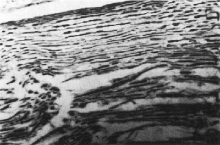
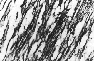
Two classifications concerning the degenerative and regenerative changes in crushed sciatic nerves and nerve grafts, on their "medium segment", were used. When investigating Hematoxilin-Eosin stained nerves we systematized: Grade 0 - complete vacuolization of the nerve segment; Grade I - vacuolization in 2/3 or more of the nerve segment; Grade II - vacuolization in less than 1/2 of the nerve segment; Grade III - vacuolization in less than 1/3 of the nerve segment and Grade IV - normal sciatic and sural nerves (Figs. 5 and 6).
When evaluating Palmgren stained nerves, we systematized: Grade 0 - absence of normal nerve fibers; Grade I - presence of nerve fibers in 1/3 or less of the nerve segment; Grade II - presence of nerve fibers in more than 1/3 of the nerve segment; Grade III - presence of nerve fibers in more than 1/2 of the nerve segment and Grade IV - normal sciatic and sural nerves ( Figs. 7 and 8).
- Grade III sciatic nerve: presence of normal nerve fibers in more than 1/2 of the nerve segment.
We validated these classifications as a pathological staging system after submitting the samples to three different physicians who used that system. Their results showed no significant differences for the same samples (P < 0.05).
The Mann-Whitney test was used to compare the crushed sciatic and the sural nerve grafts from both groups. Intermediate degrees were considered and received crescent non-parametric values. Grade I-II, for example, received a greater value than Grade I, and so on.
RESULTS
Crushed Sciatic Nerves: 30th day
Severe Wallerian degeneration with vacuolization, thinning and fragmentation of the myelin membrane occured in all crushed sciatic nerves.
Large bands of organized fibrin were often seen and vessels were dilated and filled with erythrocytes. Vigorous proliferation of fibrous tissue occurred and epineurial fibrosis was marked at 30 days.
The sciatic nerves from Group 2 were statistically better conserved than the sciatic nerves from Group 1 (P < 0.05).
Rare signs of regeneration were observed in transverse sections in Groups 1 and 2. No significant differences were observed between the sciatic nerves from Group 1 when stained either with Hematoxilin-Eosin or Palmgren. The same results were observed for sciatic nerves from Group 2.
Sural Nerve Grafts: 30th day
Sural nerve grafts from Groups 1 and 2 showed significant better degrees of conservation than crushed sciatic nerves from Groups 1 and 2 (Hematoxilin-Eosin and Palmgren).
Sural nerve grafts from Group 1 did not show significant differences when stained either with Hematoxilin-Eosin or Palmgren. The same results were observed for sural nerve grafts from Group 2.
Sural nerve grafts from Group 2 had no significant differences when compared to those from Group 1. "Bünger bands" were observed in sural nerves from both groups, demonstrating signs of regeneration in a higher percentage than in crushed sciatic nerves.
DISCUSSION
Many experiments on the degeneration and regeneration after trauma on the rat sciatic nerve are found in the literature 2,6,17.The animal is inexpensive, easy to work with and well studied .
It is well known that compression of peripheral nerves may induce impairment of nerve function and histologic changes 17,19.
The near normal return of function has been shown after crushing, if the endoneurial tubes maintain physical continuity 4. DE MEDINACELi, in 1988, refers that optimal conditions for regrowth are present whenever the traveling sprouts stay in close contact with a support tissue that is healthy 7.
The crushed sciatic nerves from Groups 1 and 2 in our experiment were severely degenerated with a high incidence of fibrosis, leaving unfavorable conditions for guidance of regenerating axons. Nevertheless, rare morphologic signs of regeneration were observed and no functional study was carried on. Two classifications were made after exhaustive light microscopy analysis of all nerve segments. The vacuolization was the most frequent histologic change found in crushed sciatic nerves and nerve grafts stained with Hematoxilin-Eosin. This nonespecific method particularly well demonstrate changes in nerves under conditions of ischemia and/or anoxia 3, 15, 18. Palmgren stains mainly nerve fibers being also of common use in nerve analysis 1, 9. This pathological staging system was considered reliable after submitting it to different physicians and comparing their results that were not significantly different. Sciatic nerves from Group 2 presented significant better degree of conservation than sciatic nerves from Group 1, indicating that the epineurial incision was less injurious to the sciatic nerve than the segmentar resection.
It is generally agreed that recuperation after crush is observed between the 16th and 25th days 6, 8, 11, although abnormalities can still be observed for several months 10, 16. In our series we removed the nerves for evaluation in the 30th post-operative day.
The choice of the "Péan-Murphy" forceps for crushing the rat sciatic nerve was due to its larger area of contact when compared to the classic use of jewlers 14, 23 and hemostatic forceps 4, 7 and to its high intensity trauma. A greater amount of crushed tissue was achieved for longitudinal and transverse sections.
Sural nerve grafts from Groups 1 and 2 showed a significant better staging grade of conservation than crushed sciatic nerves from both groups at the 30th post-operative day. Sural nerve grafts from Group 1 (segmentar resection) had no significant difference if compared to those from Group 2 (burying method).
The sural vein and artery were removed with the sural nerve segment and used as a biological marker to facilitate its identification after grafting (Fig. 9). In the pilot study it was many times difficult to differ the sural nerve graft from the other sciatic nerve fascicles, due to its excellent integration.
- Buryied sural nerve graft with is acompanying vessels used as a biological marker for identification after grafting.
Nerve grafts from Group 2 (burying method) were not sutured by means of end to end coaptation. The presence of nerve fibers in buryied nerves would be from sprouts of tubes torn open by the crushing trauma and from involuntary lesion of a sciatic nerve fascicle during epineurial incision.
The intrinsec blood supply is enough to keep the good vascularization of the rat sciatic nerve 21,22.This observation would justify the reason why nerve grafts buryied into the epineurium take well. High intensity trauma for a long period indicates that deferring repair could preclude good results because fibrosis would increase in the endoneurium space distally and retard regeneration 5, 17. Preliminary histological results are encouraging, but further functional studies will be necessary to better evaluate the usefulness of burying grafts underneath the epineurium of severely compressed and crushed nerves.
CUNHA, M.T.R.; SILVA, A.L.; FENELON, S.B. - Comparação das integrações de enxertos de nervo após ressecção segmentar versus sepultamento epineural em nervos ciáticos esmagados de rato. Acta. Cir. Bras. 12(4):221-5, 1997.
RESUMO: O objetivo desta pesquisa é comparar e correlacionar as integrações de segmento de nervo em nervos gravemente esmagados. Foram utilizados 10 ratos adultos da linhagem Wistar. Os nervos ciáticos direitos foram esmagados, por uma pinça de "Péan-Murphy", por 40 minutos. No Grupo 1 (n=20), executou-se um ressecção segmentar no nervo ciático esmagado e um segmento de nervo sural, retirado da pata contra-lateral, foi colocado na falha. No Grupo2 (n=20), uma incisão longitudinal no epineuro do nervo lesado foi realizada. Um segmento de nervo sural foi sepultado sob o epineuro. Os nervos ciáticos esmagados desenvolveram degeneração Wallerina e fibrose endoneural. Os nervos ciáticos do Grupo2 apresentaram pormenores histológicos melhores do que os do grupo 1. Os enxertos de nervo sural apresentaram melhores graus de regeneração do que os dos nervos ciáticos esmagados. Os enxertos de nervo sural do Grupo 2 (método de sepultamento) intagraram-se tão bem quanto os do Grupo 1 (ressecção segmentar).
DESCRITORES: Compressão nervosa. Regeneração nervosa. Degeneração neural. Nervo Sural. Ratos.
Adress for correspondence:
Marco Túlio Rodrigues da Cunha
Rua Alfén Paixão, 180 - ap. 202
CEP: 38060-230 - UBERABA - MG
FAC SIMILE - (34) 312-6640
Accepted for publication july, 1997
- 01. ALMGREN, K.G.- Revascularization of free peripheral nerve grafts. Acta Orthop. Scand.; 1:5-104,1974..
- 02. BAIN, J.R.; MACKINNON, S.E.; HUNTER, D.A.- Functional evaluation of complete sciatic, peroneal and posterior tibial nerve lesions in the rat. Plast. Reconstr. Surg. ; 83:129-36, 1989.
- 03. BERRY R.B.; EWART, W.R.; REEVE, D.R.E.; SOMMERLAD, B.C.-. Experimental observations on the behaviour of nerve grafts in sheaths formed around silicone rods. Br. J. Plast. Surg; 33:324-39, 1980.
- 04. BUEHLER, M.J.; SEABER, A.V.; URBANIAK, J.R.- The relationship of functional return to varying methods of nerve repair. J. Reconstr. Microsurg ; 6:61-9, 1990..
- 05. CABAUD, H.E.; RODKEY.W.G. - NEMETH, T.J.- Progressive ultrastructural changes after peripheral nerve transection and repair. J. Hand Surg. (AM) ; 7:353-65, 1982.
- 06. DE MEDINACELI, L.; FREED, W.J.; WYATT, R.J.- An index of the functional condition of rat sciatic nerve based on measurements made from walking tracks. Exp. Neurol. ; 77:634-43, 1982.
- 07. DE MEDINACELI, L.- Functional consequences of experimental nerve lesions: effects of time, location and extent of damage. Exp. Neurol. ; 100:154-65, 1988.
- 08. DEVOR, M.; SCHONFELD, D.; SELTZER, Z.; WALL , P.D.- Two modes of cutaneous reinnervation following peripheral nerve injury. J. Comp. Neurol. ; 185:211-20, 1979.
- 09. GU, L. & ZHU, J.- Repair of different sized nerve defects using degenerated muscle grafts with vascular implantation: an experimental study in the rat. J. Reconstr. Microsurg ; 8:47-52, 1992.
- 10. GUTMANN, E.- Factors affecting recovery of motor function after nerve lesions. J. Neurol. Psychiatr. ; 5:81-95, 1942.
- 11. GUTMANN, E. & GUTMAN, L. - Factors affecting recovery of sensory function after nerve lesions. J. Neurol. Psychiatr. ; 5:117-29, 1942.
- 12. HAFTECK, J. & -THOMAS, P.K.- Electron-microscope observations on the effects of localized crush injuries on the connective tissues of peripheral nerve. J. Anat. ; 103:233-43, 1968.
- 13. HRUSKA, R.E.; KENNEDY, S.; SILBERGELD, E.K.- Quantitative aspects of normal locomotion in rats. Life Sci.; 25:171-80, 1979.
- 14. JENQ, C.; JENQ, L.L.; COGGESHALL, R.E.- Numerical patterns of axon regeneration that follow sciatic nerve crush in the neonatal rat. Exp. Neurol. ; 95:492-99, 1987.
- 15.MALAMUD,N; HIRANO, A.- Atlas of Neuropathology Los Angeles: University of California Press Ltd, 1974.
- 16. MIRA, J.C.- Quantitative studies of the regeneration of rat myelinated nerve fibers: variations in the number and size of regenerating fibers after repeated localized freezings. J .Anat.; 129:77-93, 1979.
- 17. POWELL, H.C. & MYERS, R.R.- Pathology of experimental nerve compression. Lab. Invest. ; 55: 91-100, 1986..
- 18. RISITANO, G.; CAVALLARO, G.; LENTINI, M.- Autogenous vein and nerve grafts: a comparative study of nerve regeneration in the rat. J. Hand Surg. (BR) ; 14:102-4, 1989.
- 19. RYDEVIK, B. & NORDBORG, C.- Changes in nerve function and nerve fiber structure induced by acute, graded compression. J. Neurol. Neurosurg. Psychiatr. ; 43:1070-82, 1980.
- 20. SCHMALBRUCH, H.-.Fiber composition of the rat sciatic nerve. Anat. Rec. ; 215:71-81, 1986.
- 21. SLADKY, J.T.; GREENBERG, J.H.; BROWN, M.J.- Regional perfusion in normal and ischemic rat sciatic nerves. Ann. Neurol. ; 17:191-95, 1985.
- 22. STEVENS, W.G.; HALL, J.D.; YOUNG, V.L.; WEEKS, P.M. - When should nerve gaps be grafted? An experimental study in rats. Plast. Reconstr. Surg. ; 75:707-11, 1985.
- 23. WILLIAMS, P.L. & HALL, S.M.- Prolonged in vivo observations of normal peripheral nerve fibers and their acute reactions to crush and deliberate trauma. J .Anat.; 108:397-408, 1971.
Publication Dates
-
Publication in this collection
04 June 2001 -
Date of issue
Dec 1997
History
-
Accepted
July 1997