Abstracts
The aim of the present study was to compare and correlate histologically and electromyographically the effects of partial epineural burying of sural nerve segments in sectioned and sutured rat sciatic nerves. Sixty adult male Wistar rats were operated on 3 groups: Group 1, sural nerve graft, 9mm long, placed next to neurorrhaphy; Group 2, sural nerve graft, 9mm long, buryied 10mm distant from neurorrhaphy; Group 3, sural nerve graft, 18mm long, set next to neurorrhaphy. The morphological features were examined at light microscope after 3 months in 45 rats. The elements observed were: vascularization, vacuoles in nerve fibers, mastocytes and inflammatory infiltrate. The morphometry was made after 6 months in 15 rats from Group 1, 2 and 3, measuring external nerve fiber diameters and counting myelinated nerve fibers/mm². The electrophysiological study was perfomed after 6 months, registering maximum amplitude and frequency of EMG pontentials, at rest, in extensor digitorum longus muscle. Group 3 rats presented sciatic nerves better conserved morphologically and mean external nerve fiber diameters greater than those from Groups 1 and 2. There were no significant differences in density of nerve fibers/mm², and in the electrophysiological study in rats from Group 1, 2 and 3. The epineural burying of sural nerve grafts with greater length and placed next to the neurorrhaphy’s site had a significantly better regeneration of the histological features than the smaller ones distant from neurorrhaphy.
Sciatic nerve; Sural nerve; Nerve regeneration; Nerve degeneration; Rats
O objetivo foi comparar e correlacionar, histológica e eletromiograficamente, os efeitos do sepultamento parcial de segmentos de nervo sural em nervos ciáticos de rato, seccionados e suturados. Sessenta ratos adultos, da linhagem Wistar, foram operados em três Grupos: Grupo 1 , um segmento de nervo sural com 9mm. de comprimento colocado próximo à neurorrafia; Grupo 2 , um segmento de nervo sural com 9mm. de comprimento sepultado 10mm. distante da neurorrafia ; Grupo 3, um enxerto de nervo sural com 18mm. de comprimento, posicionado próximo à neurorrafia. Os pormenores histológicos foram examinados sob microscopia de luz em 45 ratos, 3 meses depois. Os elementos observados foram : vascularização, vacúolos nas fibras nervosas, mastócitos e infiltrado inflamatório. A morfometria foi realizada, após 6 meses, em 15 ratos dos Grupos 1, 2 e 3, medindo-se os diâmetros externos e contando-se as fibras nervosas mielinizadas por mm². O estudo eletrofisiológico foi feito após 6 meses, registrando a amplitude máxima e a freqüência dos potenciais eletromiográficos no músculo extensor longo dos dedos, em repouso. Os ratos do Grupo 3 apresentaram-se morfologicamente melhor conservados e com diâmetros de fibras maiores do que nos Grupos 1 e 2. Não houve diferença na densidade de fibras/mm² e no estudo eletrofisiológico entre os ratos dos Grupos 1 , 2 e 3. O sepultamento epineural de segmentos de nervo sural com maior comprimento e colocados próximos à neurorrafia apresentaram pormenores morfológicos de regeneração significantemente melhores do que os menores e posicionados distantes da sutura do nervo.
Nervo ciático; Nervo sural; Regeneração nervosa; Degeneração neural; Ratos
1 - ORIGINAL ARTICLE
PARTIAL EPINEURAL BURYING OF NERVE GRAFTS WITH DIFFERENT SIZES NEXT TO OR DISTANT FROM NEURORRHAPHYS SITE: HISTOLOGICAL AND ELECTROPHYSIOLOGICAL STUDIES IN RAT SCIATIC NERVES1 1 - Parte da Tese de Doutorado apresentada ao Curso de Pós-Graduação em Cirurgia, Universidade Federal de Minas Gerais. 2 - Prof. Dr. Disciplina de Cirurgia Plástica da Fac. Med. do Triângulo Mineiro. 3 - Prof. Titular de Cirurgia do Aparelho Digestivo da Univ. Federal de Minas Gerais. 4 - Prof. Dra. de História e Microscopia Eletrônica da Fac. Med. do Triângulo Mineiro. 5 - Prof. Dr. de Filosofia da Fac. Med do Triângulo Mineiro.
Marco Túlio Rodrigues da Cunha2 1 - Parte da Tese de Doutorado apresentada ao Curso de Pós-Graduação em Cirurgia, Universidade Federal de Minas Gerais. 2 - Prof. Dr. Disciplina de Cirurgia Plástica da Fac. Med. do Triângulo Mineiro. 3 - Prof. Titular de Cirurgia do Aparelho Digestivo da Univ. Federal de Minas Gerais. 4 - Prof. Dra. de História e Microscopia Eletrônica da Fac. Med. do Triângulo Mineiro. 5 - Prof. Dr. de Filosofia da Fac. Med do Triângulo Mineiro.
Alcino Lázaro da Silva3 1 - Parte da Tese de Doutorado apresentada ao Curso de Pós-Graduação em Cirurgia, Universidade Federal de Minas Gerais. 2 - Prof. Dr. Disciplina de Cirurgia Plástica da Fac. Med. do Triângulo Mineiro. 3 - Prof. Titular de Cirurgia do Aparelho Digestivo da Univ. Federal de Minas Gerais. 4 - Prof. Dra. de História e Microscopia Eletrônica da Fac. Med. do Triângulo Mineiro. 5 - Prof. Dr. de Filosofia da Fac. Med do Triângulo Mineiro.
Maria das Graças Reis4 1 - Parte da Tese de Doutorado apresentada ao Curso de Pós-Graduação em Cirurgia, Universidade Federal de Minas Gerais. 2 - Prof. Dr. Disciplina de Cirurgia Plástica da Fac. Med. do Triângulo Mineiro. 3 - Prof. Titular de Cirurgia do Aparelho Digestivo da Univ. Federal de Minas Gerais. 4 - Prof. Dra. de História e Microscopia Eletrônica da Fac. Med. do Triângulo Mineiro. 5 - Prof. Dr. de Filosofia da Fac. Med do Triângulo Mineiro.
Valdo José Dias da Silva5 1 - Parte da Tese de Doutorado apresentada ao Curso de Pós-Graduação em Cirurgia, Universidade Federal de Minas Gerais. 2 - Prof. Dr. Disciplina de Cirurgia Plástica da Fac. Med. do Triângulo Mineiro. 3 - Prof. Titular de Cirurgia do Aparelho Digestivo da Univ. Federal de Minas Gerais. 4 - Prof. Dra. de História e Microscopia Eletrônica da Fac. Med. do Triângulo Mineiro. 5 - Prof. Dr. de Filosofia da Fac. Med do Triângulo Mineiro.
Cunha MTR, Silva AL, Reis MG, Silva VJD. Partial epineural burying of nerve grafts with different sizes next to or distant from neurorrhaphys site: histological and electrophysiological studies in rat sciatic nerves. Acta Cir Bras [serial online] 2001 Oct-Dec;16(4). Available from: URL: http://www.scielo.br/acb.
ABSTRACT: The aim of the present study was to compare and correlate histologically and electromyographically the effects of partial epineural burying of sural nerve segments in sectioned and sutured rat sciatic nerves. Sixty adult male Wistar rats were operated on 3 groups: Group 1, sural nerve graft, 9mm long, placed next to neurorrhaphy; Group 2, sural nerve graft, 9mm long, buryied 10mm distant from neurorrhaphy; Group 3, sural nerve graft, 18mm long, set next to neurorrhaphy. The morphological features were examined at light microscope after 3 months in 45 rats. The elements observed were: vascularization, vacuoles in nerve fibers, mastocytes and inflammatory infiltrate. The morphometry was made after 6 months in 15 rats from Group 1, 2 and 3, measuring external nerve fiber diameters and counting myelinated nerve fibers/mm2. The electrophysiological study was perfomed after 6 months, registering maximum amplitude and frequency of EMG pontentials, at rest, in extensor digitorum longus muscle. Group 3 rats presented sciatic nerves better conserved morphologically and mean external nerve fiber diameters greater than those from Groups 1 and 2. There were no significant differences in density of nerve fibers/mm2, and in the electrophysiological study in rats from Group 1, 2 and 3. The epineural burying of sural nerve grafts with greater length and placed next to the neurorrhaphys site had a significantly better regeneration of the histological features than the smaller ones distant from neurorrhaphy.
KEY WORDS: Sciatic nerve. Sural nerve. Nerve regeneration. Nerve degeneration. Rats.
INTRODUCTION
Peripheral nerve repair is still a challenge to microsurgeons because there is no way to rejoin each proximal stump fiber to the relevant distal stump during anastomosis or in nerve grafting (15).
Schwann cells play a crucial role in the regeneration of peripheral axons, as regeneration in their absence is deficient(9, 10). SON e THOMPSON, 1995(17), states that Schwann cell processes lead and guide peripheral regeneration. The rate of process extension limits the rate at which axons regenerate.
Nerve autografts are commonly used for bridging large defects in injuries to peripheral nerves, but potential donor sites are limited(8,12,18).
In previous experiment we have shown that sural nerve grafts integrated well when buried underneath the epineurium of crushed rat sciatic nerves. That integration was not significantly different from those grafts placed after segmentar nerve resection, in well vascularized receptor beds(3, 4).
The purpose of this study was to examine the use of segments of sural nerve grafts buried underneath the epineurium of sectioned and sutured rat sciatic nerves at the same surgical procedure. In particular, emphasis was placed on regeneration of that sciatic nerves. Comparisions were made among sural nerve grafts placed next to or distant from the neurorraphys site and with grafts of different size.
METHODS
Animal model
Sixty 10-week-old male Wistar rats weighing 250 to 295 grams were used. All animals received water and rat chow ad libitum and were maintained in a central animal care facility in individual cages.
Surgical procedures
Ethylic ether was adiministered by mask as an anaesthetic. The right sciatic nerve of 60 animals was exposed through a gluteal muscle splitting incision. The sciatic nerve was then cross-sectioned 5mm distally to sciatic notch. A terminoterminal ( end to end) neurorrhaphy with 10-0 epi-perineural nylon sutures was made (Fig. 1).
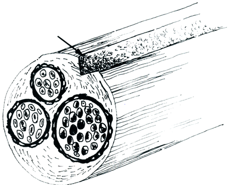
Sural nerve autografts 9mm or 18mm long were taken from the opposite hindpaw (Fig. 2).
The epineurium of the right sciatic nerve was incised using a number 11 blade. Care was taken to avoid damage to sciatic nerve fascicles (Fig. 3A). The sural nerve grafts were partially buried underneath the epineurium of sciatic nerves (Fig. 3B), and fixed by two 10-0 nylon epineural stitches, as follows: Group 1 (n=20) - sural nerve grafts, 9mm long, placed 1mm distally to the neurorrhaphys site; Group 2 (n=20) - sural nerve grafts, 9mm long, placed 10mm distally to the neurorrhaphys site; Group 3 (n=20) - sural nerve grafts, 18mm long, placed 1mm distally to the neurorrhaphys site (Fig. 4).

Skin incision was sutured with interrupted 4-0 nylon sutures.The left hindpaw served as controls.
The histological studies were made after 3 months and 6 months. The electromyographic study was perfomed after 6 months.
Morphological study
Three months after surgical procedure, part of the right sciatic nerve includind the graft was removed from the thigh of 45 rats ( 15 from each group). The excised segments of nerve were fixed with 8% paraformaldehyde and embedded in historesin. The 1m m sections were stained with toluidine blue and examined by light microscopy.
Morphometric studies
The right sciatic nerve of 15 rats, being 5 from each group, were removed after six months of the operation. The nerves were fixed with 2,5% glutaraldehyde and washed in sodium buffer (0,2M). The fixed tissues were postfixed with 2% osmium tetroxide, dehydrated in a series of alcohol baths from 50% to 100%, and embedded in Epon 812. Transverse sections 0,5m m were taken in every specimen at 2 locations and stained with toluidine blue. Under 2.500 x magnification the section through the sciatic nerve was photographed. Six photomicrographs, three proximal and three distal ones, were used to count myelinated axons and to measure external nerve fiber diameters in each nerve.
Using a computer-linked digital pen (MOP- Video Plan, Kontron Eletronik, Germany) external fiber diameters were measured. The number of myelinated axons were counted by area (mm2).
Electrophysiological study
Six months after surgical procedure 15 rats , being 5 animals from each group, were re-anaesthetized (ethylic ether). Three stainless steel electrodes were implanted, one into the belly of the extensor digitorum longus muscle, another into the distal tendon of the same muscle, and the other (earth) was laid on the surface of the gastrocnemius muscle. The same procedure was made bilaterally. The three wires were threaded under the skin and joined to a three-pin socket attached with dental cement to the surface of the skull.
Forty eight hours after recovery from surgery the animal was connected via a headset containing an amplifier to a grass polygraph (Lafayette Inst. Co.). The polygraph was calibrated in 100 mV/cm. The EMG signal was amplified, filtered and the resultant signal was recorded in paper. Only EMG potential at rest were considered.
Statistical assessment
The data with normal distribution were compared using the Students t test and ANOVA, otherwise, the data were assessed with Mann-Whitney "U" and Kruskal-Wallis "H" tests. Statistical significance was accepted at p < 0,05.
RESULTS
Morphology
Group 1 - It was not observed inflammatory infiltrate in the nerve and in the connective tissue. Mastocytes were seen in the epineurium and in the endoneurium. Blood vessells were present in large number filled with erythrocytes. Vacuolization in nerve fibers was evident in small number.
Group 2 - Mastocytes (Fig. 5) and dilated blood vessels were observed in sciatic nerve in a greater number than in those from Group 1. Vacuolization was seen in most of the nerve fibers also in a greater number than in those from Group 1 (Fig. 6). No inflammatory infiltrate was observed.
Group 3- In the endoneurium vessels with smaller diameter and mastocytes with characteristics size and form were present in a smaller number than in nerves from Groups 1 e 2 . Nerve fibers had also fewer vacuoles than in Groups 1 e 2. Most of the nerve fibers showed a normal aspect.
Morphometry
External nerve fiber diameters
Rats from Control Group (Fig. 7) showed mean external nerve fiber diameters significantly greater than those from Group 1, 2 and 3 (p < 0,001). The mean diameters in Group 3 were significantly greater than that in Group 2 (Fig. 8, 9 and 10). Rats from Group 1 presented mean diameters greater than in Group 2 (Table 1).
Axonal counts
Mean nerve fiber density by mm2 (fb/mm2) in Groups 1, 2 and 3 was significantly different from that in Control Group. On the other hand there were no significant differences among values from Groups 1, 2 and 3 (Table 2).
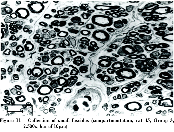
Nerve fibers were observed in some nerves from Groups 1, 2 and 3 arranged into small fascicles, containing both myelinated and unmyelinated fibers delimited by perineurium (compartmentation, Fig. 11).
Electromyography
The maximum amplitude of electromyographic (EMG) potentials did not differ significantly both in normal paws (ANOVA, F=2,15; p=0,168) and in the lesioned ones (ANOVA, F=0,82, p=0,465,Table 3). Also, the maximum frequency of EMG potentials in normal paws did not differ significantly in Groups 1, 2 and 3. The same occurred with the lesioned paws (ANOVA, F=0,14, p=0,868, Table 4).
DISCUSSION
Extensive earlier works investigating the histological aspects of regeneration in rats sciatic nerve are found in the literature(2,5,13,14).
The operating procedure used to bury the sural nerve grafts underneath the epineurium in our work was an incision with a number 11 blade(3. 4). HUDSON et al., in 1980, referred that this procedure when performed carefully did not cause histological and functional damage in 108 rat sciatic nerves studied. The Schwann cells from buried nerve grafts would act as nerve growth factors (NGF). It was suggested that neurotrophic factors would influence the guidance of regenerating axons(15).
The features observed in morphology were inflammatory infiltrate, vasculature, mastocytes and vacuolization in nerve fibers.The latter was of great value in previous work and was used in a pathological staging system(4). Sciatic nerves from Group 3 showed better histological elements of regeneration than those from Groups 1 and 2.
Mastocytes are normal components in mammalian peripheral nerves. In human beings only occasional mastocytes are seen. The number of mastocytes in the experimental Wallerian degeneration rises four to five times(7).
The raise in number of capillaries represented na attempt of revascularization in lesioned nerves (16).
The concept of "compartmentation" was introduced by MACKINNON et al., in 1985(13). It was suggested by these authors that the components of these compartments represented a functional unit as the regenerating nerve attempted to reconstitute its own endoneurial environment. The "compartmentation" was equally observed in our experiment in rats from Groups 1, 2 and 3.
Mean external fiber diameters and density of fibers/mm2 were used as parameters for morphometry in this work. Rats from Group 3 presented greater mean diameters than those from Groups 1 and 2 . Group 1 showed greater mean external diameters than those from Group 2. It was not observed significant differences in mean density of nerve fibers/mm2 in sciatic nerves from Groups 1, 2, and 3. The density of fibers/mm2 in sciatic nerves from Control Group was significantly smaller than in Groups 1, 2 and 3.
For evaluation of function recovery we used the analysis of maximum amplitude and maximum frequency of EMG potentials(1,6), at rest. EMG potentials with the animal in movement were discarded due to the uncertainty of correlating maximium intensity movements of different animals even with the same stimulus, minimizing this variant.
The development of new techniques and the understanding of the way that neurotrophic factors act influencing nerve regeneration are promising trails to be followed.
CONCLUSIONS
The epineural buring of sural nerve grafts with greater length and placed next to the neurorrhaphys site had significantly better regeneration of the histological features than those smaller ones and put distant from neurorrhaphy. Nerve grafts with the same length placed next to the neurorrhaphys site were morphologically and morphometrically better than the distant ones, although no significant differences were observed in the electromyographic study.
Cunha MTR, Silva AL, Reis MG, Silva VJD. Sepultamento epineural parcial de enxertos de nervo com tamanhos diferentes, próximos ou distantes da neurorrafia: estudos histológico e eletrofisiológico em nervos ciáticos de rato. Acta Cir Bras [serial online] 2001 Out-Dez;16(4). Disponível em: URL: http://www.scielo.br/acb.
RESUMO: O objetivo foi comparar e correlacionar, histológica e eletromiograficamente, os efeitos do sepultamento parcial de segmentos de nervo sural em nervos ciáticos de rato, seccionados e suturados. Sessenta ratos adultos, da linhagem Wistar, foram operados em três Grupos: Grupo 1 , um segmento de nervo sural com 9mm. de comprimento colocado próximo à neurorrafia; Grupo 2 , um segmento de nervo sural com 9mm. de comprimento sepultado 10mm. distante da neurorrafia ; Grupo 3, um enxerto de nervo sural com 18mm. de comprimento, posicionado próximo à neurorrafia. Os pormenores histológicos foram examinados sob microscopia de luz em 45 ratos, 3 meses depois. Os elementos observados foram : vascularização, vacúolos nas fibras nervosas, mastócitos e infiltrado inflamatório. A morfometria foi realizada, após 6 meses, em 15 ratos dos Grupos 1, 2 e 3, medindo-se os diâmetros externos e contando-se as fibras nervosas mielinizadas por mm². O estudo eletrofisiológico foi feito após 6 meses, registrando a amplitude máxima e a freqüência dos potenciais eletromiográficos no músculo extensor longo dos dedos, em repouso. Os ratos do Grupo 3 apresentaram-se morfologicamente melhor conservados e com diâmetros de fibras maiores do que nos Grupos 1 e 2. Não houve diferença na densidade de fibras/mm² e no estudo eletrofisiológico entre os ratos dos Grupos 1 , 2 e 3. O sepultamento epineural de segmentos de nervo sural com maior comprimento e colocados próximos à neurorrafia apresentaram pormenores morfológicos de regeneração significantemente melhores do que os menores e posicionados distantes da sutura do nervo.
DESCRITORES: Nervo ciático. Nervo sural. Regeneração nervosa. Degeneração neural. Ratos.
Conflito de interesses: nenhum
Fontes de financiamento: nenhuma
Address for correspondence:
Marco Túlio Rodrigues da Cunha
Rua: Alfén Paixão, 180/202
Uberaba MG
38060-230
Fax: (34) 312-0249
Data do recebimento: 26/04/2001
Data da revisão: 01/06/2001
Data da aprovação: 08/07/2001
- 1 Qing Z, Shi-Bi L, Jian S. Neurotrophism to tissue specificity in nerve regeneration. Chin Med J 1989;102:926-30.
- 2 - Gulati AK. Evaluation of acellular and cellular nerve grafts in repair of rat peripheral nerve. J Neurosurg 1988;68:117-23.
- 3 - Hall S, Berry M. Electron microscopic study of the interaction of axons and glia at the site of anastomosis between the optic nerve and cellular or acellular sciatic nerve grafts. J Neurocitol 1989;18:171-84.
- 4 - Son Y, Thompson WJ. Schwann cell processes guide regeneration of peripheral axons. Neuron 1995;14:125-32.
- 5 - Gibson K, Daniloff JK. Peripheral nerve repair. Compen Count Vet Ed 1989;11:938-45.
- 6 - Lee HK, Chung MS, Kim HJ. A comparison of the passage of regenerating axons through old degenerated nerve autografts and fresh nerve autografts in rats. Int Orthop 1993;17:193-7.
- 7 - Terzis JK, Liberson WT, Peffley C. Reinervation of peripheral nerve grafts by spinal cord fibres tested by transcranial brain stimulation: electromyogr Clin Neurophysiol 1989;29:417-23.
- 8 Cunha, MTR. Estudo microscópico das integraçőes de enxertos de nervo sural, após ressecçăo segmentar ou incisăo epineural, em nervos ciáticos esmagados de rato [Tese-Mestrado]. Universidade Federal de Minas Gerais; l993.
- 9 Cunha MTR, Silva AL, Fenelon SB. Comparison of nerve graft integration after segmentar resection versus epineural burying in crushed rat sciatic nerves. Acta Cir Bras 1997;12(4):221-5.
- 10 - Hudson AR, Gentili F, Hunter D. The role of internal neurolysis in peripheral nerve surgery. Can J Neurol Sci 1980;7:335.
- 11 Bain JR, Mackinnon SE, Hunter DA. Functional evaluation of complete sciatic, peroneal and posterior tibial nerve lesions in the rat. Plast Reconstr Surg 1989;83:129-36.
- 12 - De Medinaceli, L. Functional consequences of experimental nerve lesions: effects of time, location and extent of damage. Exp Neurol 1988;l00:l54-65.
- 13 - Mackinnon SE, Hudson AR, Hunter DA. Histologic assessment of nerve regeneration in the rat. Plast Reconstr Surg 1985;75:384-8.
- 14 - Powell HC, Myers RR. Pathology of experimental nerve compression. Lab Invest 1986;55:91-100.
- 15 - Gamble HJ, Goldby S. Mast cells in peripheral nerve trunks. Nature 1961;l89:766.
- 16 - Rydevik B, Lundborg G. Permeability of intraneural microvessels and perineurium following acute, graded experimental nerve compression. Scand J Plast Reconstr Surg 1977;11:l79-87.
- 17 - Atrakchi A, Gray SD, Carlsen RC. Development of soleus muscle in SHR: relationship of muscle to rise in blood pressure. Am J Phisiol 1994;267:C827-35.
- 18 Double KL, Crocker AD. Dopamine receptors in the substantia nigra are involved in the regulation of muscle tone. Proc Natl Acad Sci 1995;92:1669-73.
Publication Dates
-
Publication in this collection
11 Sept 2003 -
Date of issue
Dec 2001
History
-
Received
26 Apr 2001 -
Reviewed
01 June 2001 -
Accepted
08 July 2001