Abstracts
The objective of this paper was to develop an experimental model to be used in the study of Transcutaneous Electrical Nerve Stimulation (TENS) on viability of random skin flap in rats. The sample was 15 Wistar-EPM rats. The random skin flap measured 10x4 cm and a plastic barrier was placed between the flap and the donnor site. The animals were submited to TENS for 1 hour immediately after the surgery and on the two subsequent days. On the seventh postoperative day, the percentage of necrotic area was measured and calculated. The experimental model proved to be reliable to be used in the study of effects of Transcutaneous Electrical Nerve Stimulation in random skin flap in rats.
Electrical stimulation; Skin flap; Necrosis
Este artigo propõe o desenvolvimento de um modelo experimental para verificar o efeito da Estimulação Elétrica Nervosa Transcutânea (TENS) na viabilidade do retalho cutâneo randômico em ratos. A amostra constituiu-se de 15 ratos, da linhagem Wistar-EPM. O retalho cutâneo randômico foi realizado com dimensões de 10x4 cm e uma barreira plástica foi interposta entre o mesmo e o leito doador. Os animais foram submetido à TENS por 1 hora imediatamente após a operação e nos outros dois dias subseqüentes. No sétimo dia pós-operatório foram calculadas as porcentagens de área de necrose. O modelo experimental mostrou-se factível para estudo dos efeitos da Estimulação Elétrica Nervosa Transcutânea em retalho cutâneo randômico em ratos.
Estimulação elétrica; Retalho cutâneo; Necrose
EXPERIMENTAL MODELS
Experimental model for transcutaneous electrical nerve stimulation on ischemic random skin flap in rats1 1 This study is part of a thesis presented to Federal University of São Paulo - Paulista School of Medicine, for Master Degree in Basic Sciences in Plastic Surgery.
Modelo experimental para estimulação elétrica nervosa transcutânea em retalho cutâneo randômico isquêmico em ratos
Richard Eloin LiebanoI; Lydia Masako FerreiraII; Miguel Sabino NetoIII
IMaster in Basic Sciences in Plastic Surgery by Federal University of São Paulo - Paulista School of Medicine (UNIFESP-EPM). Brazil.
IIHead of Plastic Surgery Division of Surgery Department and Coordinator of Post-graduation Program in Reconstructive Plastic Surgery - UNIFESP - EPM. Brazil.
IIIProfessor of Post-graduation Program in Reconstructive Plastic Surgery of UNIFESP-EPM. Brazil.
Correspondence to Correspondence: Richard Eloin Liebano UNIFESP-EPM, Plastic Surgery Division, Surgery Division Rua Napoleão de Barros, 715, 4º andar 04024-900 São Paulo SP Tel: (11)557604118 FAX: (11) 55716579 sandra.dcir@epm.br charde@riopreto.com.br
ABSTRACT: The objective of this paper was to develop an experimental model to be used in the study of Transcutaneous Electrical Nerve Stimulation (TENS) on viability of random skin flap in rats. The sample was 15 Wistar-EPM rats. The random skin flap measured 10x4 cm and a plastic barrier was placed between the flap and the donnor site. The animals were submited to TENS for 1 hour immediately after the surgery and on the two subsequent days. On the seventh postoperative day, the percentage of necrotic area was measured and calculated. The experimental model proved to be reliable to be used in the study of effects of Transcutaneous Electrical Nerve Stimulation in random skin flap in rats.
KEY WORDS: Electrical stimulation. Skin flap. Necrosis.
RESUMO: Este artigo propõe o desenvolvimento de um modelo experimental para verificar o efeito da Estimulação Elétrica Nervosa Transcutânea (TENS) na viabilidade do retalho cutâneo randômico em ratos. A amostra constituiu-se de 15 ratos, da linhagem Wistar-EPM. O retalho cutâneo randômico foi realizado com dimensões de 10x4 cm e uma barreira plástica foi interposta entre o mesmo e o leito doador. Os animais foram submetido à TENS por 1 hora imediatamente após a operação e nos outros dois dias subseqüentes. No sétimo dia pós-operatório foram calculadas as porcentagens de área de necrose. O modelo experimental mostrou-se factível para estudo dos efeitos da Estimulação Elétrica Nervosa Transcutânea em retalho cutâneo randômico em ratos.
DESCRITORES: Estimulação elétrica. Retalho cutâneo. Necrose.
Introduction
Skin flaps are largely used in all fields of plastic surgery, especially in reconstructives1. They have been utilizated for centuries and, during this time, one preoccupation has been to develop techniques to provide more assurance in skin flap realization. The research of skin flaps survival mechanisms and your possibles curate factors have been motive for publications.2,3,4 But in spite of all of these studies related with random skin flap survival, little progress was gotten in this field in the last 50 years.5
It is know that one of main complications that occur in the creation of the flap is ischemia, which, in a large number of cases, develops to tissue necrosis taking a failure in proposed treatment.2 Due to that complication, a considerable amount of research has been done with the aim of improving the blood flow in flaps, decreasing ischemic conditions and preventing necrosis3, 6
In the literature, there have been publications about several types of drugs, such as vasodilators, calcium channel blockers, prostagalndin inhibitors, anticoagulants, antiadrenergics and antioxidants.3,7,8,9 However, many of those present undesirable adverse effects, which makes their use in clinical practice unviable.3 Therefore, a new research field using nonpharmacological agents such as acupuncture and electroacupuncture10,11, pulsed electromagnetic energy12, low-power lasers13,14,15, polarized low frequency electrical currents16,17 and nonpolarized currents18,19 has emerged.
Among these resources, the transcutaneous electrical nerve stimulation has deserved detach due to its low cost and application facility, begining to be studied as possible method in the treatment of ischemic skin flaps.
Proposition
Due to the characteristics of this kind of stimulation, associated with the methodological deficiency of few experimental studies performed in animals with relation of current parameters and operation procedure, the purpose of this study was to develop an experimental model to be used in the study of Transcutaneous Electrical Nerve Stimulation (TENS) on viability of random skin flap in rats.
Method description
The study was approved by Comission of Ethics in Research of Universidade Federal de São Paulo Escola Paulista de Medicina going in the current apt legislation.
Animals
The present study used 15 rats (Rattus norvegicus: var. albinus, Rodentia, Mammalia), adults, males, of lineage Wistar EPM 1, weighing 260 to 330 g.
Electrostimulator
The device used in this study was the Orion Tens® [Orion Aparelhos para Fisioterapia LTDA; serie number 00849], digital e controlled by microprocessor. The emitted pulses are rectangulars, biphasics and simmetricals. With electrical stimulator, it was used a cable, two silicone electrodes (4,2 x 1,5 cm), gel and adhesive tape for fix the electrodes.
Operation technique
In animals, it was realized a random skin flap with cranial base, measuring 10 cm length and 4 cm width. The animals were anesthestized with sodium thiopental (50 mg/kg) intraperitoneally during the operatory procedure and during electrostimulation sessions. After anesthesia induction, the rats were positioned in a plane surface with members extended and it was performed a digital tricotomy in their backs.
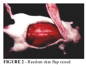
Then it was done the planning of the flap through a plastic mould [film F-1 (poliester + poliethilene)], cut out in pattern sizes (10x4 cm) in the backs of animals, taking as limits the inferior angles of the scapulae and the superior bones of pelvis (Figure 1). The random skin flap with cranial base was cut by scalpel and elevated through deep fascia, including the superficial fascia, panniculus carnosus, the subcutaneous tissue and skin20,9 (Figure 2). After flap elevation a plastic barrier (film F1), with same dimensions (10x4 cm), was placed between it and the donnor site.7 The suture was realized with simple nylon 4-0 stitches (Figure 3).9
After operatory procedure, the animals were kept anesthetized and subjected to Transcutaneous Electrical Nerve Stimulation (TENS) for 1 hour and, on the two subsequent days, in determinated time, with high intensity (± 15 mA) and high frequency (80 Hz). The electrodes were placed on the base of the flap, where the first was positioned thorough of incision beginning and the second apart 1,5 cm from first electrode, in direction of a distal portion of the flap (Figure 4). The electrical pulses had a duration of 200 microseconds. After the electrical stimulation sessions, the animals got back to their respective cage and received commercial ration and water ad libitum.
Method for estimate percentage of necrotic area in distal portion of flaps
The percentage of skin flap necrosis area was calculated on the seventh postoperative day via the paper template method. The limit between viable tissue characterized by soft skin, rosy, warm and with hair and necrotic tissue (stiff skin, dark, cool and without hair) was demarcated in the animals.
A mould of entire flap was drawn and cut in transparent paper, being checked in a precision balance (+/- 0,001g error). It was cut from this fragment just the correspondent area to flap necrosis that was also checked.
After that, it was used the following formule:
Perspectives
The experimental model proved to be reliable to be used in the study of effects of transcutaneous electrical nerve stimulation in random skin flap in rats.
Conflict of interest: none
Finantial source: CAPES
Data do recebimento: 22/ 04/2003
Data da revisão: 18/05/2003
Data da aprovação: 28/07/2003
- 1. Ferreira LM. Retalhos Cutâneos. In: Ferreira LM. Manual de Cirurgia Plástica. 1ed. Săo Paulo: Atheneu; 1995. p. 45-62.
- 2. Kerrigan CL. Skin flap failure: pathophysiology. Plast Reconstr Surg 1983;72:766-77.
- 3. Gherardini G, Lundeberg T, Cui J, Eriksson SV, Trubek S, Linderoth B. Spinal cord stimulation improves survival in ischemic skin flaps: an experimental study of the possible mediation by calcitonin gene-related peptide. Plast Reconstr Surg 1999;103:1221-8.
- 4. Salmi AM, Hong C, Futrell JW. Preoperative cooling and warming of the donor site increase survival of skin flaps by the mechanism of ischaemic preconditioning: an experimental study in rats. Scand J Plast Reconstr Hand Surg 1999;33:163-7.
- 5. Silva ER. Retalhos Cutâneos. In: Mélega JM, Zanini SA, Psillakis JM. Cirurgia plástica reparadora e estética. Rio de Janeiro: MEDSI; 1988.
- 6. Davis ER, Wachholz JH, Jassir D, PerlynCA, Agrama MH. Comparison of topical anti-ischemic agents in salvage of failing random-pattern skin flaps in rats. Arch Facial Plast Surg 1999;1:27-32.
- 7. Ugland O. Flaps and flap necrosis. Acta Chir Scand 1966;131:408-12.
- 8. Jurell G, Jonsson, CE. Increased survival of experimental skin flaps in rats following tretment with antiadrenergic drugs. Scand J Plast Reconstr Surg 1976;10:169-72.
- 9. Abla LEF. Efeito da N-Acetil-L-Cisteína sobre a necrose dos retalhos cutâneos, em ratos. [Tese Mestrado]. Universidade Federal de Săo Paulo - Escola Paulista de Medicina; 1997.
- 10. Jansen G, Lundeberg T, Samuelson UE, Thomas M. Increased survival of ischemic musculocutaneous flaps in rats after acupuncture. Acta Physiol Scand 1989;135:555-8.
- 11. Niina Y, Ikeda K, Iwa M, Sakita M. Effects of electroacupuncture and transcutaneous electrical nerve stimulation on survival of musculocutaneous flap in rats. Am J Chin Med 1997;25:273-80.
- 12. Krag C, Taudorf U, Siim E, Bolund S The effect of pulsed eletromagnetic energy (Diapulse) on the survival of experimental skin flaps. Scand J Plast Reconstr Surg 1979;13:377-80.
- 13. Kami T, Yoshimura Y, Nakajima T, Ohshiro T, Fujino T. Effects of low-power diode lasers on flap survival. Ann Plast Surg 1985;14:278-83.
- 14. Smith RJ. The effect of low-energy laser on skin-flap survival in the rat and porcine animal models. Plast Reconstr Surg 1992;89:306-10.
- 15. Amir A, Solomon AS, Giler S, Cordoba M, Hauben DJ. The influence of helium-neon laser irradiation on the viability of skin flaps in the rat. Br J Plast Surg 2000;53:58-62.
- 16. Kjartansson J, LundebergT, Samuelson UE, Dalsgaard J. Transcutaneous electrical nerve stimulation (TENS) increases survival of ischemic musculocutaneous flaps. Acta Physiol Scand 1988;134:95-9.
- 17. Im JM, Lee WPA, Hoopes JE. Effect of electrical stimulation on survival of skin flaps in pigs. Phys Ther 1990;70:37-40.
- 18. Lundeberg T, Kjartansson J. Effect of electrical nerve stimulation on healing of ischemic skin flaps. Lancet 1988;24:712-4.
- 19. Kjartansson J, Lundeberg T. Effects of electrical nerve stimulation (ENS) in ischemic tissue. Scand J Plast Reconstr Hand Surg 1990;24:129-34.
- 20. McFarlane RM, DeYoung G, Henry RA. The design of a pedicle flap in the rat to study necrosis and its prevention. Plast Reconstr Surg 1965;35:177-82.
Publication Dates
-
Publication in this collection
16 Jan 2004 -
Date of issue
2003
History
-
Accepted
28 July 2003 -
Reviewed
18 May 2003 -
Received
22 Apr 2003


