Abstracts
The study of acute arterial insufficiency of the extremities is an area of continuing interest and investigation, in light of the possible effects stemming from the evolution of the disease and the necessity for urgent treatment. PURPOSE: To analyze the effects of the interruption of the flow in the normal arterial endothelium morphology and correlate them with the ischemia duration. METHODS: We submitted 30 rabbits to the ligature of the right external iliac artery for 6 hours or 72 hours and observed specific morphological variables in the endothelial layer under optical and electronic microscopy. RESULTS: In the optical microscopic study, no statistically significant results were observed in the comparison of the groups (control, 6- and 72-hour occlusions). With electronic microscopy, we observed alterations in the endothelial cell characterized by hyperpigmentation with detachment of the same from its bed; and rupture of the internal elastic membrane, with the exposure of the subendothelial material to the vascular lumen. CONCLUSIONS: The optical microscopy was not an effective method for the determination of endothelial morphological alterations; the electronic microscopy allowed us to observe initial signals of the endothelial cell and layer injury 72 hours after the interruption of the normal arterial flow.
Endothelium; Microscopy, electron; Ischemia; Rabbits
O estudo da insuficiência arterial aguda das extremidades é área de contínuo interesse e investigação, devido possibilidade de eventos catastróficos na evolução da doença e necessidade de tratamento cirúrgico de urgência. OBJETIVO: Analisar os efeitos da ausência de fluxo na morfologia do endotélio arterial normal segmentar, como os que ocorrem na porção imediatamente abaixo da área que sofreu oclusão arterial aguda por embolia, e correlacioná-los com o tempo de isquemia. MÉTODOS: Submetemos 30 coelhos à ligadura da artéria ilíaca externa direita por 6 horas ou 72 horas e observamos variáveis morfológicas específicas da camada endotelial e subendotelial à microscopia óptica e eletrônica. RESULTADOS: No estudo da microscopia óptica não foram observados resultados estatisticamente significativos quando comparados os grupos entre si. À microscopia eletrônica observamos alterações da célula endotelial caracterizadas por hiperpigmentação e descolamento da mesma de seu leito, e ruptura da membrana elástica interna com exposição do material subendotelial para luz vascular. CONCLUSÕES: A microscopia óptica não foi procedimento eficaz na determinação das alterações morfológicas endoteliais estudadas; a microscopia eletrônica mostrou sinais iniciais de sofrimento da célula endotelial e lesão da camada endotelial após 72 horas da ausência de fluxo na artéria normal.
Endotélio; Microscopia eletrônica; Isquemia; Coelhos
ORIGINAL ARTICLE
Analysis of the effects of the interruption of the flow in the normal arterial endothelium morphology and the correlation with the ischemia duration in rabbits1 1 Research performed at Department of Surgery, Division of Vascular Surgery, Santa Casa de São Paulo, Faculty of Medical Sciences (FCMSCSP), São Paulo, Brazil.
Análise dos efeitos da interrupção do fluxo na morfologia do endotélio arterial normal e a correlação com o tempo de isquemia em coelhos
Walkíria Ciappina HuebI; Henrique Jorge Guedes NetoII; Carmem Lúcia Penteado LancelottiIII; Valter Castelli JúniorIII; Roberto Augusto CaffaroIV
IMD, Vascular Surgeon, FCMSCSP, São Paulo, Brazil
IIAssistant Professor, Division of Vascular Surgery, FCMSCSP, São Paulo, Brazil
IIIAssociate Professor, Division of Vascular Surgery, FCMSCSP, São Paulo, Brazil
IVHead of the Division of Vascular Surgery, FCMSCSP, São Paulo, Brazil
Correspondence Correspondence: Dra. Walkíria Ciappina Hueb Rua Macaia-Mirim 89/23 02013-080 São Paulo - SP Brazil Phone/Fax: (55 11)8159-2417 walkhueb@uol.com.br
ABSTRACT
The study of acute arterial insufficiency of the extremities is an area of continuing interest and investigation, in light of the possible effects stemming from the evolution of the disease and the necessity for urgent treatment.
PURPOSE: To analyze the effects of the interruption of the flow in the normal arterial endothelium morphology and correlate them with the ischemia duration.
METHODS: We submitted 30 rabbits to the ligature of the right external iliac artery for 6 hours or 72 hours and observed specific morphological variables in the endothelial layer under optical and electronic microscopy.
RESULTS: In the optical microscopic study, no statistically significant results were observed in the comparison of the groups (control, 6- and 72-hour occlusions). With electronic microscopy, we observed alterations in the endothelial cell characterized by hyperpigmentation with detachment of the same from its bed; and rupture of the internal elastic membrane, with the exposure of the subendothelial material to the vascular lumen.
CONCLUSIONS: The optical microscopy was not an effective method for the determination of endothelial morphological alterations; the electronic microscopy allowed us to observe initial signals of the endothelial cell and layer injury 72 hours after the interruption of the normal arterial flow.
Key words: Endothelium. Microscopy, electron. Ischemia. Rabbits.
RESUMO
O estudo da insuficiência arterial aguda das extremidades é área de contínuo interesse e investigação, devido possibilidade de eventos catastróficos na evolução da doença e necessidade de tratamento cirúrgico de urgência.
OBJETIVO: Analisar os efeitos da ausência de fluxo na morfologia do endotélio arterial normal segmentar, como os que ocorrem na porção imediatamente abaixo da área que sofreu oclusão arterial aguda por embolia, e correlacioná-los com o tempo de isquemia.
MÉTODOS: Submetemos 30 coelhos à ligadura da artéria ilíaca externa direita por 6 horas ou 72 horas e observamos variáveis morfológicas específicas da camada endotelial e subendotelial à microscopia óptica e eletrônica.
RESULTADOS: No estudo da microscopia óptica não foram observados resultados estatisticamente significativos quando comparados os grupos entre si. À microscopia eletrônica observamos alterações da célula endotelial caracterizadas por hiperpigmentação e descolamento da mesma de seu leito, e ruptura da membrana elástica interna com exposição do material subendotelial para luz vascular.
CONCLUSÕES: A microscopia óptica não foi procedimento eficaz na determinação das alterações morfológicas endoteliais estudadas; a microscopia eletrônica mostrou sinais iniciais de sofrimento da célula endotelial e lesão da camada endotelial após 72 horas da ausência de fluxo na artéria normal.
Descritores: Endotélio. Microscopia eletrônica. Isquemia. Coelhos
Introduction
The acute interruption of the arterial flow due to the presence of thrombi, emboli or spasms, causes the sudden loss of tissue perfusion and the functional harming the affected organ.1 After the event, a series of enzymatic metabolic processes are triggered, determining a variable clinical condition, possibly culminating in the loss of a member, when it is an ischemia in the extremities, or even in the loss of life.2 Due to the deleterious effects of the occlusion, the reperfusion must be reestablished as soon as possible, once the reversibility of the process is directly proportional to the ischemia duration.3,4 Upon analyzing specifically the segmentary arterial alterations caused by the absence of arterial flow we observed exuberant regional physiological and morphological events.5-8 The literature is vast in its documentation of physiological alterations due to the interruption of the arterial flow. However, little is known about its effects on the normal arterial endothelium morphology, such as those which occur in the normal arterial port9-12
Purpose
Analyze the effects of the interruption of the flow in the normal segmental arterial endothelium morphology to the area suffering acute arterial occlusion dute to emboly as well as correlate them to ischemia duration.
Methods
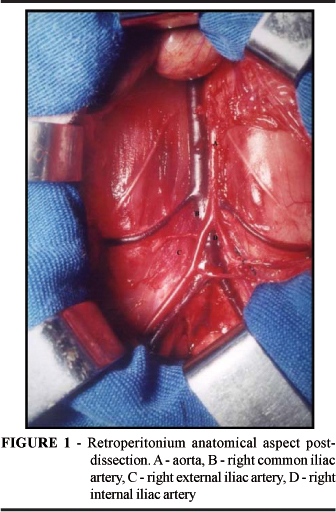
Thirty young adult rabbits of the New Zealand lineage were used. After anaesthetizing the animal with 1% Xilazine 5mg./kg and Chetamine Hydrochlorate 8 mg./kg., a median infra-umbilical abdominal incision was made. With the opening of the abdominal cavity, the splanchnic viscera was shifted to the right) and the visceral peritoneum was moved to permit the visualization of the retroperitoneal structures. The dissection and isolation of the abdominal aorta, common iliac arteries, internal and external, was performed. With the objective od interrupting the flow in the right external iliac artery, the ligature was performed with a 4.0 cotton thread on the following vessels: common iliac artery, immediately before its bifurcation; right internal iliac artery, right after its origin; right external iliac artery, immediately before its trajectory through the inguinal ligament; and small collateral branches of the right external iliac artery (Figure 1). According to the group to which they belonged, the animals were submitted to a determined procedure: control group underwent the resection of the right external iliac artery; occlusion group, 6- and 72- hour closure of the abdominal cavity and after 6 or 72 hours, arterial resection Following preparation and fixation, the material was sent to optical and electronic microscopes analysis.
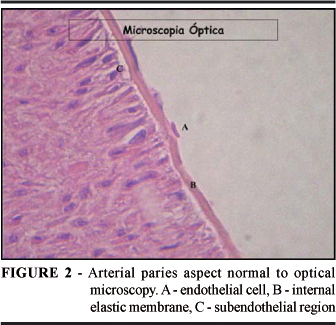
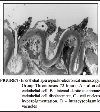
The optical microscope was used to evaluate the presence of thrombus and discontinuity of the internal elastic membrane in the endothelial layer. The electronic microscope was used to research alterations in the nucleus, presence of adhered thrombus and the presence of intracytoplasmatic pinocytotic vacuoles in the endothelial cell; discontinuity, the presence of subendothelial material, alterations in the width and hyperpigmentation in the internal elastic membrane were also evaluated (Figures 2 and 3).
Statistical analysis
We used descriptive statistical analysis to describe the qualitative variables, calculating absolute and relative frequencies. We used comparative analysis to test the homogeneity among the groups, utilizing the exact Fisher test, allowing for a significance level of 5%.
Results
Optical microscopy
The statistical analysis of the variables presence of thrombus and discontinuity of the internal elastic membrane did not reveal a statistically significant difference among the groups (Figures 4 and 5).
Electronic microscopy
The analyzed variables in the endothelial cell alterations in the nucleus, presence of adhered thrombus and presence of intracytoplasmatic pinocytotic vacuoles; and in the internal elastic membrane discontinuity, presence of subendothelial material, alteration in width and hyperpigmentation presented statistically significant differences when performed a comparative analysis among the groups. As for the characteristics of the nucleus of the endothelial cell, it was observed that in the control group all of the samples presented a normal aspect. In that of 72-hour occlusion, alterations were characterized by detachment or hyperpigmentation (p = 0.001). As for the presence of thrombus, a greater incidence was observed in the 6- and 72-hour occlusion groups (p = 0.017). Regarding the presence of intracytoplasmatic pinocytotic vacuoles, a greater incidence was observed in the control and 72-hour occlusion groups, with predominance in the later (66.7%) (p = 0.010). As for the discontinuity in the internal elastic membrane, integrity was observed in all of the control group samples, only 20 % in the 6-hour occlusion group and 0 in the 72-hour occlusion group (p < 0.001). As for the presence of subendothelial material in the internal elastic membrane, it was observed that the 6 and 72-hour occlusion groups presented 70 and 100 % presence of subendothelial content in their layers, respectively; while the control group did not present anything similar (p < 0.001). The analysis of the variables width and coloration revealed an alteration in the 6 and 72-hour occlusions, with no alteration in the control (p < 0.001) (Figures 6 to 10).


Discussion
The choice of the right external iliac artery was based on the fact that it is accessible and that it constitutes a muscular conduction artery, as those of the inferior members where the majority of the thromboembolic phenomena are located. The arterial ligature with a cotton thread is one of the simplest models of thrombosis. Its applicability is based on innumerable papers in the literature, in addition to permitting easy manipulation and representing low cost.13
In our research, we perfomed optical microscopic study, with the aim to analyze the real contribution that this method brings to the diagnosis of ischemic alterations, by comparing the results obtained with this method to those obtained with an electronic microscope. As for the use of the electronic microscope, we know that it constitutes a consecrated method and an immeasurable contribution to the evolution of knowledge in pathologies of the circulatory system. Analyzing our results, we found that the optical microscopy was not an effective method for determining the alterations in the endothelial artery observed in this study, once there are no statistically significant results from the comparison of the groups. Therefore, the discussions which follow are related to the results obtained by using the electronic microscope. We observed that even after the right external iliac artery had undergone ligature, there was no formation of thrombus in the vascular lumen in any of the samples. This interruption can be explained by the reflex contraction of the arterial wall following ligature without the formation of thrombus or reabsorption of the same. Analyzing the endothelial cell, we observed that with the evolution of the ischemia there was an increase in the number of intracytoplasmatic vesicles, appearance of subendothelial vacuoles and the presence of signs of cellular death, characterized by hyperpigmentation and detachment. The presence of the vacuoles and vesicles is shown by the increase in the endothelial cell permeability for ions and fluids, suggesting the intensification of exchanges; and the beginning of apoptosis is due to the duration of local anoxia, this being an intensifying factor in the local thrombogenisis. Analyzing the results related to the membrane, we observed that all of the samples from the control group had intact membranes, and all of the samples from the 72-hour occlusion group showed a loss in the integrity of the membrane, suggesting that the ischemia 72-hour duration factor is related to the rupture of the internal elastic membrane. This result is identical when the variable presence of subendothelial material in the internal elastic membrane is analyzed. Alterations in the internal elastic membrane coloration and width were present in some of the 6- and 72-hour occlusion samples, suggesting initial tissue duress.
Conclusions
The optical microscopy was not an effective method in determining the studied endothelial morphological alterations, while the electronic microscopy allowed us to observe the initial signals of endothelial cell duress as well as the injury in the endothelial layer 72 hours after the interruption of the normal arterial flow.
Received: November 24, 2006
Review: December 19, 2006
Accepted: January 16, 2007
Conflict of interest: none
Financial source: none
- 1. Blaisdell FW, Steele M, Allen RE. Management of acute lower extremity arterial ischemia due to embolism and thrombosis. Surgery. 1978; 84(6):822-4.
- 2. Unthank JL, Nixon JC, Dalsing MC: Acute compensation to abrupt occlusion of rat femoral artery is prevented by NO synthase inhibitors. Am J Physiol. 1994; 267 (6) 2526-30.
- 3. Burton AC: On the physical equilibrium of small blood vessels. Am J Physiol. 1951: 164(2):319-29.
- 4. Haimovici H: Arterial embolism with acute massive ischemic myopathy and myoglobinuria: evaluation of a hitherto unreported syndrome with report of two cases. Surgery. 1960; 47:739-47.
- 5. Gertz SD, Uretsky G, Wajnberg R.S, Nelson E, Kurgan A: Endothelial cell damage and thrombus formation after partial arterial constriction: relevance to the role of coronary artery spasm in the pathogenesis of myocardial infarction. Circulation. 1981; 63(3):476-86.
- 6. Buck RC: The fine structure of endothelium of large arteries. J Biophys Biochem Cytol. 1958; 4(2):189-90.
- 7. Kjeldsen K, Thomsen HK: The effect of hypoxia on the fine structure of the aortic intima in rabbits. Lab Invest. 1975; 33(5):533-43.
- 8. Yu QC, Mergner WJ, VigoritoRD, Resau JH : Postmortem viability and early changes in organ culture of human and rabbit aortic endothelial cells. Pathobiology. 1990; 58(3):138-45.
- 9. Kjeldsen K, Astrup P, Wanstrup J: Ultrastructural intimal changes in the rabbit aorta after a moderate carbon monoxide exposure. Arterosclerosis. 1972; 16(1):67-82.
- 10. Reil TD, Moore WS, Kashyap VS, Nene SS, Gelabert HA, Quinones-Baldrich WJ : The effects of thrombus, thrombectomy and thrombolysis on endothelial function. Eur J Vasc Endovasc Surg. 2000; 19(2):162-8.
- 11. Elemer G, Kerenyi T, Jellinek H: Scanning (SEM) and transmission (TEM) electron-microscopic studies on post-ischemic endothelial lesions following recirculation. Atherosclerosis. 1976; 24(1-2):219-32.
- 12. Clowes AW, Collazzo RE, Karnovsky MJ: A morphologic and permeability study of luminal smooth muscle cells after arterial injury in the rat. Lab Invest. 1978; 39(2):141-50.
- 13. MacDonald JD, Gyorke A, Jacobs JM, Mohammad SF, Sunderland PM, Reichman MV: Acute phase vascular endothelial injury: a comparison of temporary arterial occlusion using an endovascular occlusive balloon catheter versus a temporary aneurysm clip in a pig model. Neurosurgery. 1994; 34(5):876-81.
- 14. Kornowski R, Glikson M, Ohad D, Varda-Bloom N, Battler A : Battler. Electrical injury in the femoral artery of rabbits as a model for arterial thrombosis: a pilot study. Angiology. 1994; 45(4):395-400.
- 15. Margovsky AI, Lord RS, Chambers AJ: The effect of arterial clamp duration on endothelial injury: an experimental study. Aust N Z J Surg. 1997; 67(7):448-51.
- 16. Tamatani S, Ozawa T, Minakawa T, Takeuchi S, Koik T, Tanaka R : Radiologic and histopathologic evaluation of canine artery occlusion after collagen-coated platinum microcoil delivery. Am J Neuroradiol. 1999; 20(4):541-5.
- 17. Ubatuba FB: An animal model for the study of arterial thrombosis. Braz J Med Biol Res. 1989; 22(8):993-1000.
Publication Dates
-
Publication in this collection
14 Mar 2007 -
Date of issue
Apr 2007
History
-
Reviewed
19 Dec 2006 -
Received
24 Nov 2006 -
Accepted
16 Jan 2007