Abstracts
PURPOSE: To study the interatrial conduction times and atrial node performance in patients submitted to mitral valve surgery with the aid of temporary atrial epicardic electrodes. METHODS: The atriograms were carried out in the first postoperative day and before the hospital discharge of ten consecutive patients. RESULTS: Sixty percent of the patients could complete the post-operative study protocol. The main results were: a) Post-operative arrhythmias were detected in 50% of the patients; b) There were no statistical differences between the pre and post-operative 12 lead EKGs. c) The interatrial conduction time (IACT) ranged from 90 to 140ms in the first post-operative day, and from 110 to 130ms at hospital discharge; d) The sinus node recovery time (SNRT) ranged from 250 to 560 ms in the first post-operative day and from 180 to 360ms at hospital discharge; e) The sinus atrial conduction time (SACT) remained between 70 and 140ms, both in the first post-operative day and at hospital discharge, and; f) The IACT was normal in patients whose left atrium (LA) was less than 50mm in diameter but supra normal in the remaining cases. CONCLUSIONS: Sinus node function and inter-atrial conduction are not altered by mitral valve operation. Post-operative programmed epicardic atrial stimulation is easy and safe.
Epicardial; Atriograms; Atrial Arrhythmias; Mitral Valve Disease; Cardiac Surgery
OBJETIVO: Estudar os tempos de condução interatrial e a função do nó sinusal em pacientes submetidos a tratamento cirúrgico. MÉTODOS: Foram estudados 10 pacientes adultos consecutivos submetidos à operação de correção de valvopatia mitral. Registraram-se atriogramas usando eletrodos epimiocárdicos cirurgicamente implantados. Os atriogramas foram obtidos no primeiro dia do pós-operatório e antes da alta hospitalar. RESULTADOS: Os principais achados foram: a) A incidência de arritmias atriais até alta hospitalar foi de 50 %; b) O tempo de condução interatrial (TCIA) variou de 90 a 140 ms no 1°PO e 110 a130 ms antes da alta hospitalar; c) O tempo de recuperação do nó sinusal (TRNS) variou de 250 a 560 ms no 1°PO e180 a 360 ms antes da alta hospitalar; d) O tempo de condução sinoatrial (TCSA) variou de 70 a 140 ms tanto no 1ºPO, como antes da alta hospitalar e; d) O tempo de condução interatrial (TCIA) foi normal em pacientes com átrio esquerdo menores do que 50 mm e supranormal nos outros casos. CONCLUSÃO: As funções do nó sinusal e a condução interatrial não foram alteradas pelo tratamento cirúrgico da valvopatia mitral. A estimulação atrial epicárdica programada é segura de fácil realização.
Epicárdio; Atriogramas; Arritmias Atriais; Valvopatia Mitral; Cirurgia Cardíaca
21CLINICAL RESEARCH
ORIGINAL ARTICLE
CARDIOVASCULAR SURGERY
Sinus node function in patients operated for mitral valve disease. Indirect evaluation with epimyocardial electrodes1 1 . Investigation carried out at the Division of Thoracic and Cardiovascular Surgery, Department of Surgery and Anatomy, Ribeirão Preto Faculty of Medicine, University of São Paulo, Brazil.
Função do nó sinusal em pacientes submetido a correção cirúrgica da valvopatia mitral. Avaliação indireta com eletrodos epimiocárdicos
Jairo Rosa Silva JuniorI; Cesar Augusto FerreiraI; Alfredo José RodriguesI; Walter Villela de Andrade VicenteII; Paulo Roberto Barbosa EvoraIII
IMD, PhD, Division of Thoracic and Cardiovascular Surgery, Department of Surgery and Anatomy, Ribeirão Preto Faculty of Medicine, University of São Paulo, Brazil
IIMD, PhD and Head of Division Thoracic and Cardiovascular Surgery, Department of Surgery and Anatomy, Ribeirão Preto Faculty of Medicine, University of São Paulo, Brazil
IIIFull Professor and Head, Division of Thoracic and Cardiovascular Surgery, Department of Surgery and Anatomy, Ribeirão Preto Faculty of Medicine, University of São Paulo, Brazil
Correspondence Correspondence: Paulo Roberto B. Evora Rua Rui Barbosa, 367. Apt. 15 14015120 Ribeirão Preto, SP, Brazil Phone/fax 55 16 36022497 prbevora@netsite.com.br
ABSTRACT
PURPOSE: To study the interatrial conduction times and atrial node performance in patients submitted to mitral valve surgery with the aid of temporary atrial epicardic electrodes.
METHODS: The atriograms were carried out in the first postoperative day and before the hospital discharge of ten consecutive patients.
RESULTS: Sixty percent of the patients could complete the postoperative study protocol. The main results were: a) Postoperative arrhythmias were detected in 50% of the patients; b) There were no statistical differences between the pre and postoperative 12 lead EKGs. c) The interatrial conduction time (IACT) ranged from 90 to 140ms in the first postoperative day, and from 110 to 130ms at hospital discharge; d) The sinus node recovery time (SNRT) ranged from 250 to 560 ms in the first postoperative day and from 180 to 360ms at hospital discharge; e) The sinus atrial conduction time (SACT) remained between 70 and 140ms, both in the first postoperative day and at hospital discharge, and; f) The IACT was normal in patients whose left atrium (LA) was less than 50mm in diameter but supra normal in the remaining cases.
CONCLUSIONS: Sinus node function and interatrial conduction are not altered by mitral valve operation. Postoperative programmed epicardic atrial stimulation is easy and safe.
Key words: Epicardial. Atriograms. Atrial Arrhythmias. Mitral Valve Disease. Cardiac Surgery.
RESUMO
OBJETIVO: Estudar os tempos de condução interatrial e a função do nó sinusal em pacientes submetidos a tratamento cirúrgico.
MÉTODOS: Foram estudados 10 pacientes adultos consecutivos submetidos à operação de correção de valvopatia mitral. Registraramse atriogramas usando eletrodos epimiocárdicos cirurgicamente implantados. Os atriogramas foram obtidos no primeiro dia do pósoperatório e antes da alta hospitalar.
RESULTADOS: Os principais achados foram: a) A incidência de arritmias atriais até alta hospitalar foi de 50 %; b) O tempo de condução interatrial (TCIA) variou de 90 a 140 ms no 1°PO e 110 a130 ms antes da alta hospitalar; c) O tempo de recuperação do nó sinusal (TRNS) variou de 250 a 560 ms no 1°PO e180 a 360 ms antes da alta hospitalar; d) O tempo de condução sinoatrial (TCSA) variou de 70 a 140 ms tanto no 1ºPO, como antes da alta hospitalar e; d) O tempo de condução interatrial (TCIA) foi normal em pacientes com átrio esquerdo menores do que 50 mm e supranormal nos outros casos.
CONCLUSÃO: As funções do nó sinusal e a condução interatrial não foram alteradas pelo tratamento cirúrgico da valvopatia mitral. A estimulação atrial epicárdica programada é segura de fácil realização.
Descritores: Epicárdio. Atriogramas. Arritmias Atriais. Valvopatia Mitral. Cirurgia Cardíaca.
Introduction
Susceptibility to new onset postoperative atrial fibrillation (AF) after major, and in particular, heart surgery is related to different risk factors. In the general population, old age stands up as the strongest preoperative risk factor, while degenerative tissue alterations including nodal fiber loss, increased sinus node fatty and fibrous tissue, atrial dilatation, atrial fibrosis and focal amyloidal interstitial deposit in the atria constitute other important predisposing markers.13
When heart valve disease is considered, the structural and atrial electromechanical alterations generated by dilation, fibrosis, loss of muscular mass and normal tissue architecture secondary to the hemodynamic overload account for an increased risk for postoperative AF4. In addition, the diagnosis of mitral stenosis emerges as an independent risk factor for postoperative AF3.
Investigation of the propensity for postoperative atrial arrhythmias, although very important, is challenging mostly on account of the multi factorial mechanisms involved and patient selection.1,5 In this study, temporary atrial epicardic electrodes were employed to gather electrophysiological information concerning the interatrial conduction times and atrial node performance in mitral stenosis patients submitted to mitral valve surgery.
Methods
Patients: Ten consecutive adult patients (6 males), operated on for mitral valve disease treatment, over a 14 month period, at the Hospital das Clinicas of the Ribeirão Preto School of Medicine of the University of São Paulo were prospectively evaluated. Inclusion criteria were adult age, elective, first heart operation for isolated mitral valvulopathy; predominant sinus rhythm or intermittent AF or atrial flutter preoperatively; left ventricle ejection fraction higher than 35%; end diastolic left ventricle diameter less than 55mm; avoidance of vasoactive amines and antiarrhythmic drugs for the last 48h preoperatively, and no electrolytic imbalance. Informed consent according to the Ethics Committee was always obtained.
The preoperative evaluation and anesthetic protocols, as well as the operative technique and the postoperative treatment were standardized.
Surgery: The patients were operated on by the same surgeon (WVAV), via median sternotomy, with bicavalascending aorta full cardiopulmonary bypass at 32ºC. The caval veins were snared, intermittent antegrade cold blood cardioplegia was given at 20min intervals and the mitral valve was exposed by subseptal atriotomy. After bypass discontinuation, two right atrium (RA) electrodes were implanted one cm apart, and perpendicular to the interatrial groove, one cm below the superior vena cavaright atrium connection, in order to avoid damaging the sinus node (Figure 1). Another similar pair of electrodes was implanted in the midlateral border of the left atrium. One additional electrode was implanted in the right ventricle diaphragmatic wall.
The surgical procedure, and in hospital morbidmortality was evaluated.
Atriogram record protocol: Twelve lead surface EKGs were registered preoperatively, after chest closure, on admission to the intensive care unit (ICU), and when arrhythmias were detected on continuously monitoring in the ICU, and daily, until discharge from the hospital.
Sinus node function and interatrial conduction times were evaluated according to a modified Narula cardioestimulation test protocol with a cardiostimulator device.
The first test was performed in the first postoperative day with the patient extubated, hemodinamically stable and in sinus rhythm. The surface EKG was recorded at 25mm/s and the basal rate was calculated by measuring the PP interval with a calibrated rule. The right and left arm EKG cables were then hooked to the RA electrodes in order to register the bipolar right atriogram, and the same procedure was repeated with the LA electrodes. While recording the left atriogram, right atrial stimulation was started at a frequency 10 bpm higher than the patient's basal heart rate, and interrupted two min later. Care was taken to keep the stimulation amplitude and pulse width just above atrial capture in order to warrant good patient's tolerance. The largest of the first eight PP intervals was considered the test sinus node recovery time (SNRT). The test was repeated at 20 bpm increments until a limit frequency of 110 bpm. The largest PP interval in the test series was considered the patient's SNRT68 (Figure 2).
The left atriogram recording speed was then increased to 50mm/s. RA stimulation was reinitiated at a rate 10 bpm higher than the patient's and the interatrial conduction time (IACT) was measured as the interval between the RA stimulus artifact and the onset of the LA atriogram second convexity. This procedure was repeated thrice (Figure 3).
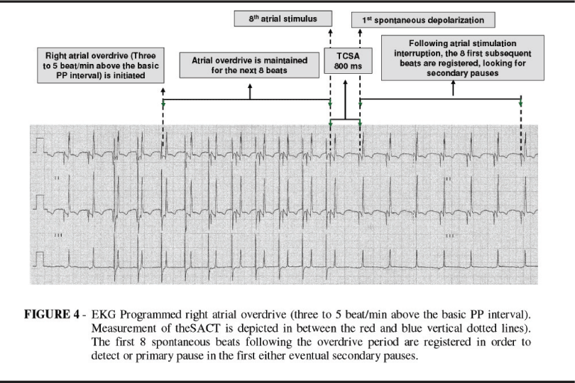
In order to determine the sinus atrial conduction time (SACT), RA overdrive was initiated and maintained for 8 beats with a frequency three to 5 bpm higher than baseline. Once pacing was turned off, the eight subsequent pp intervals were measured and the longest one was taken as the SACT (Figure 4).
Corrected SNRT and SACT were calculated by subtracting the respective pp intervals. A final EKG recording was made to document the cardiac rhythm. The study was repeated one day before hospital discharge, on postoperative day 6.
Statistical analysis: The mean and standard deviation, as well as the median and lower and higher limits were analyzed by the Wilcoxon test a significance level of 5%.
Results
The mitral valve was preserved in 70% of the cases, and a prosthetic valve was implanted in the remaining patients. There were no deaths. Postoperative complications comprised one case of each, seizures, hypertensive pneumothorax and gout crisis, in different patients.
Postoperative arrhythmias were detected in 50% of the patients (Table 1). Four patients needed temporary RA stimulation for junctional rhythm management. One patient needed electric cardioversion for AF on postoperative day one.
At hospital discharge, 8 patients were in sinus rhythm, seven of them continued to show EKG pattern of left atrial overload, two patients presented new onset first degree atrioventricular block. Two patients presented in hospital postoperative AF. The arrhythmia was transient in one patient and persistent in the other.
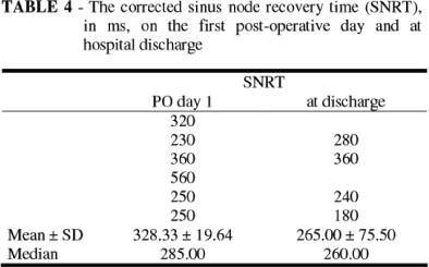
Sixty percent of the patients could complete the postoperative study protocol. There were no statistical differences between the pre and postoperative 12 lead EKGs (Table 2). The IACT ranged from 90 to 140ms in the first postoperative day, and from 110 to 130ms at hospital discharge (Table 3). The SNRT ranged from 250 ms to 560 ms in the first postoperative day and from 180 ms to 360ms at hospital discharge (Table 4).
The SACT remained between 70 and 140ms, both in the first postoperative day and at hospital discharge (Table 5), while the IACT was normal in patients whose LA was less than 50mm in diameter but supra normal in the remaining cases.
Discussion
Rheumatic heart disease is associated with, respectively, 9.9 to 27.5 times and 7.6 to 24.3 times greater incidence of chronic AF and paroxystic AF than in the normal population. This is explained by the presence of intermixed long and short refractory period atrial myocardium areas known to fragment the front wave of depolarization thus providing the substratum for incessant wave propagation or reentry that deflagrates AF.9,10 As this background is particularly found in the atrial myocardium of longstanding mitral valve stenosis, a sound pre and postoperative arrhythmogenic basis is present in these patients.
The clinical applicability of postoperative cardiostimulation studies like herein reported relates to their potential to identify heart surgery patients at risk for postoperative arrhythmias, particularly the sustained ones, like persistent AF. This would open the possibility to improve patient prognosis by starting prophylactic measures. In this investigation, preoperative EKG findings were characteristic of mitral valvulopathy.11 Although supraventricular extrasystoles are considered a frequent first postoperative day finding post mitral valve operations4,12 this is in odds with the present study for no such arrhythmias were found. Notwithstanding, our patients p wave and pR interval duration were increased, findings that according to PASSMAN et al.13 are markers for postoperative supraventricular arrhythmias. On the other hand, the 50% incidence of cardiac rhythm disorders in the first postoperative week was similar to previous reported data.
These conflicting findings certainly resulted from a weakness in the study design. As only one EKG recordings was obtained from each patient pre operatively, they probably failed to detect the usually great number of atrial electric disturbances present preoperatively.
Even though our test protocol had relatively low sensitivity, it is considered highly specific for sinus node disease detection.8,10 On other words, a normal test does not exclude the diagnosis, but an abnormal one is strongly suggestive of sinus node disease.8 In fact, HOGUE et al., in 2000,10 used a transesophageal electrode and a protocol similar to ours to demonstrate that a normal SNRT along with an altered SACT predispose to postoperative AF.
Interestingly, the sensitivity and specificity were both similar to those obtained by STEINBERG, 199314 and KLEIN, 199515 for p wave duration measured by high resolution EKG. Among the four patients presenting with SACT longer than 96ms in this study, only one had documented postoperative AF. On the other hand, one patient with intermittent preoperative AF who evolved in sinus rhythm with augmented SATCc (140 ms) during the first postoperative day had the AF recurring five days later. In this patient the FA was irresponsive to drug and electric cardioversion, and the patient was discharged home on postoperative day 10.
There was significant positive correlation between LA size and IACT in our study. The increased interatrial conduction time of these patients, most certainly a surrogate for the atrial dilation should have potentialized the preexisting atrial refractory dispersion thus providing the electrophysiological substratum for sustained reentry pathways and AF.1620
Conclusion
Sinus node function and interatrial conduction are not altered by mitral valve operation. Postoperative programmed epicardic atrial stimulation is easy to perform, well tolerated and does not induce arrhythmias on its own.
Acknowledgments
FAEPA Fundação de Apoio ao Ensino, Pesquisa e Assistência HCFMRP/USP
Conflict of interest: none
Sources of funding: none
Comments:
Postoperative atrial arrhythmias after cardiac surgical procedures are common, with a reported overall incidence of approximately 50%. Postoperative cardiostimulation studies aim to identify patients at risk for postoperative arrhythmias which would open the possibility to start prophylactic measures and to improve patient prognosis. In spite of the small number of patients investigated, the results reported by the authors suggest that conduction delay has a low sensitivity and a high specificity for prediction of AF as reported previously by others using alternative methods. Based on these data the altered SACT should not be used as a marker to start preventive measures for AF in patients undergoing elective, first heart operation for isolated mitral valvulopathy, with predominant sinus rhythm or intermittent AF.
1 Cox JL. A perspective on postoperative atrial fibrillation. Sem Thorac Cardiovasc Surg. 1999; 11:299302
2 Kalus JS, White CM, Caron MF, Coleman CI, Takata H, Kluger J. Indicators of atrial fibrillation risk in cardiac surgery patients on prophylactic amiodarone. Ann Thorac Surg. 2004; 77:128892.
3 Basko A, Simon RDB, Rinaldi AR, Gill JS. Occurence of atrial fiblillation after flutter ablation: the significance of intraatrial conduction and atrial vulnerability. J Electrocardiology 2003; 36:21925.
Antonio Carlos Pereira Martins
Full Professor and Head, Division of Urology, Department of Surgery and Anatomy, Ribeirão Preto Faculty of Medicine, University of São Paulo, Brazil.
- 1. Creswell LL, Damiano RJ. Posoperative atrial fibrilation an old problem crying for new solutions. J Thorac Cardiov Surg. 2001;121:63664.
- 2. Butler J, Rocker GM, Westaby S. Inflammatory response to cardiopulmonary bypass. Ann Thorac Surg. 1993;55:5529.
- 3. Asher CR, Miller DP, Grimm RA, Cosgrove DM 3rd, Chung MK. Analysis of risk factors for development of atrial fibrillation early after cardiac valvular surgery. Am J Cardiol. 1998;82:8925.
- 4. Kligfield P, Hochreiter C, KramerH, Devereux RB, Niles N, KramerFox R, Borer JS. Complex arrhythmias in mitral regurgitation with and without mitral valve prolapse: contrast to arrhythmias in mitral valve prolapse without mitral regurgitation. Am J Cardiol. 1985;55(13Pt1):15459.
- 5. Yamada T, Fukumanami M, Shimonagata T, Kumagai K, Asano Y, Hirata A, Asai M, Makino N, Hoki N. Effects of atrial septal pacing on P wave duration dispersion and atrial late potentials in patients with paroxysmal atrial fibrillation. Am J Cardiol. 2001;88:7958.
- 6. Ferrer MI. The sick sinus syndrome in atrial disease. JAMA 1968;206:6456.
- 7. Narula OS, Shantha N, Vasquez M, Towne WD, Linhart JW. A new method for measurement of sinoatrial conduction time. Circulation. 1978;58:70614.
- 8. Bigger Jr JT, Reiffel JA. Sick sinus syndrome. Ann Rv Med. 1979;30:91118.
- 9. Cox JL. A perspective of posoperative atrial fibrillation in cardiac operations. Ann Thorac Surg. 1993;56:4059.
- 10. Hogue CW, Hyder ML. Atrial fibrillation after cardiac operation: risks, mechanisms and treatment. Ann Thorac Surg. 2000;69:3006.
- 11. Acar J, Michel PL, Cormier B, Vahanian A, Iung B. Features of patients with severe mitral stenosis with respect to atrial rhythm. Atrial fibrillation in predominant and tight mitral stenosis. Acta Cardiol. 1992;47:11524.
- 12. Waldo AL. Perioperative arrhythmias. Use of Epicardial Wire Electrodes for the Diagnosis and Tratment. In: Utley JR(Ed). Perioperative cardiac dysfunction. Willians & Wilkins, USA. 1985, vol. III, p. 18099.
- 13. Passman R, Beshai J, Pavri B, Kimmel S. Predicting post coronary bypass surgery atrial arrhythimias from the preoperative electrocardiogram. Am Heart J. 2001;142:80610.
- 14. Steimberg JS, Selenkofske S, Wong SC, Gelernt M, Sciacca R, Menchavez E. Value of the Pwave signal averaged EKG for predicting atrial fibrillation after cardiac surgery. Circulation. 1993;88:261822.
- 15. Klein M, Evans SJL, Blumberg S, Cataldo L, Bodenheimer MM. Use of P wave triggered, P wave signal averaged eletrocardiogram to predict atrial fibrillation after coronary artery bypass surgery. Am Heart J. 1995;129:895901.
- 16. Josephenson ME, Kastor JA, Morganroth J. Eletrophysiologic left atrial enlargement: electrophysiologic, echocardiografic and hemodynamic correlates. Am J Cardiol. 1977;39:96771.
- 17. Kalil RAK, Maratia CB, Davilla A, Ludwig FB. Predictive factors for persistence of atrial fibrlillation after mitral valve operation. Ann Thorac Surg. 1999;67:6147.
- 18. Creswell LL. Posoperative atrial arrhythmias: risk factors and associated adverse outcome. Semin Thorac Cardiov Surg. 1999;11:3037.
- 19. Buxton AE, Josephenson ME. The role of P wave duration as a predictor of posoperative atrial arrhythmia. CHEST. 1981;80:6873.
- 20. Goyal SB, Spodick DH. Eletromechanical dysfuntion of the left atrium associated with interatrial block. A Heart J. 2001;142:8237.
Publication Dates
-
Publication in this collection
20 May 2008 -
Date of issue
2008