Abstracts
PURPOSE: To evaluate the deposition of collagen fibers at pig's vocal folds after topical use of mitomycin or 5-fluorouracil, when partial exeresis of mucosa layer had been promoted by CO2 laser. METHODS: There were used 18 Larger white pigs which were anesthetized and submitted to mucosa fragment's exeresis, bilaterally, at its free border. The animals were divided into 3 groups, each one with 6 animals: control group, without topical drug application; mitomycin group; and 5-fluorouracil group. After 30 days, the animals were subjected to euthanasia, and samples of the vocal folds were collected and stained by picrosirius red technique with polarization for quantification of total collagen deposition. RESULTS: In control group, the mean rate of right vocal fold's collagen deposition at submucosa consisted in a 3428.66 micrometers area. There was found an area whose size had, in average, 2196.36 micrometers, in mitomycin group, and 2269.19 micrometers, in 5-fluorouracil group. CONCLUSION: Mitomycin and 5-fluorouracil had promoted beneficial change in vocal fold's cicatrization with less collagen deposition, but there was no significant statistically difference when they were compared between themselves.
Mitomycin; Fluorouracil; Wound Healing; Vocal Cords; Laser Therapy; Swine
OBJETIVO: Avaliar a deposição das fibras de colágeno total em pregas vocais suínas após o uso tópico de mitomicina ou 5-fluorouracil nas exéreses parciais de mucosa com laser de CO2. MÉTODOS: Foram utilizados 18 porcos da raça Larger white anestesiados e submetidos à exérese de fragmento de mucosa de borda livre da prega vocal direita e prega vocal esquerda. Os animais foram divididos em 3 grupos com 6 animais cada: grupo controle, sem aplicação de medicação tópica; grupo mitomicina, com uso tópico dessa substância; grupo 5-fluorouracil, uso tópico. Após 30 dias do experimento os animais foram submetidos à eutanásia, coletadas amostras das pregas vocais e coradas pela técnica do picrosirius red com polarização para a quantificação computadorizada da deposição do colágeno total. RESULTADOS: No grupo controle, a média da área do colágeno depositado na submucosa da prega vocal direita foi de 3428,66 micrômetros. No grupo mitomicina a média foi de 2196,36 micrômetros. No grupo 5-fluorouracil, a média foi de 2269,19 micrômetros. CONCLUSÃO: A mitomicina e o 5-fluorouracil promoveram mudança benéfica na cicatrização da prega vocal, com menor deposição de colágeno, porém, quando comparados entre si, eles não apresentaram diferença estatisticamente significante.
Mitomicina; Fluoruracila; Cicatrização de Feridas; Cordas Vocais; Terapia a Laser; Suínos
4 - ORIGINAL ARTICLE
WOUND HEALING
Comparative study in swines' vocal cords healing after excision of fragment with CO2 laser with mitomycin and 5-fluorouracil postoperative topical application1 1 Research performed at Research Medical Institute, University Evangelic Hospital/ Evangelic Paraná Faculty, Curitiba, Brazil.
Estudo comparado da cicatrização da prega vocal de suínos após exérese de fragmento com laser de CO2 e aplicação tópica pós-operatória de mitomicina e 5-fluorouracil
Eduardo BaptistellaI; Osvaldo MalafaiaII; Nicolau Gregori CzeczkoIII; Jurandir Marcondes Ribas-FilhoIII; Paulo Afonso Nunes NassifIII; Marcelo Mazza do NascimentoIII; Jan Pawel Andrade PachnickiIV
IMaster in Sciences, Principles of Surgery Post-Graduation Program, University Evangelic Hospital/Evangelic Faculty of Parana, Curitiba, Brazil
IIPhD, Coordinator of Principles of Surgery Post-Graduation Program and Full Professor of Surgery, University Evangelic Hospital/Evangelic Faculty of Parana, Curitiba, Brazil
IIIPhD, Associate Professor, University Evangelic Hospital/Evangelic Faculty of Parana, Curitiba, Brazil
IVFellow Master degree, Principles of Surgery Post-Graduation Program, University Evangelic Hospital/Evangelic Faculty of Parana, Curitiba, Brazil
Correspondence Correspondence: Eduardo Baptistella Alameda Augusto Stellfeld, 1980 80730-150 Curitiba - PR Brazil Phone: (55 41) 3240-5488 ipem@evangelico.org.br
ABSTRACT
PURPOSE: To evaluate the deposition of collagen fibers at pig's vocal folds after topical use of mitomycin or 5-fluorouracil, when partial exeresis of mucosa layer had been promoted by CO2 laser.
METHODS: There were used 18 Larger white pigs which were anesthetized and submitted to mucosa fragment's exeresis, bilaterally, at its free border. The animals were divided into 3 groups, each one with 6 animals: control group, without topical drug application; mitomycin group; and 5-fluorouracil group. After 30 days, the animals were subjected to euthanasia, and samples of the vocal folds were collected and stained by picrosirius red technique with polarization for quantification of total collagen deposition.
RESULTS: In control group, the mean rate of right vocal fold's collagen deposition at submucosa consisted in a 3428.66 micrometers area. There was found an area whose size had, in average, 2196.36 micrometers, in mitomycin group, and 2269.19 micrometers, in 5-fluorouracil group.
CONCLUSION: Mitomycin and 5-fluorouracil had promoted beneficial change in vocal fold's cicatrization with less collagen deposition, but there was no significant statistically difference when they were compared between themselves.
Key words: Mitomycin. Fluorouracil. Wound Healing. Vocal Cords. Laser Therapy. Swine.
RESUMO
OBJETIVO: Avaliar a deposição das fibras de colágeno total em pregas vocais suínas após o uso tópico de mitomicina ou 5-fluorouracil nas exéreses parciais de mucosa com laser de CO2.
MÉTODOS: Foram utilizados 18 porcos da raça Larger white anestesiados e submetidos à exérese de fragmento de mucosa de borda livre da prega vocal direita e prega vocal esquerda. Os animais foram divididos em 3 grupos com 6 animais cada: grupo controle, sem aplicação de medicação tópica; grupo mitomicina, com uso tópico dessa substância; grupo 5-fluorouracil, uso tópico. Após 30 dias do experimento os animais foram submetidos à eutanásia, coletadas amostras das pregas vocais e coradas pela técnica do picrosirius red com polarização para a quantificação computadorizada da deposição do colágeno total.
RESULTADOS: No grupo controle, a média da área do colágeno depositado na submucosa da prega vocal direita foi de 3428,66 micrômetros. No grupo mitomicina a média foi de 2196,36 micrômetros. No grupo 5-fluorouracil, a média foi de 2269,19 micrômetros.
CONCLUSÃO: A mitomicina e o 5-fluorouracil promoveram mudança benéfica na cicatrização da prega vocal, com menor deposição de colágeno, porém, quando comparados entre si, eles não apresentaram diferença estatisticamente significante.
Descritores: Mitomicina. Fluoruracila. Cicatrização de Feridas. Cordas Vocais. Terapia a Laser. Suínos.
Introduction
Endolaringeous surgery has concentrated efforts in removing benign diseases with highest preservation of normal tissue around the injury, in order to improve phonactory function by preserving the mucosa wave1. This wave is generated by the sliding of the vocal cord mucosa over ligaments and muscles, which provide perfect glottic vibration2.
The lamina propria mucosa has a key role in the viscosity, contractility and formation of the vocal cord mucosa wave. Its proper functioning is essential to initiate and to maintain the vibration sustained by Bernoulli's phenomenon.
Since many years, enlightening new knowledge concerning the physiopathology of vocal cord benign lesions, laryngologists developed more conservative surgical techniques to optimize their results in surgery3.
Techniques using mucosal retail were developed, allowing dissection and excision of cysts, nodules and polyps at the lamina proprias' surface layer, whereas normal adjacent mucosa and subjacent vocal ligament are preserved3,4,5. This technique aims to minimize scar formation and subsequent mucosal hardening and synechia, which commonly results in injury on vocal cord vibration. However, vocal cord mucosa excision leads to scar formation2 at its lamina propria, resulting abnormalities of body and coverage relationship and decreasing the mucosa wave. If the scar worsens, it may increase segment without mucosa wave and also the dysphonia, present in 35% of the cases6.
Adjuvant therapies have been investigated to help modulation of the healing process at the vocal cord after its mucosa excision. Systemic or topic corticosteroid7, prior cooling before surgical procedure, fibrin glue, among other methods have been used to improve mucosas' cicatrisation after the procedures2.
Mitomycin is a antimetabolic agent, isolated from Streptomyces caespitosus. It is able to inhibit tumor growth by acting directly on cellular DNA synthesis, besides its inhibitory effects on fibroblasts proliferation8. In one experiment where this drug was topically applied in canine model of mucosal retail, it resulted in decreased collagen deposition at vocal fold's wound, but there was functional deficit observed by videolaryngostroboscopy2. It was successful when experimentally used for subglottic stenosis and posterior glottic stenosis treatment. In these studies, abnormal cicatrisation and stenosis were modulated and replaced by new mucosa coating9.
In ophthalmological surgeries, 5-fluorouracil is commonly used, mainly in trabeculectomies. In these operations, using antimitotics, as well as mitomycin prevents fibrosis and increased intraocular pressure10.
CO2 laser utilization, associated with other topical medications, inhibits the proliferation of cells involved in laryngeal vocal cord cicatrisation, decreasing collagen formation and contributing to postoperative vocal quality11.
The purpose of this study was to microscopically analyze the healing of pigs vocal cords after partial exeresis with CO2 Laser, evaluating total collage quantification at the injury and its deposition on this area, with and without the use of topical mitomycin or 5-fluorouracil.
Methods
Eighteen Larger white pigs (13 males and 5 females) were utilized, weighting an average of 10.1 kg (8.7 kg to 11.5 kg), and aged 27 to 31 days. All pigs were medically evaluated by veterinarians and were considered healthy, presenting normal weight for age distribution, using Embrapa's model.
The animals were numbered using 1 to 6 cuts at ears' tips: right ear for the experimental group and left for the control group. They were housed in groups of 6 animals, randomly divided into 3 groups (Table 1).
Stalls with 1.2 m x 1.8 m, were used, were floors were composed of woods strips for waste collection. The feed trough was always filled up, before and after procedure, same as the water recipient.
All animals were anesthetized by the same veterinarian who was specialized in anesthesia for medium and large animals. Before anesthetic induction, they received intra-muscular midazolam (Dormonid® 1 mg/mL) at dose of 0.1 mg/kg, intra-muscular acepromazine 1% (Acepran® 1%) at dose of 0.1 mg/kg, intra-muscular ketamine (Ketalar® 50 mg/mL) at a dose of 10 mg/kg and intra-muscular atropine 0.5 mg/mL at dose of 0.06 mg/kg. These procedures were followed by venous accession in femoral veins. After pre-anesthetic medication, anesthesia with Propofol® at dose of 6 mg/kg was performed and maintained with 0.4 mg/kg. Pigs were monitored through cardioscope.
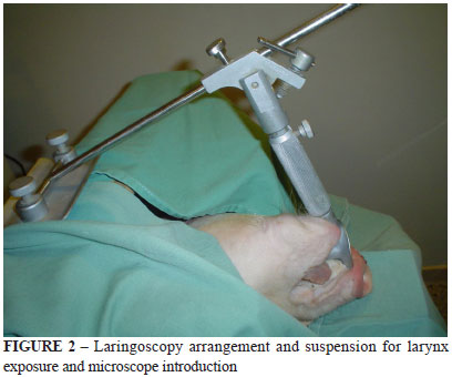
Once anesthetized, animals were placed by dorsal position, at a particular table. For adjustment of the larynx, it was placed a pad (Figures 1 and 2).
A D. F. Vasconcelos 6700 microscope, with attached camera system, was used for glottis visualization and surgical procedure (Figure 2). All surgical procedures were recorded on VHS tape.
The procedure started with the seizure of vocal cord freeboard, at its anterior part, with a Bouchayer. Then CO2 laser was used, in microspot way, carrying up a fragment's excision of about 2 mm.
After excision, in mitomycin group, a 5 mm cotton was used, soaked in mitomycin 0.04%, and let in contact with the surgical wound for exactly 3 minutes. After this period, the cotton was removed and excess mitomycin was cleaned. The same occurred in the 5-fluorouracil group, using this substance at 1% dilution.
Euthanasia was held 30 days after the surgical procedure, through electrocution, with the same anesthetic procedure. It was removed the entire larynx, making a longitudinal incision and exposing the vocal folds' region (Figure 3). It was also held an incision nearby thyroid cartilage for vocal fold's withdrawal and preservation of the mucosa coverage.
Histological material was set with pins on a paper's rectangle in order to keep it well stretched. After sample's identification in group, number and vocal side, it was sent to a randomized, double-blind, microscopic analysis. Later, it was fixed at 10% formalin for 24 hours, and the 2 rectangular samples were removed for histology and stained in picrosirius red.
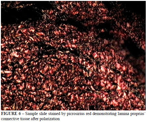
Slides were read through Pro-image-plus 4.5 program for Windows® in a computer attached to a microscope and video camera. Tissues stained with picrosirius red were read, first examining epithelium and submucosa microscopic characteristics, then, with image polarization, reading the collagen deposition based on tissues' colors. The results were transferred to the Windows® Excel program.
Fibroplasia, which is collagen deposition at mucosal surface in squared micrometers, occurring during the healing process, was measured. Two measures in each field were analyzed.
After performing the image's polarization, the collagen tissue, object of the search, could be differentiated from the non-collagen tissue, noting its bi-refringent character, orange and red colored, when compared with non-collagen substance, dark colored (Figures 4, 5 and 6).
For comparisons between vocal cords within each group, the non-parametric Wilcoxon test was used. For comparison of groups, the variable on collagens' percentage was used in the right fold related to collagen in the left fold. Groups' differences were evaluated considering the non-parametric test Kruskal-Wallis. For comparison in pairs, the non-parametric Mann-Whitney test was used. Values of P < 0.05 indicated statistical significance.
Results
All eighteen pigs survived the 30 days experiment, with appropriate weights for age. After subjective evaluation by an experienced veterinarian, they were considered healthy. The larynx macroscopic analysis had not shown any change when compared with a normal adult pig. No synechia or adhesions were present and histological materials' dimension was fit for analysis.
For each group, a null hypothesis was tested. The amount of collagen in the right fold is equal to the amount of collagen in the left fold. Table 2 presents the values of mean rate, median, minimum value, maximum value and root-mean-square (RMS) of collagen's amount. The values of P statistical tests are also presented.
For 5-fluorouracil and mitomycin's groups, the amount of collagen in the left side was significantly different from its quantity at the right side. In Table 2, for the 2 treated groups, medians were higher on the left side (without using medication) than on the opposite side. However, the control group had another result: collagen amount was higher at the right side.
Comparison between groups
This assessment was achieved by variable ratio between the amount of collagen in the right fold and the amount of collagen in left fold, multiplied by 100. Thus, the null hypothesis was tested, being that an equal collagen percentage reasons at the 3 groups versus an alternative hypothesis with a different percentage reason at least in 1 group. Table 3 presents the mean rate, median, minimum value, maximum value and RMS of collagen.
Comparisons were made between: mitomycin x 5-fluorouracil (P = 0.065); mitomycin x control group (P = 0.002), and 5-fluorouracil x control group (P = 0.002); it was used the non-parametric test of Mann-Whitney. As seen, the control group was significantly different when compared to the others groups. Five-fluorouracil and mitomycin's groups had no significantly difference between themselves.
Discussion
Only one research, comparing animal models for endolaryngeal surgery was found in literature. Garrett et al.2, compared dog, pig and monkey's vocal folds, noting the deposition of collagen, elastin fibers and amorphous tissue, and concluded that pig and dog's vocal fold show a great concentration of elastic fibers and collagen, in lamina propria deep layers, resembling the histologic characteristics of man's vocal fold more than monkey's. Pig and dog's vocal folds show a dissection plan for mucosa retail that most resembles humans'.
In this study, the experimental model called a midsize animal with histological featuring vocal folds, without needing observation of the function by videostroboscopy. For experiment's results being useful in larynx' surgical practice, it was opted for pig's achievement, with standardization of weight and age, accommodations and diet.
In recent decades, technological innovations have led to substantial gains in laryngeal surgery of the vocal fold. These innovations increased the understanding of physiological principles that concern laryngeal sound's production and the strobe's use for analysis of the vocal fold's fluctuations in laser operations.
Santos et al.12, compared incisions in canine's vocal cords performed by instruments with that performed by CO2 laser evaluating, through histological sections, the fibroplasia amount. Results showed that the collagen's amount was higher when treated instrumentally than in time used of CO2 laser. Therefore, CO2 laser method is safer in collagen's deposition as compared to instruments, when following up the fundamental principles of phonomicrosurgery.
Picrosirius red is the standard staining for studying and quantifying collagen deposition12,13. It is preferred because, besides being specific, it can differentiate between maturation stages of collagen fibers after polarization, coloring green and yellow-green the youngest fibers (thinner) and orange and red the oldest ones (thicker)14,15,16,17,18.
The computer morphometric quantification, by adding the stained area after polarization, is a method that is well established in the literature for evaluation of the collagen's deposition amount in studies that aims evaluating the wound healing12,13.
Mitomycin is a chemotherapy drug used systemically in conjunction with other anti-neoplasic drugs for the treatment of various malignancies. In the 80's, it was discovered that mitomycin in low doses inhibits healing. After several animals' studies, it started to be used in eye operations subjected to a failure risk of secondary healing (trabeculectomy and pterygium surgery) with good results19.
Since 1997, the otolaryngologists were interested in using mitomycin in situations that required scars' treatment on structural tubes as larynx, trachea and nose, reporting success in these applications20,21,9,22.
In this study, when using mitomycin compared with the control group, there was a significant difference with lower collagen's deposition in the medicated vocal fold, what corroborates with the mentioned studies.
Camargo et al.11, when studying a pigs' group with laser exeresis and later application of mitomycin, had a mean area of 2648.03 µm2 concerning to the vocal fold's total collagen in the control group, while the experimental group's total collagen was 2200.30 µm2 (P = 0.0043), demonstrating a significant difference when using mitomycin.
In this study, the control group showed an average of 3428.66 µm2, while the total collagen mean rate in the mitomycin group was 2196.36 µm2 (P = 0.002). This result agrees with the other studies findings11, showing lower collagen's deposition when using mitomycin.
Five-fluorouracil is widely used in treatment for many carcinomas. Its use in primary head and neck tumors is done through intravenous infusion associated with others chemotherapy drugs. This using allows that, even in some cases of larynx tumors, occurs an almost total remission of the disease, not being necessary a surgical procedure. When accomplished local use of the substance, in tumors, for a long time, it ends up appearing an inflammatory reaction (mucosite), and because of this, its use is recommended intravenously in a dose of 600 mg per period, maximum of 5 days23.
Topical use of 5-fluorouracil had not relevant side effects in this study. Animals tolerability was good, without any absorption in other regions, and there were no cases of diarrhea or weight loss by some type of mucosa inflammatory reaction.
In ophthalmology, 5-fluorouracil is used in the treatment of glaucoma, beholding trabeculectomy. This drug is able to inhibit the proliferation of fibroblasts in vitro and in vivo, reducing the synthesis of collagen and the scar formation. In this operation, its utilization is accomplished on topic conjunctival or injections in the subconjunctival space at the postoperative period, leading to a well induced conjunctival layer without closing the drainage hole10.
This research tried to measure mitomycin against 5-fluorouracil related to the collagen's deposition in vocal folds. It was found better response on using mitomycin, similar to the trabeculectomy's study. There was, nevertheless, no statistical significance comparing 5-fluorouracil and mitomycin.
Statistical difference was found, in this study, after 30 days, when comparing the instillation of 5-fluorouracil to the group that received no medication. However, no differences were found between the groups medicated.
It was not found research comparing topical use of 5-fluorouracil and mitomycin in vocal cords, in literature.
Conclusion
When vocal cords are submitted to partial exeresis with CO2 laser, making use of topical mitomycin or 5-fluorouracil, there can be a significant decrease of total collagen deposition. The comparison between 5-fluorouracil and mitomycin is not significantly different on the vocal cord cicatrisation.
Received: August 19, 2008
Review: October 21, 2008
Accepted: November 18, 2008
Conflict of interest: none
Financial source: none
How to cite this article
Baptistella E, Malafaia O, Czeczko NG, Ribas-Filho JM, Nassif PAN, Nascimento MM, Pachnicki JPA. Comparative study in swines' vocal cords healing after excision of fragment with CO2 laser and mitomycin and 5-fluorouracil postoperative topical application. Acta Cir Bras. [serial on the Internet] 2009 Jan-Feb;24(1). Available from URL: http://www.scielo.br/acb
- 1. Coleman Jr JR, Smith S, Reinisch L. Histomorphometric and laryngeal videostroboscopic analysis of the effects of corticosteroids on microflap healing in the dog larynx. Ann Otol Rhinol Laryngol. 1999;108(2):119-27.
- 2. Garrett CG, Soto J, Riddick J, Billante CR, Reinisch L. Effect of mitomycin-C on vocal fold healing in canine model. Ann Otol Rhinol Laryngol. 2001;110(1):25-30.
- 3. Courey MS, Gardner GM, Stone RE. Endoscopic vocal fold microflap: a thre-year experience. Ann Otol Rhino Laryngol. 1995;104:267-73.
- 4. Sataloff RT, Spiegel JR, Heuer RJ. Laryngeal minimicroflap: a new technique and reassessment of microflap saga. J Voice. 1995;9:198-204.
- 5. Courey MS, Garrett CG, Ossoff RH. Medial microflap excision of benign vocal fold lesions. Laryngoscope. 1997;107(3):340-4.
- 6. Woo P, Casper J, Colton R, Brewer D. Diagnosis and treatment of persistent dysphonia after laryngeal surgery: a retrospective analysis of 62 patients. Laryngoscope. 1994;104:1084-91.
- 7. Sheppard LM. Effect of steroids or precooling on edema and tissue thermal coagulation after CO2 laser impact. Lasers Surg Med. 1992;12:137-46.
- 8. Villaescuda A, Santana M, Sierra N, Vizoto A, Rodríguez R. Producción de mitomicina por vía fermentativa: estudio inicial. Rev Farm Cuba;1994;28(1):9-13.
- 9. Eliashar R, Eliashar I, Esclamado R, Gramlich T, Strome M. Can topical mitomycin prevent laryngotracheal stenosis? Laryngoscope. 1999;109(10):1594-600.
- 10. Susanna JR, Takahashi WY, Carvalho RC. Perda do campo visual em pacientes trabeculemizados. Rev. Bras Oftalmol. 1995;54:23-6.
- 11. Camargo PAM, Campos ACL, Matias JEF, Rispoli DZ, Przysiezny PE, Fonseca VR. Topical mitomycin C effect on swine vocal folds healing. Rev Bras Otorrinolaringol. 2006;72(5):601-4.
- 12. Santos FCC, Grellet M, Junior AR, Jamur MC, Pinto JA, Fomim DS. Estudo comparativo histológico na prega vocal após incisão com instrumental a frio e com laser de CO2 em modelo animal. Rev Bras Otorrinolaringol. 2003;69:753-9.
- 13. Ribeiro FAQ, Guaraldo L, Borges JP, Zacchi FFS, Eckley CA. Clinical and histological healing of surgical wounds treated with mitmycin C. Laryngoscope. 2004;114:148-52.
- 14. Junqueira LCU, Bignolas G, Brentani RR. Pricrosirius staining plus polarization microscopy, a specific method for collagen detection in tissue sections. Histochem J. 1979;11:447-55.
- 15. Szendröi M, Vajta G, Kovács L, Schaff Z, Lapis K. Polarization colours of collagen fibres: a sign of collagen production activity in fibrotic processes. Acta Morphol Hung. 1984;32:47-55.
- 16. Rabau MY, Dayan D. Polarization microscopy of picrosirius red stained sections: A useful metod for qualitative evaluation of intestinal wall collagen. Histol Histopathol. 1994;9:525-8.
- 17. Hirshberg A, Buchner A, Dayan D. The central odontogenic fibroma and the hyperplastic dental follicle: study with Picrosirius red and polarization microscopy. J Oral Pathol Med. 1996;25(3):125-7.
- 18. Hirshberg A, Buchner A, Dayan D. Collagen fibres in the wall of odontogenic keratocysts: a study with picrosirius red and polarizing microscopy. J Oral Pathol Med. 1999;28:410-2.
- 19. Frucht PJ, Rozenmann Y. Mitomycin C therapy for corneal intraepithelial neoplasia. Am J Ophthalmol. 1994;117(2):164-8.
- 20. Ward RF, April MM. Mitomycin-C in the tratment of tracheal cicagtrix after tracheal reconstruction. Int J Pediatr Otorhinolaryngol. 1998;44:221-6.
- 21. Spector JE, Werkhaven JA, Spector NC. Preservation of function and histological appearenci in the injured glottis with topical mitmycin-C. Laryngosocpe. 1999;109:1125-9.
- 22. Estrem SA, Batra PS. Preventing myringotomy closue qith topical mitomycin C in rats. Otolaryngol Head Neck Surg. 1999;120:794-8.
- 23. Vokes EE, Stenson K, Rosen FR, Kies MS, Rademaker AW, Witt ME, Brockstein BE, List MA, Fung BB, Portugal L, Mittal BB, Pelzer H, Weichselbaum RR, Haraf DJ. Weekly Carboplatin and paclitaxel followed by concomitant paclitaxel, fluorouracil, and hydroxyurea chemoradiotherapy: curative and organ-preserving therapy for advanced head and neck cancer. J Clin Oncol. 2003;21(2):320-6.
Publication Dates
-
Publication in this collection
14 Jan 2009 -
Date of issue
Feb 2009
History
-
Accepted
18 Nov 2008 -
Reviewed
21 Oct 2008 -
Received
19 Aug 2008