Abstracts
PURPOSE: To compare sutures with polypropylene and poliglecaprone 25 after partial cecotomy in rats. METHODS: Thirty six rats divided into two groups, A and B, of 18 animals; each group was also divided into three subgroups of six animals sacrificed at 4th, 7th and 14th days after surgery. Were studied the mortality, morbidity, complications attributable to sutures, macroscopy, optical microscopy and measurement of hydroxyproline at the level of the suture. RESULTS: There were no deaths or wound complications such as hematoma, seroma, abscess, evisceration or eventration. On microscopic evaluation reepithelization, coaptation and inflammation in both groups did not differ significantly. The average rate of tissue hydroxyproline found in the samples on the 4th day after surgery, was 21.38 mg/g tissue for group A and 16.68 mg/g for group B; on day 7 after surgery, the average was 15.64 mg/g tissue for group A and 26.53 mg/g for group B; on day 14, the average was 8.09 mg/g tissue for group A and 25.07 mg/g for group B. CONCLUSION: There were no differences on clinical evolution, macroscopic aspect, microscopic data and hydroxyproline concentration on both sutures.
Suture Techniques; Anastomosis, Surgical; Cecum; Polypropylenes; Rats
OBJETIVO: Comparar a sutura com fio de polipropileno e poliglecaprone 25 após cecotomia parcial em ratos. MÉTODOS: Trinta e seis ratos foram distribuídos em dois grupos A e B de 18 animais, e cada grupo foi dividido em três subgrupos de seis, sacrificados no 4º, 7º e 14º dias do pós-operatório. Estudou-se a mortalidade, morbidade, complicações atribuíveis às suturas, macroscopia, microscopia ótica e dosagem de hidroxiprolina no nível da sutura. RESULTADOS: Não houve mortalidade ou complicações da ferida operatória como hematoma, seroma, abscesso, evisceração ou eventração. Na avaliação microscópica os critérios de re-epitelização, coaptação e processo inflamatório ambos os grupos não apresentaram diferença significativa. A taxa tecidual média da hidroxiprolina encontrada nas amostras no 4º dia de pós-operatório foi de 21,38 mg/g de tecido para o grupo A e 16,68 mg/g para o grupo B; no 7º dia a média foi de 15,64 mg/g de tecido para o grupo A e 26,53 mg/g para o grupo B; no 14º dia ela foi de 8,09 mg/g de tecido para o grupo A e 25,07 mg/g para o grupo B. CONCLUSÃO: Não houve diferença estatística entre a evolução clínica, avaliação macroscópica, microscopia e dosagem de hidroxiprolina entre as suturas realizadas com os fios estudados.
Técnicas de Sutura; Anastomose Cirúrgica; Ceco; Polipropilenos; Ratos
8 - ORIGINAL ARTICLE
WOUND HEALING
Cecorraphy in single layer using polypropylene and poliglecaprone 25 threads. Comparative study in rats1 Correspondence: Fernando Issamu Tabushi Avenida Três Marias, 1175, Casa 41 82310-000 Curitiba - PR Brasil malafaia@evangelico.org.br
Cecorrafia em plano único com fios de polipropileno e poliglecaprone 25. Estudo comparativo em ratos
Fernando Issamu TabushiI; Luiz Masakatso NomuraI; Osvaldo MalafaiaII; Jurandir Marcondes Ribas-FilhoIII; Benur PolonioIV; João Carlos Domingues RepkaIV; Sergio IoshiiV; Octavio Antonio Azevedo da Costa-FilhoVI
IMaster in Surgery, FEPAR, Curitiba-PR, Brazil. Responsible for intellectual, scientific content of the study and critical revision
IIFull Professor, Head, Department of Surgery, FEPAR and Federal University of Parana (UFPR), Coordinator of Principles of Surgery Postgraduate Program of FEPAR, Curitiba-PR, Brazil. Mentor of Master degree Fernando Issamu Tabushi and Luiz Masakatso Nomura. Responsible for conception of the scientific content, supervision of all phases of the study, manuscript writing and critical revision
IIIFull Professor of Surgery, FEPAR, Curitiba-PR, Brazil. Manuscript writing and critical revision
IVPhD in Surgery, Clinics Hospital, UFPR, Curitiba-PR, Brazil. Helped with technical procedures and acquisition of data
VPhD in Surgery, Assistant Professor of Pathology, UFPR, Curitiba-PR, Brazil. Macroscopic and histopathological examinations
VIMaster in Surgery, FEPAR, Curitiba-PR, Brazil. Helped with technical procedures and acquisition of data
Correspondence Correspondence: Fernando Issamu Tabushi Avenida Três Marias, 1175, Casa 41 82310-000 Curitiba - PR Brasil malafaia@evangelico.org.br
ABSTRACT
PURPOSE: To compare sutures with polypropylene and poliglecaprone 25 after partial cecotomy in rats.
METHODS: Thirty six rats divided into two groups, A and B, of 18 animals; each group was also divided into three subgroups of six animals sacrificed at 4th, 7th and 14th days after surgery. Were studied the mortality, morbidity, complications attributable to sutures, macroscopy, optical microscopy and measurement of hydroxyproline at the level of the suture.
RESULTS: There were no deaths or wound complications such as hematoma, seroma, abscess, evisceration or eventration. On microscopic evaluation reepithelization, coaptation and inflammation in both groups did not differ significantly. The average rate of tissue hydroxyproline found in the samples on the 4th day after surgery, was 21.38 mg/g tissue for group A and 16.68 mg/g for group B; on day 7 after surgery, the average was 15.64 mg/g tissue for group A and 26.53 mg/g for group B; on day 14, the average was 8.09 mg/g tissue for group A and 25.07 mg/g for group B.
CONCLUSION: There were no differences on clinical evolution, macroscopic aspect, microscopic data and hydroxyproline concentration on both sutures.
Key words: Suture Techniques. Anastomosis, Surgical. Cecum. Polypropylenes. Rats.
RESUMO
OBJETIVO: Comparar a sutura com fio de polipropileno e poliglecaprone 25 após cecotomia parcial em ratos.
MÉTODOS: Trinta e seis ratos foram distribuídos em dois grupos A e B de 18 animais, e cada grupo foi dividido em três subgrupos de seis, sacrificados no 4º, 7º e 14º dias do pós-operatório. Estudou-se a mortalidade, morbidade, complicações atribuíveis às suturas, macroscopia, microscopia ótica e dosagem de hidroxiprolina no nível da sutura.
RESULTADOS: Não houve mortalidade ou complicações da ferida operatória como hematoma, seroma, abscesso, evisceração ou eventração. Na avaliação microscópica os critérios de re-epitelização, coaptação e processo inflamatório ambos os grupos não apresentaram diferença significativa. A taxa tecidual média da hidroxiprolina encontrada nas amostras no 4º dia de pós-operatório foi de 21,38 mg/g de tecido para o grupo A e 16,68 mg/g para o grupo B; no 7º dia a média foi de 15,64 mg/g de tecido para o grupo A e 26,53 mg/g para o grupo B; no 14º dia ela foi de 8,09 mg/g de tecido para o grupo A e 25,07 mg/g para o grupo B.
CONCLUSÃO: Não houve diferença estatística entre a evolução clínica, avaliação macroscópica, microscopia e dosagem de hidroxiprolina entre as suturas realizadas com os fios estudados.
Descritores: Técnicas de Sutura. Anastomose Cirúrgica. Ceco. Polipropilenos. Ratos.
Introduction
The digestive suture represents fertile field of medical research. The large intestine, due to its content and its peculiar conditions - such as the presence of large amounts of bacteria in its lumen and the absence of peritoneal lining in some segments - is more prone to anastomotic dehiscence compared to stomach and small intestine1-8.
Among the factors reported as associated with large intestine anastomotic dehiscence and septic complications are: bad preparation of the colon for surgery, bad patient's nutritional status, associated diseases, suture in diseased intestine, intestinal ischemia, tension on the suture line, inadequate points of suture, trauma, perforation, use of drains and construction with anastomosis below the peritoneal reflection1,3,9,10.
The occurrence of anastomotic dehiscence appears to be most frequent in surgery of the colon in contrast to the small intestine, where this complication is rarely mentioned. Anastomotic dehiscence rates in the large intestine reported in the literature range from 0% to 35%10.
Several authors agree that an ideal anastomosis should have no foreign body, be easy, fast and safe, even in cases of increased intraluminal pressure1,11.
The sutures had significant evolution over the past 50 years. Initially available as catgut, cotton, silk, linen and steel, nowadays is offered with many different threads. Until the 70s, the only absorbable suture was the catgut obtained from the intestine of animals, rich in collagen fibers. However, even the chromic catgut, is absorbed with an intense inflammatory process and variable absorption time. Were precisely these features - variable absorption time, intense inflammatory process for absorption and the fact that the thread was based on animal proteins -, which led researchers to look for new synthetic absorbable ones12.
In modern times, were created the multifilamentar absorbable synthetic threads, such as polyglycolic acid and polyglactin 910. They had the advantages of predictable absorption time with a mild inflammatory reaction. Afterwards, was produced a monofilament poliglecaprone 25 with advantages of absorption time be predictable and relatively short13-20. Other absorbable monofilament threads, as polydioxanone and poligliconate, require medium or long time for its absorption12,21,22.
The polypropylene threads are characterized by being synthetic, nonabsorbable monofilament. It features excellent maneuverability, safety in the knots and low inflammatory tissue reaction. For these good qualities, the suture is widely used in surgery23,24.
The aim of this study was to compare the suture with polypropylene and poliglecaprone 25 after partial cecotomy in rats
Methods
Were used 36 rats Rattus norvegicus albinus - Wistar EPM-1, males, weighing between 250 and 328 grams. They were operated at the Institute of Medical Research (IPEM) from Post-Graduate Program in Principles of Surgery of the University Evangelic Hospital/Evangelic Faculty of Paraná, Curitiba-PR, Brazil. The animals were divided into two groups of 18, with group A (animals 1 to 18) defined as the control group and group B (animals 19 to 36) experimental group. The surgical procedures were identical in both groups: partial cecorraphy at the same level. The suture were different using in group A polypropylene and in group B poliglecaprone 25.
The animals were housed during an observation period of 15 days, where they were fed with balanced feed for the species and had free access to water. At the end of this period they were submitted to surgery with 12 hours fasting time prior the procedure, but with free access to water.
The rats were anesthetized using ether inhalation and prepared for surgery in the routine way. It was not use bowel preparation or antibiotics.
An abdominal median xiphopubic incision with a scalpel 15 was done interesting all layers of the parietal wall. After inspecting the abdominal cavity and revision of the hemostasis, was identified the cecum, vermiform appendix and ileum. Cecotomy included the entire anterior surface of the cecum at the site of implantation of the vermiform appendix. Was also identified the posterior wall of the cecum, and proximal and distal ends of the lumen (Figure 1).
The cecorraphy was done identically in both groups. Was used continuous suture leaving the fixation knots outside the cecum and producing an overlapping on cecal wall edges, 2 mm from the borders. Abdominal closure was realized with an aponeurotic continuous suture with 5-0 nylon thread with cylindrical needle of 1.5 cm. The skin was approximated with a continuous 5-0 nylon intradermal suture.
In group A the cecum wall was sutured with 5-0 polypropylene using cylindrical needle of 1.5 cm. In group B the same suture was done with poliglecaprone 25.
During the postoperative period, the animals were maintained in box with six rats, identified with the day of surgery and which suture was used.
The animals were examined daily, morning and evening, observing the conditions of the wound and its complications. The animals in each group were divided into three subgroups of six animals; in group A they were named as subgroups A1, A2, A3, and group B, named B1, B2, B3. The ones in subgroups A1 and B1, A2 and B2, A3 and B3 were sacrificed at 4th, 7th and 14th day after the operation, respectively. They were sacrificed by administering a lethal dose of inhaled ether.
The abdominal cavity was opened far from the surgical incision, removing the operated intestinal segment in a single block. Was observed the presence of abdominal wall seroma, hematoma and integrity of the suture. In the peritoneal cavity, was evaluated the presence of infection, peritoneal adhesions, the integrity of the suture in the cecum and the presence/absence of fistula. Structures or organs that adhered to the cecal suture and/or wall of the cecum, were also removed en-bloc.
The specimen was opened by the middle of the posterior wall of the cecum, and was observed the presence of macroscopic changes in the suture on luminar surface (Figure 2).
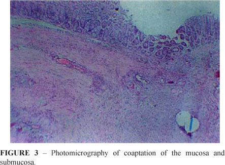
Before placing the specimens in formaldehyde, was removed a fragment of 0.5x0.5 cm of cecal wall to be used in hydroxyproline dosage. Then, the fragments were processed by usual histological technique for hematoxylin-eosin. From each paraffin block were obtained two slides for histological examination studying the coaptation and inflammation of the tunica serosa, muscle, submucosa and mucosa (Figure 3).
Hydroxyproline concentration of the fragments was done with a spectrophotometer, with the technique proposed by Kivirikko and Laitinen in 1967 and modified by Ibbott in 197425,26.
The statistic analyze was based on non-parametric tests, taking into account the nature of the variables. Were used Fisher's exact test, Kruskal-Wallis and Mann-Whitney test for two independent groups. This analysis was done for each day of sacrifice, separately. In all tests was set at 0.05 or 5% (α < 0.05) level for rejecting the null hypothesis.
Results
All animals recovered well from anesthesia. During the postoperative course there was no mortality and no wound complications, such as hematoma, seroma, abscess, evisceration or eventration.
The weight variation preoperatively and on the respective dates of sacrifice had no differences.
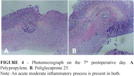
Microscopic assessment studying separately the serous and muscular coats, mucosa and submucosa according to the criteria of re-epithelialization, inflammation and coaptation, showed similar results (Figure 4).
Note: An acute moderate inflammatory process is present in both.
Regarding the dosage of hydroxyproline, the average rate found in tissue samples on the 4th day after surgery, was 21.38 mg/g tissue for group A and 16.68 mg/g tissue for group B; on day 7 after surgery, the average was 15.64 mg/g tissue for group A and 26.53 mg/g tissue for group B; on the 14th day after surgery the average was 8.09 mg/g tissue for group A and 25.07 mg/g tissue for group B. After comparative calculations by applying the analysis of variance by ranks of Kruskal-Wallis the results were not statistic different between the groups. Applying the Mann-Whitney test to compare to the subgroups of group A with group B, also no differences were found among all groups (Table 1).
Discussion
The period of 15 days of preoperative observation was necessary for animals to get used to the environment and food testing. It also served as an observation period to check whether the animals were healthy. It was established no use of bowel preparation and antibiotics due to the fact that rats are successfully resistant to surgical contamination.
Although the good development of intestinal suture in recent years, the large intestine maintain the highest rates of complications (such as fistulas), when compared with small intestine or stomach3,10.
The suture of the intestine with the polypropylene is safe, ease to be done and have low inflammatory reaction. These qualities put it near the ideal suture. One of its features that is not consistent with this proposition is that it is nonabsorbable and, therefore, remains indefinitely in the suture line when it is not eliminated in the bowel lumen.
Among the absorbable threads, the catgut was used before the Christian era. It is animal in origin, characterized by large inflammatory response during absorption, which is highly variable in time. The synthetic absorbable polyglycolic acid and polyglactin 910 have absorbable time more uniform, less inflammatory reaction during its absorption, but are multifilament and, so, have the possibility to host bacteria among fibers, a fact that may contribute to the formation and/or maintenance of microabscesses. The absorbable polydioxanone/poligliconate is monofilament and need long absorption time12,27.
Poliglecaprone 25 is a synthetic, absorbable monofilament which is proposed to have lower absorption time10,13,17,18,21,24,28-32.
Was chosen the 4th, 7th and 14th days after surgery to have the evaluation because the inflammatory process is particularly acute in these phases and the overall rate of degradation of collagen is greater than the rate of synthesis, mainly by the 5th postoperative day. The 7th postoperative day was chosen because it is around this period of time that occur most enterocutaneous fistulas. In 14 days high collagen rates happen almost similar to the ones in pre-operative period, and also in this period all complications that might exist, occur 10,13,14,15,17,21,33.
The amount of hydroxyproline follows the collagen presence during the healing process. It is observed that around the 4th day after surgery its rate get the lowest point, with a tendency for growing from the 7th day after surgery6,10,28.
Conclusion
There were no differences on clinical evolution, macroscopic aspect, microscopic data and hydroxyproline concentration on both sutures using polypropylene and poliglecaprone 25 threads.
Received: October 18, 2011
Review: December 14, 2011
Accepted: January 20, 2012
Conflict of interest: none
Financial source: none
1 Research performed at Postgraduate Program, Department of Surgery, Evangelic Faculty of Parana (FEPAR), Curitiba-PR, Brazil.
- 1. Ballantyne GH. The experimental basis of intestinal suturing: effect of surgical technique, inflammation, and infection on enteric wound healing. Dis Colon Rectum. 1984;27:61-71.
- 2. Chung RS. Blood flow in colonic anastomosis: effect of stapling and suturing. Ann Surg., 1987;206:335-9.
- 3. Fielding LP, Stewart-Brown S, Blesovsky L, Kearney G. Anastomotic integrity after operations for large-bowel cancer: a multicentre study. Br Med J. 1980;281:411-4.
- 4. Ho UH, Ashour MAT. Techniques for colorectal anastomosis. World J Surg. 2010;16:1610-21.
- 5. Irvin TT, Hunt KK. Reappraisal of the healing process of anastomosis of the colon. Surg Gynecol Obstet. 1974;138:741-6.
- 6. Krasniqi A, Gashi-Luci L, Krasniqi S, Jakupi M, Hashani S, Limani D, Dreshaj IA. A comparison of three single layer anastomotic techniques in the colon of the rat. Int J Surg. 2009;7:31-5.
- 7. Quilici FA, Cordeiro F, Faria Jr PC, Reis Neto JA. Mechanical and manual anastomoses of the extraperitoneal rectum: experimental comparative study in dogs. Arq Bras Cir Dig. 1990;5:41-50.
- 8. Subhas G, Brullar JS, Cook J, Shah A, Silberberg B, Andrus L, Decker M, Mittal VK. Topical gentamicin does not provide any additional anastomotic strength when combined with fibrin glue. Am J Surg. 2011;201:339-43.
- 9. Ballantyne GH. Intestinal suturing: review of the experimental foundations for traditional doctrines. Dis Colon Rectum. 1983;26:836-43.
- 10. Hendriks T, Mastboom WJB. Healing of experimental intestinal anastomoses: parameters for repair. Dis Colon Rectum. 1990;33:891-901.
- 11. Rosenberg D, Nasser A, Regen JB, Behmer OA. Suturas intestinais: estudo comparativo entre a sutura clássica em dois planos e a sutura extramucosa num plano único com emprego de um novo fio absorvível, o ácido poliglicólico. Rev Assoc Med Bras. 1973;19:249-58.
- 12. Fagundes DJ, Kharmandayan P. O fio cirúrgico. Acta Cir Bras. 1991;6:177-81.
- 13. Bezwada RS, Jamiokowski DD, Lee IY, Agarwal V, Persivale J, Trenka-Benthin S, Erneta M, Suryadevara J, Yang A, Liu S. Monocryl suture, a new ultra-pliable absorbable monofilament suture. Biomaterials. 1995;16:1141-6.
- 14. Biondo-Simões MLP, Sech M, Adur RC, Marques LO, Corbellini M, Canalli LS, Veronese M, Cabrera P, Vaz LI. A comparative study of the performance of catgut and polyglecaprone 25 sutures in rat abdominal walls, contaminated or not. Acta Cir Bras. 1997;12:163-8.
- 15. Labagnara J. A review of absorbable suture materials in head & neck surgery and introduction of monocryl: a new absorbable suture. Ear Nose Throat J. 1995;74:409-15.
- 16. Lara EG, Martinez BM, Ayala HBM. Valoración de un nuevo material de sutura, poliglecaprone 25 em ginecología y obstetricia. Ginecol Obstet Mex. 1996;64:40-2.
- 17. Molea G, Tirone L, Schonauer F. Sutura cutanea intradermica a punti staccati con monofilamento riassorbibile (poliglecaprone 25). Minerva Chir. 1997;52:1261-5.
- 18. Trimbus JB, Niggebrugge A, Trimbus R, van Rijssel EJC. Knotting abilities of a new absorbable monofilament suture: poliglecaprone 25 (Monocryl). Eur J Surg. 1995;161:319-22.
- 19. Rey SD, Czeczko NG, Nassif PAN, Ribas-Filho JM, Malafaia O, Grisa L, Torres OJM, Czeczko AEA. Perose drain influence in cecum healing. Experimental study in rats. ABCD Arq Bras Cir Dig. 2004;17(1):29-33.
- 20. Nomura LM, Ribas-Filho JM, Malafaia O, Dietz UA, Skare TL, Kume MH. Processo cicatricial de sutura em ceco com os fios polipropilene, poliglecaprone 25 e glicomer 60 em ratos. ABCD Arq Bras Cir Dig. 2009;22(2):82-8.
- 21. Braghetto IM, Rappoport JS. Evaluación prospectiva de sutura poliglecaprone 25 (Monocryl) en cirurgia general. Rev Chil Cir. 1994;46:299-305.
- 22. Ray JA, Doddi N, Regula D, Williams JA, Melveger A. Polydioxanone (PDS), a novel monofilament synthetic absorbable suture. Surg Gynecol Obstet. 1981;153:497-507.
- 23. Miller JM. Evaluation of a new surgical suture (Prolene). Am Surg. 1973;39:31-9.
- 24. Orringer MB, Appleman HD, Bove E, Cimmino V. Polypropylene suture in esophageal and gastrointestinal operations. Surg Gynecol Obstet. 1977;144:67-70.
- 25. Ibbott FA. Amino acids and related substances. In: Henry RJ, Cannon DC, Winkelman JW. Clinical chemistry principles and technics. New York: Harper & Row Publishers; 1974. p.608-14.
- 26. Kleiman I, Simões MJ, Goldenberg S. Aspectos atuais do processo de reparação tecidual. Acta Cir Bras. 1987;2:19-21.
- 27. Edlich RF, Panek PH, Rodeheaver GT, Turnbull VG, Kurtz LD, Edgerton MT. Physical and chemical configuration of sutures in the development of surgical infection. Ann Surg. 1973;177:679-88.
- 28. Cronin K, Jackson DS, Dunphy JE. Changing bursting strength and collagen content of the healing colon. Surg Gynecol Obstet. 1968;126:747-53.
- 29. Jiborn H, Ahonen J, Zederfeldt B. Healing of experimental colonic anastomoses: IV. Effect of suture technique on collagen metabolism in the colonic wall. Am J Surg. 1980;139:406-13.
- 30. Miller JM. A new era of non-absorbable sutures. Exp Med Surg. 1970;28:274-80.
- 31. Ettinger JEMTM, Amaral PCG, Ázaro-Filho E, Ettinger Junior E, Santos-Filho PV, Fahel E.Reparo de hérnia de Grynfeltt com uso de tela de polipropileno sob anestesia local. ABCD Arq Bras Cir Dig. 19(1):36-8, 2006.
- 32. Koruda MJ, Rolandelli RM. Current research review:experimental studies on the healing of colonic anastomosis. J Surg Res. 1990;48:504-15.
Publication Dates
-
Publication in this collection
26 Mar 2012 -
Date of issue
Mar 2012
History
-
Received
18 Oct 2011 -
Accepted
20 Jan 2012 -
Reviewed
14 Dec 2011