Abstracts
PURPOSE: Compare two new methods with the traditional end-to-side neurorrhaphy. METHODS: Rats were divided into four groups. In A-L group the peroneal nerve was sectioned and the distal stump was connected to the lateral of the tibial nerve (donor) with two 10-0 nylon points. In A-R group two perineurium flaps embraced the donor nerve. In the B-R group a suture embraced the donor nerve. Group B-L was the control. After six months tibial cranial muscle mass and morphometry of the distal stump of the peroneal nerve were evaluated. RESULTS: Muscle mass in groups A-R, A-L and B-R were lower than B-L group (p<0.0001) an equal between themselves (p>0.05). Groups A-R, B-R and A-L had a lower number of nerve fibers when compared with B-L (p=0.0155, p=0.016, p=0.0021). CONCLUSION: The three types of neurorrhaphy showed no differences related to muscle mass and number of nerve fibers suggesting that the embracing with a single suture has great potential due its simplicity and usefulness in deep areas.
End-to-side neurorrhaphy; Embracing suture; Rats
OBJETIVO: Comparar dois novos métodos com o método tradicional da neurorrafia término-lateral. MÉTODOS: Os ratos foram separados em quatro grupos. No grupo A-E o nervo peroneal foi seccionado e o coto distal foi suturado à lateral do nervo tibial com dois pontos de nylon 10-0. No grupo A-D duas abas de epi-perineuro abraçaram o nervo doador. No grupo B-D foi realizada sutura com um único ponto abraçando o nervo doador. O grupo B-E foi o controle. Após seis meses foram observados massa do músculo tibial cranial e morfometria do coto distal do nervo peroneal. RESULTADOS: Foi encontrada menor massa muscular nos grupos A-D, A-E e B-D quando comparados com o grupo B-E (p<0.0001) e mesma massa quando comparados entre si (p>0,05). Os grupos A-D, A-E e B-D apresentaram menor número de fibras nervosas quando comparados ao grupo B-E (p=0,0155; p=0,016; p=0,0021) e mesmo número quando comparados entre si. CONCLUSÃO: Os três tipos de neurorrafia não apresentaram diferenças relacionadas à massa muscular e número de fibras nervosas sugerindo que a sutura abraçante com apenas um ponto apresente grande potencial em áreas cirúrgicas mais profundas.
Neurorrafia término-lateral; Sutura abraçante; Ratos
10 - ORIGINAL ARTICLE
EXPERIMENTAL NEUROLOGY
The embracing end-to-side neurorrhaphy in rats1 Correspondence: Fausto Viterbo Rua Domingos Minicucci Filho, 587 18607-030 Botucatu - SP Brasil Tel.: (55 14)3882-5414 fv@faustoviterbo.com.br
A neurorrafia término-lateral abraçante em ratos
Fausto ViterboI; Ana Gabriela SalvioII; Beatriz Lotufo GrivaIII; Fábio Oliveira MacielIV
IAssociate Professor and Head of Plastic Surgery Division, Botucatu School of Medicine, UNESP, Brazil. Main author. Conception and design, intellectual and scientific content of the study, interpretation of data, critical revision
IIGraduate student, Botucatu School of Medicine, UNESP, Brazil. Acquisition of data
IIIAssistant Professor, Head of Department of Tropical Diseases and Diagnostic Imaging, Discipline of Nuclear Medicine, Botucatu School of Medicine, UNESP, Brazil. Statistical analysis
IVAssistant Professor of Physiotherapy, Institute for Health and Biotechnology, Federal University of Amazonas (UFAM). Fellow Master degree, Postgraduate Program of General Basis of Surgery, Botucatu School of Medicine, UNESP, Brazil. Manuscript writing
Correspondence Correspondence: Fausto Viterbo Rua Domingos Minicucci Filho, 587 18607-030 Botucatu - SP Brasil Tel.: (55 14)3882-5414 fv@faustoviterbo.com.br
ABSTRACT
PURPOSE: Compare two new methods with the traditional end-to-side neurorrhaphy.
METHODS: Rats were divided into four groups. In A-L group the peroneal nerve was sectioned and the distal stump was connected to the lateral of the tibial nerve (donor) with two 10-0 nylon points. In A-R group two perineurium flaps embraced the donor nerve. In the B-R group a suture embraced the donor nerve. Group B-L was the control. After six months tibial cranial muscle mass and morphometry of the distal stump of the peroneal nerve were evaluated.
RESULTS: Muscle mass in groups A-R, A-L and B-R were lower than B-L group (p<0.0001) an equal between themselves (p>0.05). Groups A-R, B-R and A-L had a lower number of nerve fibers when compared with B-L (p=0.0155, p=0.016, p=0.0021).
CONCLUSION: The three types of neurorrhaphy showed no differences related to muscle mass and number of nerve fibers suggesting that the embracing with a single suture has great potential due its simplicity and usefulness in deep areas.
Key words: End-to-side neurorrhaphy. Embracing suture, Rats.
RESUMO
OBJETIVO: Comparar dois novos métodos com o método tradicional da neurorrafia término-lateral.
MÉTODOS: Os ratos foram separados em quatro grupos. No grupo A-E o nervo peroneal foi seccionado e o coto distal foi suturado à lateral do nervo tibial com dois pontos de nylon 10-0. No grupo A-D duas abas de epi-perineuro abraçaram o nervo doador. No grupo B-D foi realizada sutura com um único ponto abraçando o nervo doador. O grupo B-E foi o controle. Após seis meses foram observados massa do músculo tibial cranial e morfometria do coto distal do nervo peroneal.
RESULTADOS: Foi encontrada menor massa muscular nos grupos A-D, A-E e B-D quando comparados com o grupo B-E (p<0.0001) e mesma massa quando comparados entre si (p>0,05). Os grupos A-D, A-E e B-D apresentaram menor número de fibras nervosas quando comparados ao grupo B-E (p=0,0155; p=0,016; p=0,0021) e mesmo número quando comparados entre si.
CONCLUSÃO: Os três tipos de neurorrafia não apresentaram diferenças relacionadas à massa muscular e número de fibras nervosas sugerindo que a sutura abraçante com apenas um ponto apresente grande potencial em áreas cirúrgicas mais profundas.
Descritores: Neurorrafia término-lateral. Sutura abraçante. Ratos.
Introduction
Since Viterbo et al.1-3 introduced the End-to-Side Neurorraphy (ETS) concept without lesion in the donor nerve, many studies were published4,5.
When there is the proximal and distal stump after a nerve section it is possible to perform the surgical repair with the End-to-End Neurorraphy (ETE). In this process there is the union of the endoneurium conduits, making the nerve fibers regeneration easier6.
In many clinical situations the proximal stump is not available, making the end-to-end neurorraphy impossible. In these cases, an option is the end-to-side neurorraphy, introduced by Viterbo et al.1, without lesion of the donor nerve. In experimental studies in rats, the peroneal nerve was sectioned and its distal stump was sutured to the lateral face of the intact tibial nerve, with and without the epi-perineurium removal. The proximal stump was put away and buried in the subjacent muscle. It was noticed growth of the axons from an intact nerve into the distal stump of a receptor nerve. For the first time they obtained muscle reinnervation without lesion of the donor nerve. The importance of this study was that any nerve can be a potential axonal donor. Later studies proved these initial findings7,8.
Today we know that after some months of ETS, there is the absorption of the conjunctiva layers and lateral sprouting of axons coming from the intact nerve to inside of the receptor nerve, maybe due to the liberation of enzymes or nerve growth factors, still unknown. The direct contact between the donor and receptor nerve seems to be a prerequisite to the reinnervation, because it allows the interaction of the Schwann cells between the two nerves, maybe increasing the production of growth factors, inducing and leading to the axonial sprouting inside the sectioned nerve9.
Currently the ETS is performed with two to four suture stitches in the lateral surface of the donor nerve. This is time consuming and a more delicate procedure. In an attempt of simplify this technique, two simple methods were tested, the "embracing" with epineurium flaps or just one "embracing" stitche.
The aim of this study was to compare two new methods to perform the ETS with the traditional one.
Methods
Twenty Wistar rats were divided between two groups, A and B. Each side was considered a sub-group, right (R) and left (L), constituting the groups A-R, A-L, B-R and B-L.
The rats had their mass measured and anesthetized with ketamine and xylazine at a dose of 70 mg/kg and 30 mg/kg respectively, intramuscularly. The posterior members had a 2 to 3 centimeters longitudinal incision, compromising skin and subcutaneous with posterior section of the biceps femoris muscle. After that, the procedure was performed, according to the experimental group.
In the group A-L a traditional end-to-side neurorrhaphy (TETS) was performed. The peroneal nerve was sectioned and the distal stump was sutured to the lateral surface of the tibial nerve with two 10-0 nylon stitches (Figure 1).
In the group A-R an embracing end-to-side neurorrhaphy with epi-perimeurium flaps (ETSF) was done. The extremity of the distal stump of the peroneal nerve was divided longitudinally in two parts and the axons were removed, resulting in two epi-perineural flaps. These flaps surrounded the tibial nerve and were fixed with one 10-0 nylon stitche (Figure 2).
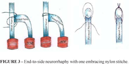
In the group B-R an embracing end-to-side neurorrhaphy was performed with one embracing 10-0 nylon stitche (ETSS). The procedure was almost the same than in the group A-R, although without the epineurum flaps. A single nylon 10-0 point went through the epineurum of the peroneal nerve, passed behind the tibial nerve, came back and passed in the front of this nerve, and then went through the epineurum of the peroneal nerve again like a loop, in a way that when fixed, this point approximated the extremity of the peroneal nerve distal stump to the tibial nerve (Figure 3).
The group B-L (Control) was the normal control and did not received any procedure (Figure 4).
The epineurum of the tibial nerve were intact in all groups. The proximal stump of the peroneal nerve was inverted, buried in the subjacent muscle and fixed with one 5-0 nylon sticthe.
The incision was sutured by plans with nylon 5-0.
The animals were housed in appropriate cages of the Animal Laboratory of Surgical Technique and Experimental Surgery, Department of Surgery and Orthopedics, with temperature of 24 degrees (+/- 0.5), with water and ration ad libitum, respecting the 12 hours dark/clear cycle.
After six months the rats were sacrificed and the cranial tibial muscle and the distal stump of the peroneal nerve were harvested. The muscles had their mass measured and the nerves were submitted to fibers counting.
For statistic analysis, since the variable answers did not show adherence to the normal distribution of probabilities, the Mann-Whitney non-parametric technique was used. All the comparisons were performed with the significance level of 5%.
Results
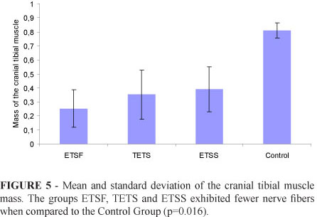
Table 1 and Figure 5 refers to the average of the cranial tibial muscle mass. All groups were different from the control but not between themselves.
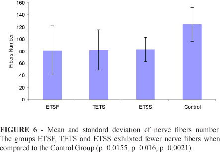
Table 2 and Figure 6 refers to the average number of nerve fibers per microscopic field of 0.175 mm2. The experimental groups also showed lower results from the control group, but not between themselves.
Discussion
In 1895 Ballance (apud Ballance et al.10) performed for the first time the suture of sectioned nerve to the lateral of a donor nerve, after cutting partially the donor. These lesions, determined by incision10 or by scarification11 in the lateral aspect of the donor nerve, allow the sectioned axons to grow inside the receptor nerves, providing, in fact, an end-to-end union between the endoneural tubes from both nerves.
These methods invariably showed functional disadvantage to the muscles innervated by the donor nerves. This observation leads Babcock12 to suggest the definitive abandon of the neurorhaphy. After that, except for the Gatta13 and Pozzan14 publications, there was a gap in the literature, without any report about this subject. For these reasons plus the operative microscope and microsurgical techniques advent, the end-to-end neurorraphy (ETE) became the standard treatment for peripheral nerve lesion, and the end-to-side neurorraphy (ETS) was forgotten6.
In 19921-3, with the introduction of the new concept of the end-to-side neurorraphy (ETS) without lesion on the donor nerve, new surgical possibilities appeared. Since the donor nerve doesn't suffer any lesion, any nerve can be used as donor nerve.
During the end-to-end neurorraphy usually the suture is made with 8-0, 9-0, 10-0 monofilament nylon, or even thinner, like 11-015. It's performed the epineural suture6-10, the perineural suture, also called interfascicular or funicular15-18, and even both associated in the same procedure, the epi-perineural suture. In this way, first the fascicles are sutured and then the epineurium6.
The fibrin glue, already used in the ETE19-23, can also be used in the ETS4. An important disadvantage of this method is it high cost24. However it's use became very interesting, especially in some situations where the nerve to be repaired are deep, making it more difficult to do the sutures.
In this study it was tried a most simple alternative, that could also be applied in the depth. The result of this study showed that both proposed new techniques, epineurium flap and embracing suture were similar to the traditional ETS.
In the embracing suture it is obvious that the suture can't be neither very tight, because it would cause stenosis in the donor nerve, nor very floppy, because it wouldn't provide appropriate contact between both nerves. However the ideal tension still was not determined and it's a subjective factor.
An interesting and contradictory aspect is the opinion of some authors who advocates lesion on the donor nerve, even minimum, like the ones caused by the needle during the suture in the end-to-side neurorraphy5,25-27. These small lesions would be important to provide bigger contact between the nerves and also for the liberation of growth and neurotrophic factors, related to the development, maintenance and regeneration of the axonal sprouts28. For this reason and also to reduce the dehiscence, some authors advocate the association of some anchorage microsurgical points in the ETS with fibrine glue29.
In the present study no donor nerve lesion was done in both embracing ETS groups, and the axonal sprouting occurred similarly to the traditional ETS, where needle was used. Other authors, like Akeda et al.30 an Matsumoto et al.31 also noticed axonal regeneration with no donor nerve lesion, using a silicone chamber in "T" and without suture. These authors understand that the minimum lesion caused in the donor nerve can harm it, so it must be avoided5,31.
The results obtained in the ETS with epi-perineural embracing flap and ETS with embracing suture support the theory that the two proposed techniques can be used in the ETS, even without harming the donor nerve.
The axonal sprouting exact mechanism through ETS still was not established5,30,32. The macrophages, related to the Walerian regeneration, seem to be the responsible for the perineurum absorption, allowing the neurotrophins from the receptor nerve to initiate the axonal sprouting5,33. This would explain the axonal sprouting after the embracing ETS, even without harming the donor nerve.
Liu et al.5 questioned the reproducibility of experimental studies in human begins. The authors affirm that the bigger perineurium, epineurium e endoneurium thickness from the human begins could prevent the axonal sprouting with techniques that doesn't cut the donor nerve. However, Samii et al.32 reported success in a clinical case using ETS with fibrin glue, that, in the same way than the embracing from this study, didn't have any lesion on the donor nerve.
Many experimental and clinical studies met good results with the ETS9,10,34-37. Some, however, were not success or the results were limited4,38-40. Several technical aspects in the ETS performance are being studied and maybe can explain the bad results found by some authors.
In the present study was proved that the similar results between the three types of ETS, traditional, embracing with a single suture or two epineurum flaps. This leads us to believe in the big potential of the embracing ETS with a single suture because it's simpler to be done, especially in deeper places.
Conclusion
The three types of neurorrhaphy showed no differences related to muscle mass and number of nerve fibers suggesting that the embracing with a single suture has great potential due its simplicity and usefulness in deep areas.
Received: October 18, 2011
Review: December 14, 2011
Accepted: January 20, 2012
Conflict of interest: none
Financial source: none
1 Research performed at Experimental Surgery Laboratory, Department of Surgery and Orthopedics, Botucatu School of Medicine (FMB), State University of Sao Paulo (UNESP), Brazil.
- 1. Viterbo F, Trindade JC, Hoshino K, Mazzoni Neto A. Latero-terminal neuroraphy without removal of the epineural sheath. Experimental study in rats. Rev Paul Med. 1992;110(6):267-75.
- 2. Viterbo F, Trindade JC, Hoshino K, Mazzoni Neto A. End-to-side neurorrhaphy with removal of the epineural sheath. Experimental study in rats. Plast Reconstr Surg. 1994;94(7):1038-47.
- 3. Viterbo F, Trindade JC, Hoshino K, Mazzoni Neto A. Two end-to-side neurorraphies and nerve graft with removal of the epineural sheath. Experimental study in rats. Br J Plast Surg. 1994;47(2):75-80.
- 4. Bertelli JA, dos Santos ARS, Calixto JB. Is axonal sprouting able to transverse the conjunctival layers of the peripheral nerve? A behavioral, motor, sensory study of end-to-side nerve anastomosis. J Reconstr Microsurg. 1996;12(8):559-63.
- 5. Liu K, Chen LE, Seaber AV, Goldner RV, Urbaniak JR. Motor functional and morphological findings following end-to-side neurorrhaphy in the rat model. J Orthop Res. 1999;17(2):293-300.
- 6. Rouleau M, Crepeau J, Tetreault L, Lamarche J. Facial nerve sutures: epineural versus perineural sutures. J Otolaryngol. 1981;10(5):338-42.
- 7. Ayan I, Bora A, Karakaplan M, Inan M, Bostan B, Germen B, Zorludemir S, Atik E. Effect of end-to-side repair of proximal nerve stumps of transected peripheral nerves on the development of neuroma (experimental study). Hand (N Y). 2007;2(4):199-205.
- 8. Matsuda K, Kakibuchi M, Fukuda K, Kubo T, Madura T, Kawai K, Yano K, Hosokawa K. End-to-side nerve grafts: experimental study in rats. J Reconstr Microsurg. 2005;21(8):581-91.
- 9. Brenner MJ, Dvali L, Hunter DA, Myckatyn TM, Mackinnon SE. Motor neuron regeneration through end-to-side repairs is a function of donor nerve axotomy. Plast Reconstr Surg. 2007;120(1):215-23.
- 10. Ballance CA, Ballance HA, Stewart P. Remarks on the operative treatment of chronic facial palsy of peripheral origin. Br Med J. 1903;1(2209):1009-13.
- 11. Krivolutskaia EG, Chumasov EI, Matina VN, Mel'tsova GM, Kirillov AL. End-to-side type of plastic repair of the facial nerve branches. Stomatologiia (Mosk). 1989;68(6):35-8.
- 12. Babcock W. A standard technique for operations on peripheral nerves with special reference to the closure of large gaps. Surg Gynecol Obstet. 1927;45:364-78.
- 13. Gatta R. Sulla anastomosi latero-terminale dei tronchi nervosi. Arch Ital Chir. 1938;48:155-71.
- 14. Pozzan, D.A. Contributo alla tecnica dell'anastomosi latero-laterale dei nervi periferici. Arch Ital Chir. 1939;57:458-82.
- 15. Braun RM. Comparative studies of neurorrhaphy and sutureless peripheral nerve repair. Surg Gynecol Obstet. 1966;122(1):15-8.
- 16. Hakstian RW. Funicular orientation by direct stimulation. An aid to peripheral nerve repair. J Bone Joint Surg Am. 1968;50(6):1178-86.
- 17. Grabb WC, Bement SL, Koepke GH, Green RA. Comparison of methods of peripheral nerve suturing in monkeys. Plast Reconstr Surg. 1970;46(1):31-8.
- 18. Hakstian RW. Perineural neurorrhaphy. Orthop Clin North Am. 1973;4(4):945-56.
- 19. Young JZ, Medawar PB. Fibrin suture of peripheral nerves. Lancet. 1940;2:126-8.
- 20. Tarlov IM, Boernstein W. Nerve regeneration; a comparative experimental study following suture by colt and thread. J Neurosurg. 1948;5(1):62-83.
- 21. Epstein JA. Neurography following experimental nerve suture and crush injury. J Neuropathol Exp Neurol. 1949;8(4):419-27.
- 22. Da-Silva CF, Gama SAM, Mattar Jr R, Pereira FC. Influence of highly purified preparations of hyaluronic acid in peripheral nerve regeneration in vivo. Braz J Morphol Sci. 2003;20(2):121-4.
- 23. Vicente EJD, Rodrigues AC, Vicente PC, Marmora CHC, Chagas PSC, Santos SMR. Regeneração de nervo periférico por meio da coaptação com cola de fibrina. HU Rev. 2007;33(2):53-6.
- 24. Bertelli JA, Mira JC. Nerve repair using freezing and fibrin glue: immediate histologic improvement of axonal coaptation. Microsurgery. 1993;14(2):135-40.
- 25. Pondaag W, Gilbert A. Results of end-to-side nerve coaptation in severe obstetric brachial plexus lesion. Neurosurgery. 2008;62(3):656-63.
- 26. Pannucci C, Myckatyn TM, Mackinnon SE, Hayashi A. End-to-side nerve repair: review of the literature. Restor Neurol Neurosci. 2007;25(1):45-63.
- 27. Bontioti E, Dahlin LB, Kataoka K, Kanje M. End-to-side nerve repair induces nuclear translocation of activating transcription factor 3. Scand J Plast Reconstr Surg Hand Surg. 2006;40(6):321-8.
- 28. Johnson EO, Charchanti A, Soucacos PN. Nerve repair: experimental and clinical evaluation of neurotrophic factor in peripheral nerve regeneration. Injury. 2008;39 Suppl 3:S37-42.
- 29. Menovsky T, Beek JF. Laser, fibrin glue, or suture repair of peripheral nerves: a comparative functional, histological and morfometric study in the rat sciatic nerve. J Neurosurg. 2001;95(4):694-9.
- 30. Akeda K, Hirata H, Matsumoto M, Fukuda A, Tsuji M, Nagakura T, Yoshida SOT, Uchida A. Regenerating axons emerge far proximal to the coaptation site in end-to-side nerve coaptation without a perineurial window using a T-Sharped chamber. Plast Reconstr Surg. 2006;117(4):1194-203.
- 31. Matsumoto M, Hirata H, Nishiyama M, et al. Schwann cells can induce collateral sprouting from intact axons: experimental study of end-to-side neurorrhaphy using a Y-chamber model. J Reconstr Microsurg. 1999;15(4):281-6.
- 32. Samii M, Koerbel A, Safavi-Abbasi S, Di Rocco F, Samii A, Gharabaghi A. Using an end-to-side interposed sural nerve graft for facial nerve reinforcement after vestibular schwannoma resection. Technical note. J Neurosurg. 2006;105(6):920-3.
- 33. Robillard BG, Myckatyn MT, Mackinnon SE, Hunter A. End-to-side neurorraphy and lateral axonal sprouting in a long graft rat model. Laryngoscope. 2002;112(5):899-905.
- 34. Millesi H, Schmidhammer R. Nerve fiber transfer by end-to-side coaptation. Hand Clin. 2008;24(4):461-83.
- 35. Kalbermatten DF, Wettstein R, von Kanel O, Erba P, Pierer G, Wiberg M, Haug M. Sensate lateral arm flap for defects of the lower leg. Ann Plast Surg. 2008;61(1):40-6.
- 36. Amr SM, Essam AM, Abdel-Meguid AM, Kholeif AM, Moharram AN, El-Sadek RE. Direct cord implantation in brachial plexus avulsions: revised technique using a single stage combined anterior (first) posterior (second) approach and end-to-side side-to-side grafting neurorrhaphy. J Brachial Plex Peripher Nerve Inj. 2009;19:4-8.
- 37. Viterbo F, Amr AH, Stipp EJ, Reis FJ. "End-to-side neurorrhaphy: past, present, and future". Plast Reconstr Surg. 2009;124(6 Suppl):351-8.
- 38. Fernandez E, Lauretti L, Tufo T, D'Ercole M, Ciampini A, Doglietto F. End-to-side nerve neurorrhaphy: critical appraisal of experimental and clinical data. Acta Neurochir Suppl. 2007;100:77-84.
- 39. Bertelli JA, Ghizoni MF. Nerve repair by end-to-side coaptation or fascicular transfer: a clinical study. J Reconstr Microsurg. 2003;19(5):313-8.
- 40. Dvali LT, Myckatyn TM. End-to-side nerve repair: review of the literature and clinical indications. Hand Clin. 2008;24(4):455-60
Publication Dates
-
Publication in this collection
26 Mar 2012 -
Date of issue
Mar 2012
History
-
Received
18 Oct 2011 -
Accepted
20 Jan 2012 -
Reviewed
14 Dec 2011