Abstracts
PURPOSE: To study the macro and microscopic evaluation of the damage caused by clamping or section of cervical sympathetic nerve in rabbits, quantifying the collagen in the lesions. METHODS: Twenty rabbits were divided into two groups of ten, doing in group 1 (section) section of the right cervical sympathetic nerve, while in group 2 (clipping) clipping of the nerve. All rabbits were induced to death on the seventh day after surgery. The macroscopic variables were: consequences of nerve lesion, clip appearance, presence of infection and adhesions around the nerve. Microscopy used hematoxylin-eosin staining to evaluate the stages and the degree of inflammation and necrosis, and F3BA Picrosirius red staining to quantify collagen. Mann-Whitney test was used for comparisons of collagen types I and III between groups. Fisher exact test analyzed the macroscopic variables, the degree of inflammation and necrosis. RESULTS: There was no discontinuity of nerve injury in the clipping group, as well as the clip was closed in all animals. The presence of severe adhesions was significantly higher in the clipping group (p<0.05). There was no significant difference on other variables macroscopically analyzed. There was no significant difference between groups regarding the type of inflammatory process and its intensity, as well as the presence of necrosis and collagen deposition in the nerves. CONCLUSIONS: In the macroscopic evaluation, the section caused discontinuity, which did not occur in the clamping group; there was no development of local infection; the clipping of the cervical sympathetic nerve was linked to the presence of a greater number of adhesions in comparison to the section group. Microscopically, no difference existed in relation to the type and intensity of inflammation reaction between the groups; occurred predominance of chronic and severe inflammation on the specimens; the necrosis was noticed equally in both groups; there was predominance of type I collagen deposition in relation to type III in both groups.
Sympathetic Nervous System; Surgical Stapling; Autonomic Nerve Block; Rabbits
OBJETIVO: Estudar a lesão provocada pela secção e pela clipagem no nervo simpático cervical de coelhos, avaliando-se a macroscopia, microscopia e quantificando-se o colágeno nas lesões. MÉTODOS: Foram utilizados 20 coelhos, distribuídos em dois grupos de dez, sendo nos animais do grupo 1 (secção) realizada secção do nervo simpático cervical direito, enquanto nos do grupo 2 (clipagem) realizada a clipagem desse nervo. Todos os coelhos foram induzidos à morte no sétimo dia de pós-operatório. As variáveis macroscópicas avaliadas foram: presença de lesão de descontinuidade do nervo, aspecto do clipe, presença de infecção e de aderências ao redor do nervo. A microscopia com hematoxilina-eosina foi feita para avaliar as fases, o grau do processo inflamatório e a presença de necrose; a coloração de Picrosirius red F3BA quantificou o colágeno. Utilizou-se o teste de Mann-Whitney nas comparações dos colágenos tipo I e tipo III entre os grupos. As variáveis macroscópicas, o grau do processo inflamatório e presença de necrose foram analisadas pelo teste de Fisher. RESULTADOS: Não houve lesão de descontinuidade do nervo no grupo clipagem, assim como o clipe encontrou-se fechado em todos os animais desse grupo. A presença de aderências intensas foi significativamente maior no grupo clipagem (p<0,05). Não houve diferença significativa quanto às demais variáveis analisadas macroscopicamente. Não houve diferença significativa entre os grupos quanto ao tipo de processo inflamatório e sua intensidade, assim como quanto à presença de necrose e ao depósito de colágeno nos nervos. CONCLUSÕES: Na avaliação macroscópica, a secção causou lesão de descontinuidade, o que não ocorreu na clipagem; não houve desenvolvimento de infecção local; a clipagem do nervo simpático cervical foi associada à presença de maior quantidade de aderências em relação à secção. Na avaliação microscópica reconheceu-se não haver diferença no tipo e na intensidade do processo inflamatório entre os grupos; ocorreu predomínio de processo inflamatório crônico e acentuado; as necroses ocorreram igualmente em ambos os grupos; houve predomínio de depósito de colágeno tipo I em relação ao tipo III em ambos os grupos.
Sistema Nervoso Simpático; Grampeamento Cirúrgico; Bloqueio Nervoso Autônomo; Coelhos
4 - ORIGINAL ARTICLE
WOUND HEALING
Evaluation of the damage caused by clamping or section of cervical sympathetic nerve in rabbits1 1 Research performed at Postgraduate Program in Principles of Surgery, Evangelic Faculty of Parana (FEPAR), Evangelic University Hospital, Curitiba-PR, Brazil.
Avaliação do dano causado pela clipagem ou secção do nervo simpático cervical em coelhos1 1 Research performed at Postgraduate Program in Principles of Surgery, Evangelic Faculty of Parana (FEPAR), Evangelic University Hospital, Curitiba-PR, Brazil.
Carlos Hespanha Marinho JuniorI; Jurandir Marcondes Ribas FilhoII; Osvaldo MalafaiaIII; Carmen Australia Paredes Marcondes Ribas FilhoII; Celia Toshi YamamotoIV; Orlando TorresV; Octavio Antonio Azevedo da Costa FilhoVI; Andrea Mendes de Oliveira NaufelVI; Fernando Todt CarbonieriVII; Lucas Wagner GortzVII
IMaster in Surgery, FEPAR, Curitiba-PR, Brazil. Main author, conception, design and critical review
IIPhD in Surgery, Full Professor, FEPAR and Federal University of Parana (UFPR), Curitiba-PR, Brazil. Supervised all phases of the study, manuscript writing and critical revision
IIIFull Professor, Head, Department of Surgery, FEPAR and UFPR, Coordinator of Principles of Surgery Postgraduate Program, FEPAR, Curitiba-PR, Brazil. Supervised all phases of the study, manuscript writing and critical revision
IVPhD in Surgery, UFPR, Curitiba-PR, Brazil. Conception, design, intellectual and scientific content of the study
VPhD in Surgery, Federal University of Maranhao (UFMA), MA, Brazil. Conception, design, intellectual and scientific content of the study
VIFellow Master degree, Postgraduate Program in Principles of Surgery, FEPAR, Curitiba-PR, Brazil. Involved in technical procedures
VIIGraduate student, FEPAR, Curitiba-PR, Brazil. Involved in technical procedures
Correspondence Correspondence Carlos Hespanha Marinho Junior Alameda Augusto Stellfeld, 1980 80730-150 Curitiba - PR Brasil Tel.: (55 41)3240-5488 ipem@evangelico.org.br
ABSTRACT
PURPOSE: To study the macro and microscopic evaluation of the damage caused by clamping or section of cervical sympathetic nerve in rabbits, quantifying the collagen in the lesions.
METHODS: Twenty rabbits were divided into two groups of ten, doing in group 1 (section) section of the right cervical sympathetic nerve, while in group 2 (clipping) clipping of the nerve. All rabbits were induced to death on the seventh day after surgery. The macroscopic variables were: consequences of nerve lesion, clip appearance, presence of infection and adhesions around the nerve. Microscopy used hematoxylin-eosin staining to evaluate the stages and the degree of inflammation and necrosis, and F3BA Picrosirius red staining to quantify collagen. Mann-Whitney test was used for comparisons of collagen types I and III between groups. Fisher exact test analyzed the macroscopic variables, the degree of inflammation and necrosis.
RESULTS: There was no discontinuity of nerve injury in the clipping group, as well as the clip was closed in all animals. The presence of severe adhesions was significantly higher in the clipping group (p<0.05). There was no significant difference on other variables macroscopically analyzed. There was no significant difference between groups regarding the type of inflammatory process and its intensity, as well as the presence of necrosis and collagen deposition in the nerves.
CONCLUSIONS: In the macroscopic evaluation, the section caused discontinuity, which did not occur in the clamping group; there was no development of local infection; the clipping of the cervical sympathetic nerve was linked to the presence of a greater number of adhesions in comparison to the section group. Microscopically, no difference existed in relation to the type and intensity of inflammation reaction between the groups; occurred predominance of chronic and severe inflammation on the specimens; the necrosis was noticed equally in both groups; there was predominance of type I collagen deposition in relation to type III in both groups.
Key words: Sympathetic Nervous System. Surgical Stapling. Autonomic Nerve Block. Rabbits.
RESUMO
OBJETIVO: Estudar a lesão provocada pela secção e pela clipagem no nervo simpático cervical de coelhos, avaliando-se a macroscopia, microscopia e quantificando-se o colágeno nas lesões.
MÉTODOS: Foram utilizados 20 coelhos, distribuídos em dois grupos de dez, sendo nos animais do grupo 1 (secção) realizada secção do nervo simpático cervical direito, enquanto nos do grupo 2 (clipagem) realizada a clipagem desse nervo. Todos os coelhos foram induzidos à morte no sétimo dia de pós-operatório. As variáveis macroscópicas avaliadas foram: presença de lesão de descontinuidade do nervo, aspecto do clipe, presença de infecção e de aderências ao redor do nervo. A microscopia com hematoxilina-eosina foi feita para avaliar as fases, o grau do processo inflamatório e a presença de necrose; a coloração de Picrosirius red F3BA quantificou o colágeno. Utilizou-se o teste de Mann-Whitney nas comparações dos colágenos tipo I e tipo III entre os grupos. As variáveis macroscópicas, o grau do processo inflamatório e presença de necrose foram analisadas pelo teste de Fisher.
RESULTADOS: Não houve lesão de descontinuidade do nervo no grupo clipagem, assim como o clipe encontrou-se fechado em todos os animais desse grupo. A presença de aderências intensas foi significativamente maior no grupo clipagem (p<0,05). Não houve diferença significativa quanto às demais variáveis analisadas macroscopicamente. Não houve diferença significativa entre os grupos quanto ao tipo de processo inflamatório e sua intensidade, assim como quanto à presença de necrose e ao depósito de colágeno nos nervos.
CONCLUSÕES: Na avaliação macroscópica, a secção causou lesão de descontinuidade, o que não ocorreu na clipagem; não houve desenvolvimento de infecção local; a clipagem do nervo simpático cervical foi associada à presença de maior quantidade de aderências em relação à secção. Na avaliação microscópica reconheceu-se não haver diferença no tipo e na intensidade do processo inflamatório entre os grupos; ocorreu predomínio de processo inflamatório crônico e acentuado; as necroses ocorreram igualmente em ambos os grupos; houve predomínio de depósito de colágeno tipo I em relação ao tipo III em ambos os grupos.
Descritores: Sistema Nervoso Simpático. Grampeamento Cirúrgico. Bloqueio Nervoso Autônomo. Coelhos.
Introduction
It took more than 17 centuries between the first anatomical reference on the sympathetic trunk, made by Claudius Galen in the II century, until the first operation on the sympathetic nervous system held by Alexander of Liverpool in 18891.
Based on obscure observations in eighteenth century, the section of the sympathetic trunk was indicated for the treatment of various diseases such as epilepsy, goiter, glaucoma, vasospastic disorders, and only in the second decade of the twentieth century it was used to avoid the symptoms of hyperhidrosis2..
From the 90s, with the advent of thoracoscopy, the operation on the sympathetic chain gained prominence and preoccupation about its side effects. Several series were published for the treatment of hyperhidrosis of the upper limb3-11. One of the problems encountered in all surgical techniques was the individuality, tending to justify the side effects by saying: "it seems to work on my hands"12. Experienced surgeons perform different techniques, always believing that the operation employed by him is the most appropriate. Unfortunately it is not possible to standardize them and, consequently, its impossible to conclude which is the best.
The lack of concrete answers in the literature regarding the anatomical lesion caused by clipping of the sympathetic nervous system in humans and experimental animals, stimulated this authors to clarify facts that may be good to better understand the issue.
The objective of this paper was to study the macroscopic and microscopic lesions of the cervical sympathetic nerve caused by section and clipping in rabbits on 7th postoperatively day.
Methods
This study was conducted at the Institute of Medical Research of Evangelic University Hospital/Evangelic Faculty of Parana and the Positive University, Curitiba, PR, Brazil and approved by the Ethics Committee of the Evangelic Beneficent Society of Curitiba.
Were used 20 rabbits (Oryctolagus cuniculus) males, weighing between 1900g and 2100g fed with commercial Nuvital® ration and kept in air conditioned rooms with digital temperature control ranging from 18°C and 22°C, relative humidity of 65% and photoperiod of 12 hours of darkness. After the acclimation, the animals were weighed, identified and kept in individual cages. The identification was performed using numbers in the ears. Throughout the experimental period, the environmental conditions of light, temperature and humidity of the rooms were the same with pre-programmed control.
The 20 animals were divided into two groups of 10: group 1 or section group in witch took place the section of the right cervical sympathetic nerve and group 2 or clamping group, which clamping was carried out at the same nerve with 5 mm endoclip (SLS -CLIP®, International Vitalitec). Was used a single clip per animal in this group.
As premedication, was applied 1 mg/kg of midazolam (Dormonid®, Roche) intramuscularly. After sedation, the animal received combination of 35 mg/kg of 10% ketamine hydrochloride (Ketamin-S®, Cristalia) with 5 mg/kg xylazine 2% (Xilazin®, Syntec) intramuscularly. After obtaining the anesthesia, was proceeded the venous access in a superficial vein of the ear. Then the animal was positioned on the operating table where he received isoflurane (Sevocris®, Cristalia) by face mask for maintenance of anesthesia13. During the peri-operative period were controlled heart rate and oxygen saturation in pulse oximetry. Once positioned supine with slight neck extension, was made pullout epilation after application of a cream (Veet®, Reckiite Benckiser) and skin disinfection with iodine polyvinylpyrrolidone iodine (1% Povidine®, Johnson Diversey).
The surgical technique consisted of: 1) cervical longitudinal median incision of approximately 1.5 cm using scalpel number 3 and blade 15; 2) blunt dissection with scissors between the platysma muscle and pre-tracheal muscles; 3) opening the pretracheal muscles from its median line; 4) identification of the trachea; 5) identification of vascular-nervous bundle in the paratracheal right cervical topography and individualization of the sympathetic nerve (Figure 1). The nerve had a section or clipping between his caudal third and two cranial thirds in group 1. Figure 2 shows group 2 clipping level with a titanium 5 mm endoclip, whose compression force was approximately 150 gf14 with non-disposable applicator Edlo-Exatech (Edlo Company, Canoas-RS, Brazil).
After this procedure was closed the incision with simple stitches of nylon 3/0 (Mononylon®, Johnson & Johnson). All rabbits underwent the same surgical technique related to the group.
At the end of surgery was applied 0.8 mg/kg butorphanol tartrate (Torbugesic®, Fort Dodge) subcutaneously every eight hours for two days and a single dose of 0.5 ml pentabiotic broad-spectrum (Veterinary pentabiotic®, Fort Dodge). During the postoperative period, was controlled the quantities of water and food intakes in order to determine the level of well-being13.
The rabbits were killed on 7th postoperatively day by rapid intravenous injection of 10 mg/kg of 2.5% sodium thiopental (Pentothal®, Abbott).
Once completed the time of the experiment, the animals were sacrificed and evaluated, and the right cervical sympathetic chains dissected. Were marked the cranial and the caudal end with cotton thread, and the left caudal end chosen as the area to be studied microscopically. The pieces were placed in Bouin's solution and identified with the number of the animal. In group 2, nerve segments had the removal of the clip in the laboratory.
Macroscopic
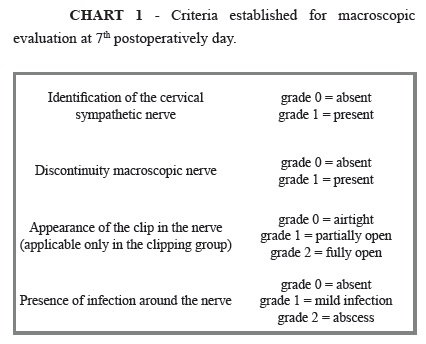
To evaluate the sympathetic nerves, clinical analysis and photographs were taken with Nikon Coolpix 4500 Digital Camera considering nerve identification, discontinuity point of the clip and the presence of infection in numerical degrees (Chart 1).
For evaluation of adherence, was adopted the methodology proposed by Jenkins15: grade 0=absent, grade 1=minimal adhesions, grade 2=moderate adhesions, grade 3=severe adhesions.
Microscopic evaluation
Microscopy was carried out at the edges of the nerve where the lesion was done group 1 and at the clamping point in group 2. After a routine procedure, 5 mm cuts were made in microtome and later mounted on 75x25 mm blades, and identified with the number of animal and stained, if hematoxylin-eosin (HE) and Picrosirius Red F3BA (PSR).
Hematoxylin-eosin staining
The slides were examined under an optical microscope binocular. This method aimed to assess the type and quantity of the predominant inflammatory cells (polymorphonuclear infiltrate and monomorfonuclear) and necrosis. Edema, congestion, hemorrhage and neutrophilic cells were indicative of acute inflammation, whereas inflammatory infiltration of chronic inflammatory process. The absence of necrosis characterized regeneration and the end result of a degenerative process.
The data obtained by the HE technique were classified into the histopathologic features according to the intensity of acute inflammation (Chart 2)16 and, according to Chart 3, in the presence of chronic inflammatory process17. By assigning numerical indices and histological features, they were transformed into quantitative variables.
F3BA Picrosirius Red (PSR)
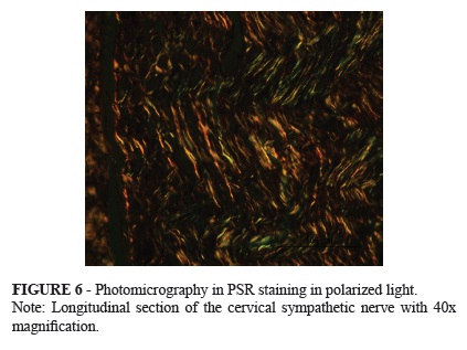
The histological sections stained by PSR aimed at the identification and quantification of collagen, both mature and immature, using the technique of polarized light microscopy and computerized morphometric analysis.
Was used the software-image-Pro Plus® 4.5 for Windows on a computer coupled to the optical microscope previously calibrated in pixels with 40-fold objective. With camera Olympus® DP71 (Sony, Tokyo, Japan), the images were sent to a color monitor, frozen and scanned. Three measurements were performed in each field of collagen longitudinal and transverse measurements in each field. Measurements were transferred to Excel for Windows statistical evaluation.
With the same application Image-Pro Plus, was analyzed the total area (in pixels) and the percentages of collagen type I and type III. The collagen fibers are thicker and highly birefringent dyed with orange and red (type I), and the thinner birefringent weakly with green (type III). Was obtained an average of these percentages in each case.
Statistical analysis
The procedure was statistically processed according to the nature of the analyzed data and the size of the groups. Was used the Mann-Whitney test for comparisons of collagen type I and type III between groups. To compare the presence of peri-neural adhesions, degree of inflammation and necrosis, was used the Fisher exact test (p=0.05 or 5% for statistical significance).
Results
Macroscopic
At autopsy, the cervical sympathetic nerve was not identified in only one animal in the group section, being excluded from the analysis of other variables.
In all animals of section group was observed discontinuity of nerve lesion, unlike the clamping group (Figures 3 and 4).
In all animals of the clipping group, the clips were securely closed. Was not observed in any case, in both groups, infection or abscess around the nerve.
Presence of adhesions around the right cervical sympathetic nerve
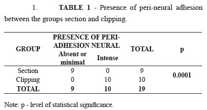
While no or minimal adhesions were found in all animals of section group, they were intense in clamping group (Table 1 and Figure 5)
Microscopic evaluation
Assessment of inflammation and necrosis
In one animal of each group was found necrosis (not significant p=0.74), whereas in the remaining animals there was chronic inflammatory infiltrate with predominance of monomorfonuclear.
Concerning the degree of inflammation in nine animals of clamping group, it was classified as strong, as well as in seven animals of section group. Only one animal in the section group had moderate inflammation. There was no significant difference in the degree of inflammation between the groups (p=0.47).
Collagen evaluation
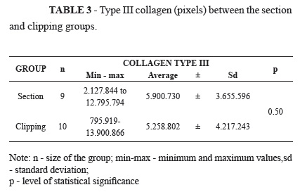
Tables 2 and 3 show in pixels the collagen type I and III, respectively, and compare them between groups.
There was no significant difference between the areas of type I collagen, in pixels, between the groups (p=0.60), as well as between areas of collagen type III (p=0.5). Figure 6 shows one staining PSR case.
Discussion
Studies comparing the anatomy of the cervical sympathetic chain in animals are scarce. Kalsey et al.18 compared the cervical sympathetic chain in dog, rabbit, guinea pig, albino rat, chicken and lizard. In all except the dog, the sympathetic nerve crossed the caudal half of the cervical vagus nerve alone. At the sympathetic nerve topography has no sub-divisions in both the rat and the guinea pig and rabbit. Was chosen the rabbits in this study due to easiness of getting them, standardization of weight and age, housing conditions, feeding and caliber identification that make them easy to be find.
During the experiment, the animal proved to be docile and easy to maintain. Although cited as too fragile to surgical procedures and anesthetics, no animal died.
Quantitative healing studies in the sympathetic nerve are scarce in the literature. In Brazil, Oliveira et al.19 developed an experimental model in pigs aiming to identify the presence of regeneration in communicating branches of the sympathetic trunk after his section and concluded that until the 15th postoperative day inflammatory reaction is intense and there is no anatomical neural regeneration. However from the 45th day after surgery, began anatomical neural regeneration. This paper confirms that there is no inflammatory reaction on day 7, postoperatively.
The present study confirms also the classical paper of Guth20, Fawcett and Keynes21, and Nicoli Aldini et al.22 demonstrating that the inflammatory activity after nerve damage is severe, associated with the process of Wallerian degeneration.
There are several observational studies on the effect of clamping of the sympathetic chest chain3,4,7,10,11. They show block conduction but without showing the histological lesion. The present study identified no significant differences with respect to the inflammatory process of clipping compared to section. It is speculated the possibility of not triggering the process of Wallerian degeneration19, what this paper does not show. Here was no significant the difference between the groups regarding the inflammatory process and its degree. However, due to lack of continuity in time in the clamping lesion, it is believed that future studies comparing the inflammatory process at different times after clip removal can demonstrate regression of an inflammatory process with or without anatomical regeneration.
Regarding to collagen, Oliveira et al.19 noted elevated levels of it in both types, I and III, increasing up to its peak point, on 135th day, postoperatively.
This paper demonstrates that there is deposition of collagen in the lesion area with greater presence of collagen type I than type III.
The less deposition of collagen type III, or immature, in relation to type I, or mature, in a short period of nerve injury is consistent with the Junqueira et al.23 findings. After peripheral nerve injury, they showed occur absorption part of the endoneurium - consisting of reticular fibers - whose main composition is collagen type III in the early stages of Wallerian degeneration.
Conclusions
In the macroscopic evaluation, the section caused discontinuity, which did not occur in the clamping group; there was no development of local infection; the clipping of the cervical sympathetic nerve was linked to the presence of a greater number of adhesions in comparison to the section group. Microscopically, no difference existed in relation to the type and intensity of inflammation reaction between the groups; occurred predominance of chronic and severe inflammation on the specimens; the necrosis was noticed equally in both groups; there was predominance of type I collagen deposition in relation to type III in both groups.
Received: January 18, 2012
Review: March 14, 2012
Accepted: April 16, 2012
Conflict of interest: none
Financial source: none
- 1. Royle JP. A history of sympathectomy. Aust N Z J Surg. 1999;69(4):302-7.
- 2. Hashmonai M, Kopelman D. History of sympathetic surgery. Clin Auton Res. 2003;13 Suppl 1:I6-9.
- 3. Lin CC, Mo LR, Lee LS, Ng SM, Hwang MH. Thoracoscopic T2-sympathetic block by clipping--a better and reversible operation for treatment of hyperhidrosis palmaris: experience with 326 cases. Eur J Surg Suppl. 1998;(580):13-6.
- 4. Lin TS, Huang LC, Wang NP, Lai CY. Video-assisted thoracoscopic T2 sympathetic block by clipping for palmar hyperhidrosis: analysis of 52 cases. J Laparoendosc Adv Surg Tech A. 2001;11(2):59-62.
- 5. Drott C, Göthberg G, Claes G. Endoscopic procedures of the upper-thoracic sympathetic chain. A review. Arch Surg. 1993;128(2):237-41.
- 6. Doolabh N, Horswell S, Williams M, Huber L, Prince S, Meyer DM, Mack MJ. Thoracoscopic sympathectomy for hyperhidrosis: indications and results. Ann Thorac Surg. 2004;77(2):410-4.
- 7. Reisfreisfeld R. Sympathectomy for hyperhidrosis: should we place the clamps at T2-T3 or T3-T4? Clin Auton Res. 2006;16(6):384-9.
- 8. Whitson BA, Andrade RS, Dahlberg PS, Maddaus MA. Evolution of clipping for thoracoscopic sympathectomy in symptomatic hyperhidrosis. Surg Laparosc Endosc Percutan Tech. 2007;17(4):287-90.
- 9. Sugimura H, Spratt EH, Compeau CG, Kattail D, Shargall Y. Thoracoscopic sympathetic clipping for hyperhidrosis: long-term results and reversibility. J Thorac Cardiovasc Surg. 2009;137(6):1370-6.
- 10. Fibla JJ, Molins L, Mier JM, Vidal G. Effectiveness of sympathetic block by clipping in the treatment of hyperhidrosis and facial blushing. Interact Cardiovasc Thorac Surg. 2009;9(6):970-2.
- 11. Coelho Mde S, Silva RF, Mezzalira G, Bergonse Neto N, Stori Wde S Jr, dos Santos AF, El Haje S. T3T4 endoscopic sympathetic blockade versus T3T4 video thoracoscopic sympathectomy in the treatment of axillary hyperhidrosis. Ann Thorac Surg. 2009;88(6):1780-5.
- 12. Rennie JA, Lin CC, Cameron AE. The technique of endoscopic thoracic sympathectomy: resection, clipping and cautery. Clin Auton Res. 2003;13 Suppl 1:I22-5.
- 13. Massone F. Neuroleptoanalgesia e anestesia dissociativa. In: Massone F (editor). Anestesiologia veterinária farmacologia e técnicas. 4ed. Rio de Janeiro: Guanabara Koogan; 2003. p.89-93.
- 14. Beltran KA, Foresman PA, Rodeheaver GT. Quantitation of force to dislodge endoscopic ligation clips: EndoClip II vs. Ligaclip ERCA. J Laparoendosc Surg. 1994;4(4):253-6.
- 15. Jenkins SD, Klamer TW, Parteka JJ, Condon RE. A comparison of prosthetic materials used to repair abdominal wall defects. Surgery. 1983;94(2):392-8.
- 16. de Sousa JB, Soares EG, Aprilli F. Effects of diclofenac sodium on intestinal anastomotic healing. Experimental study on the small intestine of rabbits. Dis Colon Rectum. 1991;34(7):613-7.
- 17. Luz-Veronez DA. Influencia da membrana basal juncional na redistribuiçăo topográfica das junçőes neuromusculares em fibras musculares regeneradas (Dissertaçăo). Instituto de Biologia, Universidade Estadual de Campinas; 1999.
- 18. Kalsey G, Mukherjee RN, Patnaik VVG. A comparative study of cervical sympathetic chain. J Anat Soc India. 2000;49(1):26-30.
- 19. Oliveira HA, Ximenes M 3rd, Filho FB, Carvalho PH, Gamafilho JB, Parra ER, Capelozzi VL, Milanez DeCampos JR. Experimental selective sympathicotomy (ramicotomy) and sympathetic regeneration. Interact Cardiovasc Thorac Surg. 2009;9(3):411-5.
- 20. Guth L. Regeneration in the mammalian peripheral nervous system. Physiol Rev. 1956;36(4):441-78.
- 21. Fawcett JW, Keynes RJ. Peripheral nerve regeneration. Annu Rev Neurosci. 1990;13:43-60.
- 22. Nicoli Aldini N, Fini M, Rocca M, Giavaresi G, Giardino R. Guided regeneration with resorbable conduits in experimental peripheral nerve injuries. Int Orthop. 2000;24(3):121-5.
- 23. Junqueira LC, Bignolas G, Brentani RR. Picrosirius staining plus polarization microscopy, a specific method for collagen detection in tissue sections. Histochem J. 1979;11(4):447-55.
Correspondence
Publication Dates
-
Publication in this collection
04 June 2012 -
Date of issue
June 2012
History
-
Received
18 Jan 2012 -
Accepted
16 Apr 2012 -
Reviewed
14 Mar 2012