Abstracts
PURPOSE: To compare the role of transitory latex and sylastic® implants in tympanoplasty on the closure of tympanic perforations. METHODS: A randomized double-blind prospective study was conducted on 107 patients with chronic otitis media submitted to underlay tympanoplasty and divided at random into three groups: control with no transitory implant, latex membrane group, and sylastic® membrane group. RESULTS: Greater graft vascularization occurred in the latex membrane group (p<0.05). Good biocompatibility was obtained with the use of the latex and silicone implants, with no effect on the occurrence of infection, otorrhea or otorragy. CONCLUSION: The use of a transitory latex implant induced greater graft vascularization, with a biocompatible interaction with the tissue of the human tympanic membrane.
Tympanic Membrane; Tympanoplasty; Wound Healing; Latex
OBJETIVO: Investigar os efeitos da biomembrana de látex e silicone no fechamento de perfurações timpânicas. METODOS: Foram estudados 107 pacientes com otite média cronica simples submetidos à cirurgia de timpanoplastia. Os pacientes foram classificados aleatoriamente, por sorteio, em três grupos. Até o final da cirurgia, os cirurgiões não tomavam conhecimento se seria ou não utilizado qualquer material externamente ao enxerto de fáscia temporal. Neste momento, o paciente que fora previamente classificado por uma ordem aleatória de entrada como participante do estudo foi identificado como participante do grupo 1, 2 ou 3, sendo, respectivamente, ausência de implante transitório no primeiro, membrana de látex no segundo e membrana de sylastic® no terceiro grupo. RESULTADOS: Houve uma maior vascularização do enxerto no grupo em que foi utilizado implante transitório de membrana de látex (p<0,05). Obteve-se boa biocompatibilidade com uso de implantes de látex e silicone sem afetar as taxas de ocorrência de infecção, otorreia ou otorragia. A porcentagem de cicatrização da membrana timpânica foi equivalente nos três grupos, assim como a melhora auditiva (p<0,01). CONCLUSÃO: O uso do implante transitório de látex provocou um maior processo de vascularização do enxerto, com interação satisfatória com os tecidos da membrana timpânica humana.
Membrana Timpânica; Timpanoplastia; Cicatrização; Látex
9 - ORIGINAL ARTICLE
CLINICAL INVESTIGATION
Anatomical and functional evaluation of tympanoplasty using a transitory natural latex biomembrane implant from the rubber tree Hevea brasiliensis1 1 Research performed at Postgraduate Program in Oftalmology, Otorrhinolaryngology and Head Neck Surgery, Department of Oftalmology, Otorrhinolaryngology and Head Neck Surgery, Faculty of Medicine of Ribeirao Preto, University of Sao Paulo (USP), Brazil.
Avaliação anatômica e funcional da timpanoplastia com a utilização de implante transitório de biomembrana natural de látex proveniente da seringueira Hevea brasiliensis
Marcos Miranda AraujoI; Eduardo Tanaka MassudaII; Miguel Angelo HyppolitoIII
IFellow PhD degree. Postgraduate Program in Oftalmology, Otorrhinolaryngology and Head Neck Surgery, Department of Oftalmology, Faculty of Medicine of Ribeirao Preto, USP, Ribeirao Preto-SP, Brazil. Interpretation of data, collection of study informations, manuscript writing
IIPhD, Assistant Professor, Department of Oftalmology, Otorrhinolaryngology and Head Neck Surgery, Faculty of Medicine of Ribeirao Preto, USP, Clinical Hospital of Ribeirao Preto, São Paulo-SP, Brazil. Interpretation of data, collection of study informations, manuscript writing
IIIAssistant Professor, Head of Division, Postgraduate Program in Oftalmology, Otorrhinolaryngology and Head Neck Surgery, Department of Oftalmology, Otorrhinolaryngology and Head Neck Surgery, Faculty of Medicine of Ribeirao Preto, USP, Ribeirao Preto-SP, Brazil. Main author. Conception, design, intellectual and scientific content of the study, acquisition, analysis and interpretation of data, manuscript writing and critical revision
Correspondence Correspondence: Prof. Dr. Miguel Angelo Hyppolito Departamento de Oftalmologia, Otorrinolaringologia e Cirurgia de Cabeça e Pescoço Faculdade de Medicina de Ribeirão Preto, USP Avenida Bandeirantes, 3900/12ºandar 14040-030 Ribeirão Preto - SP Brasil Tel.: (55 16)3602-2862 Fax: (55 16)3602 2860 mahyppo@fmrp.usp.br mhyppolito@uol.com.br
ABSTRACT
PURPOSE: To compare the role of transitory latex and sylastic® implants in tympanoplasty on the closure of tympanic perforations.
METHODS: A randomized double-blind prospective study was conducted on 107 patients with chronic otitis media submitted to underlay tympanoplasty and divided at random into three groups: control with no transitory implant, latex membrane group, and sylastic® membrane group.
RESULTS: Greater graft vascularization occurred in the latex membrane group (p<0.05). Good biocompatibility was obtained with the use of the latex and silicone implants, with no effect on the occurrence of infection, otorrhea or otorragy.
CONCLUSION: The use of a transitory latex implant induced greater graft vascularization, with a biocompatible interaction with the tissue of the human tympanic membrane.
Key words: Tympanic Membrane. Tympanoplasty. Wound Healing. Latex.
RESUMO
OBJETIVO: Investigar os efeitos da biomembrana de látex e silicone no fechamento de perfurações timpânicas.
METODOS: Foram estudados 107 pacientes com otite média cronica simples submetidos à cirurgia de timpanoplastia. Os pacientes foram classificados aleatoriamente, por sorteio, em três grupos. Até o final da cirurgia, os cirurgiões não tomavam conhecimento se seria ou não utilizado qualquer material externamente ao enxerto de fáscia temporal. Neste momento, o paciente que fora previamente classificado por uma ordem aleatória de entrada como participante do estudo foi identificado como participante do grupo 1, 2 ou 3, sendo, respectivamente, ausência de implante transitório no primeiro, membrana de látex no segundo e membrana de sylastic® no terceiro grupo.
RESULTADOS: Houve uma maior vascularização do enxerto no grupo em que foi utilizado implante transitório de membrana de látex (p<0,05). Obteve-se boa biocompatibilidade com uso de implantes de látex e silicone sem afetar as taxas de ocorrência de infecção, otorreia ou otorragia. A porcentagem de cicatrização da membrana timpânica foi equivalente nos três grupos, assim como a melhora auditiva (p<0,01).
CONCLUSÃO: O uso do implante transitório de látex provocou um maior processo de vascularização do enxerto, com interação satisfatória com os tecidos da membrana timpânica humana.
Descritores: Membrana Timpânica. Timpanoplastia. Cicatrização. Látex.
Introduction
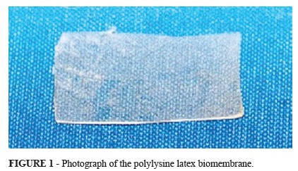
Tympanoplasty is one of the surgeries most frequently performed by otorhinolaryngologists for the correction of tympanic membrane perforations and the reconstruction of the mechanism of sound transmission in the middle ear. The graft used, such as a temporalis muscle fascia, functions as a support on which epithelialization currents slide in order to repair a perforation of the tympanic membrane1. Preliminary retrospective studies have indicated greater vascularization of tympanic membrane grafts when a natural latex membrane is used as a transitory implant in tympanoplasty2. A natural biomembrane prepared with latex extracted from the rubber tree Hevea brasiliensis and polymerized with 0.1% polylysine was developed by the Department of Biochemistry of the Faculty of Medicine, of Ribeirao Preto, University of Sao Paulo. The membrane was used experimentally as a cicatrization-inducing material on the injured esophageal walls of dogs and was shown to lead to a marked increase in vascularization and epithelialization3,4. When similar material was used clinically it led to a significant increase in the granulation tissue of phlebopathic ulcers in the legs of patients and to a concomitant reduction of pain during treatment5,6.
In order to assess the effects of transitory implants on the process of regeneration of the tympanic membrane in tympanoplasty, we carried out a prospective double-blind randomized study using the natural latex biomembrane and the Sylastic silicone membrane, another type of transitory implant already used in otorhinolaryngology practice. We also observed the interaction of the latex membrane with human tympanic tissue, its biocompatibility, possible toxicity or allergic reactions to it, and compared the success of surgery with the use of the two materials and the functional hearing recovery provided by cicatrization of the tympanic membrane.
Methods
This was a double-blind randomized study in which 107 patients followed up at the tertiary otorhinolaryngology clinic of the University Hospital, Faculty of Medicine of Ribeirao Preto, USP, were selected at random from May 2005 to November 2007. The patients had chronic perforation of the tympanic membrane and a clinical history of simple chronic otitis media, with a dry ear and no otorrhea for more than three months. All were submitted to preoperative tonal and vocal audiometry and impedanciometry. No patient had a history of traumatic rupture of the tympanic membrane and none had chronic cholesteatomatous otitis media. The study was approved by the Human Research Ethics Committee (protocol number 4593/2005) and all patients gave written informed consent to participate in the study, the only difference between groups being the use or not of the natural latex membrane or the sylastic® membrane at the end of surgery.
The patients were assigned to one of the following three groups by drawing lots: Group 1: 40 patients submitted to the standard underlay technique (without the latex or sylastic® membrane); Group 2: 39 patients submitted to the standard underlay technique in combination with the latex membrane; Group 3: 28 patients submitted to the standard underlay technique in combination with the sylastic® membrane.
The main aspects evaluated were: a successful take of the graft, with comparison of the healing ability between the control, latex and sylastic® groups; surgical access (endoaural and retroauricular); graft vascularization (hypervascularized, normally vascularized, hypovascularized); postoperative complications (infection, otorrhea, otorragy), and postoperative hearing function.
Excluded from the study were patients lost to postoperative follow-up, or followed up for less than 6 months, patients who were not submitted to the postoperative audiometry exam, patients submitted to a previous otologic surgery, patients with alterations of the ossicular chain, and syndromic patients (Down syndrome, Apert syndrome).
Surgical technique and postoperative care
The patients were submitted to the classical underlay myringoplasty technique of type I Wullstein tympanoplasty under general anesthesia and with the use of a free temporalis muscle fascia graft resting on gel foam placed in the middle ear and under the scarified margins of the perforation. Endoaural access was chosen in cases in which it was possible to visualize all the margins of the perforation by transcanal vision, and retroauricular access was chosen when these margins were not fully visible. Otorhinolaryngologists indicated surgery, chose the access option and performed the surgical procedure. The surgeons did not know until the end of surgery whether the latex membrane or the sylastic® membrane would be used externally to the temporalis fascia graft or whether no material would be applied over the graft. At that moment, the patient previously classified in a random manner as a participant in the study was identified as belonging to group 1, 2 or 3, respectively corresponding to absence of a transitory implant, use of the latex membrane and use of the sylastic® membrane. The natural latex membrane for group 2 or the sylastic membrane for group 3 was then placed externally on the remaining margins of the tympanic membrane in contact with the temporalis fascia graft. The transitory implant rested on gelfoam and an otologic pomade containing polymixin and bacitracin. In group 1 patients, the graft was placed on gelfoam and the otologic pomade containing polymixin and bacitracin according to the standard tympanoplasty or myringoplasty technique (Figures 1 and 2).
The patients were discharged from the hospital on the first postoperative day after otomicroscopic evaluation for an early diagnosis of infection (presence of otorrhea). Eardrops containing antibiotics were not routinely used for these patients, but were only used in cases in which signs of infection were noted. On the seventh postoperative day the patients were again evaluated by otomicroscopy of the operated ear for the determination of the occurrence of infection (otorrhea), of the presence of pomade in the outer auditory conduit, and of the positioning of the latex or sylastic® membrane (groups 2 and 3). When the examination revealed the presence of otorrhea, topical ciprofloxacin eardrops were used for treatment.
The patients were then evaluated on the 20th postoperative day to determine graft cicatrization (Figure 3). At that time the latex or sylastic® membrane had already been removed by the physician who had performed the tympanoplasty and the examiner was another otorhinolaryngologist who had not participated in the surgery and who was unaware of whether the latex or sylastic® membrane had been used. Vascularization of the graft was assessed by otomicroscopy and scored qualitatively as hypervascularization, normal vascularization or hypovascularization by examiners who did not know to which group the patient belonged.
The patients returned again 45 days after surgery when the aspect of the graft and the successful closure of the tympanic perforation were again evaluated by otomicroscopy. The patients with unsuccessful closure of the tympanic perforation were scheduled for a new surgical treatment upon medical indication and the otomicroscopy data were compared to those obtained 20 days after surgery. The patients with successful closure of the tympanic perforation were submitted to hearing tests to determine the functional result of the surgical procedure and the results of the three groups were compared. The patients were then followed up at six month intervals.
Statistical analysis
The data regarding the taking of the graft, the type of surgical success, the postoperative complications and the vascularization of the graft were analyzed statistically by the Fisher exact test using the PROC FREQ feature of the SAS 9.1 software. The data concerning the postoperative hearing function were analyzed using a mixed-effects (random and fixed effects) linear regression model adjusted with the SAS 9.1 software.
Results
A total of 107 patients (47 men and 60 women) were submitted to tympanoplasty from May 2005 to November 2007. Mean age was 24.7 years (range: seven to 56 years. All 107 tympanoplasty procedures were performed according to classical type I underlay technique. Mean patient follow-up was 16 months (range: 10 to 24 months). Regarding laterality, 57.7% procedures were performed on the right and 42.3% on the left. The surgical indication was due to simple chronic otitis media and no patient had cholesteatomas.
Group 1, with no application of a latex or sylastic® membrane, consisted of 40 patients, with 40 surgeries being performed. The access was retroauricular in 28 surgeries (70%) and endoaural in 12 (30%). Surgical success with complete closure of the tympanic membrane occurred in 55% (n=22) of cases, 14 of them with retroauricular access and eight with endoaural access. Group 2, with conventional underlay plus a latex membrane, consisted of 39 patients, with 39 surgeries being performed. The access was retroauricular in 28 surgeries (74.4%) and endoaural in ten (25.6%). Surgical success with complete closure of the tympanic membrane occurred in 66.6% (n=26) of cases, 19 of them with retroauricular access and seven with endoaural access. Group 3, with conventional underlay plus a sylastic® membrane, consisted of 28 patients, with 28 surgeries being performed. The access was retroauricular in 15 surgeries (53.6%) and endoaural in 13 (46.4%). Surgical success with complete closure of the tympanic membrane occurred in 57.1% (n=16) of cases, nine of them with retroauricular access and seven with endoneural access. There were no cases of blunting, graft lateralization, sensorineural hearing loss or atelectasia.
Table 1 compares the three groups regarding complete cicatrization of the tympanic membrane. The type of surgical access (endoaural and retroauricular) was also compared within each group regarding the taking of the graft. There was no significant difference between the three groups (p>0.05). Table 2 compares graft vascularization in the three groups. Vascularization was significantly greater in Group 2 in which the transitory latex implant was used (p=0.03). Table 3 compares the occurrence of complications such as infection, otorrhea and otorragy between groups, with no significant difference being observed between them (p>0.05).
Tables 4 and 5 respectively present the values of the preoperative and postoperative air-bone gap (ABG) distributed into audiometric intervals of 0-10, 11-20, 21-30, and > 30 dB. The audiometric data follow the classifications and directives recommended by the American Academy of Otorhinolaryngology.
The mean preoperative and postoperative ABG was 25.22 ± 7.21 and 11.90 ± 4.9 for Group 1, 23.51 ± 8.39 and 12.93 ± 10.63 for Group 2, and 28.05 ± 9.13 and 10.08 ± 7.35 for Group 3, respectively (see Table 6). The mean ABG closure was 13.32 ± 5.78 (P<0.01) for Gtoup 1, 10.58 ± 8.33 (Pr<0.01) for Group 2, and 17.97 ± 10.77 (Por<0.01) for Group 3 (Table 6). All values were significant for each individual group regarding ABG closure, with no significant differences between groups (Table 6).
Discussion
The rate of surgical success, which was considered to be complete graft cicatrization for at least six months after surgery and improved hearing acuity, was relatively higher in Group 2 treated with the latex membrane (66.6%) compared to Group 1, control (58.8%) and to Group 3 treated with the sylastic® membrane (55.17%). This variation, however, was not statistically significant. Also, no significant difference in surgical success was observed with the use of the endoaural or retroauricular access (Table 1). Surgical success regarding the closure of the tympanic membrane varies among reports, with rates from 51.4% to 81.8% being reported in the literature for type I tympanoplasty in training services for otologic surgery, and increasing to 88.8% to 96.43% as otologic surgeons acquire more experience2,7-10. In the present study, the use of the latex biomembrane as a transitory dressing tended to induce statistically significant vascularization (p<0.03) (Table 2). Vascularization was assessed by otomicroscopy, with a qualitative classification being attributed to the graft and compared by examiners who had not participated in the surgical procedure and who were unaware of whether a latex or sylastic® membrane had been used or not.
Preliminary studies have shown the presence of a possible vascular growth factor in Hevea brasiliensis which acts on human tissues, permitting improved vascularization of the temporalis muscle fascia graft from the remaining tympanic membrane by means of its ability to induce angiogenesis11,12. The use of the latex biomembrane as a transitory implant in addition to the temporalis muscle fascia graft proved to be biocompatible, causing no increase in infections, otorrhea or otorragy compared to the control group or the silicone membrane group (Table 3). This biocompatibility and the tissue regeneration properties of the natural latex have led to the recognition and liberation of the use of the biomembrane by the Brazilian health regulatory authority ANVISA5,11,13,14.
The three groups were homogeneous regarding the preoperative and postoperative hearing levels (Tables 4 and 5). Hearing function improved in a statistically significant manner in each group with the closure of the ABG (p<0.01, Table 6). There were no significant differences between groups regarding the audiometric data.
Conclusions
The natural latex membrane prepared from the rubber tree Hevea brasiliensis showeed satisfactory interaction with tissues of the human tympanic membrane and good biocompatibility, with no signs of toxicity or allergic manifestations being noted. The use of the transitory latex implant induced greater graft vascularization with no direct effect on the percentage of healing of the tympanic membrane. Hearing was similarly improved in all groups, with no difference regarding the use of a transitory latex or silicone implant.
Acknowledgment
We wish to thanks Clinical Hospital of the Faculty of Medicine of Ribeirao Preto, University of Sao Paulo for permitting the use of its facilities.
Received: March 29, 2012
Review: May 28, 2012
Accepted: June 25, 2012
Conflict of interest: none
Financial sources: FAPESP and FAEPA
- 1. Moberly AC, Fritsch MH. The evolution of mastoidectomy and tympanoplasty. Laryngoscope. 2010;120 Suppl 4:S213.
- 2. Oliveira JAA, Hyppolito MA, Coutinho-Netto J, Mrue F. Myringoplasty using a new biomaterial allograft. Braz J Otorhinolaryngol. 2003;69:649-55.
- 3. Andrade TA, Iyer A, Das PK, Foss NT, Garcia SB, Coutinho-Netto J, Jordão-Jr AA, Frade MA. The inflammatory stimulus of a natural latex biomembrane improves healing in mice. Braz J Med Biol Res. 2011;44(10):1036-47
- 4. de Sousa LH, Ceneviva R, Coutinho Netto J, Mrué F, de Sousa Filho LH, de Castro e Silva O. Morphologic evaluation of the use of a latex prosthesis in videolaparoscopic inguinoplasty: an experimental study in dogs. Acta Cir Bras. 2011;26 Suppl 2:84-91.
- 5. Frade MAC, Valverde RV, de Assis RVC, Coutinho-Netto J, Foss NT. Chronic phlebopathic cutaneous ulcer: a therapeutic proposal. Int J Dermatol. 2001;40:238-40.
- 6. Frade MAC, Salathiel AM, Mazzucato EL, Coutinho-Netto J, Foss NT. A natural biomembrane as a new proposal for the treatment of pressure ulcers. Med Cutánea Ibero-Latino-Americana. 2006;34:133-8.
- 7. Fukuchi I, Cerchiari DP, Garcia E, Rezende CEB, Rapoport PB. Tympanoplasty: surgical results and a comparison of the factors that may interfere in their success. Braz J Otorhinolaryngol. 2006;72:267-71.
- 8. Wang M, Yu E, Shiao A, Liao W, Liu C. The costs and quality of operative training for residents in tympanoplasty type I. Acta Otolaryngol. 2009;129:512-4.
- 9. Rizer FM. Overlay versus underlay tympanoplasty. Part II: the study. Laryngoscope. 1997;107(12):26-36.
- 10. Schraff S, Dash N, Strasnick B. Window shade tympanoplasty for anterior marginal perforations. Laryngoscope. 2005;115:1655-9.
- 11. Ferreira M, Mendonca RJ, Coutinho-Netto J, Mulato M. Angiogenic properties of natural rubber latex biomembranes and the serum fraction of Hevea brasiliensis. Braz J Physics. 2009;39:564-9.
- 12. Mendonca RJ, Mauricio VB, Teixeira LD, Lachat JJ, Coutinho-Netto J. Increased vascular permeability, angiogenesis and wound healing induced by the serum of natural latex of the rubber tree Hevea brasiliensis. Phytother Res. 2010;24:764-8.
- 13. Mrue F, Coutinho-Netto J, Ceneviva R, Lachat JJ, Thomazini JA, Tambellini H. Evaluation of the biocompatibility of a new biomembrane. Materials Res. 2004;7:277-83.
- 14. Balabanian CA, Coutinho-Netto J, Lamano.Carvalho TL, Lacerda SA, Brentegani LG. Biocompatibility of natural latex implanted into dental alveolus of rats. J Oral Sci. 2006;48:201-5.
Correspondence:
Publication Dates
-
Publication in this collection
25 July 2012 -
Date of issue
Aug 2012
History
-
Received
29 Mar 2012 -
Accepted
25 June 2012 -
Reviewed
28 May 2012