Abstracts
PURPOSE: Evaluate the influence of aging on the quality of the skin of white women, analyzing the dermal collagen. METHODS: Pre-auricular flaps were collected for histological and morphometric analysis of 218 white women who underwent spontaneous facial aesthetic plastic surgery. Picrosirius ultrared stain was used for analysis and quantification of collagen in five age groups (<40 years, 40 to 49 years, 50 to 59 years, 60 to 69 years and 70 to 79 years) . RESULTS: Histological analysis showed changes suggestive of skin aging (fragmentation and disorganization of collagen fibers), especially in patients over 60 years. There were no significant changes in the relationship of age with the thickness of the dermis and epidermis, but there was with the percentage of the collagen I, III and total (p<0.001), which decreased with increasing aging. CONCLUSION: There is reduction in collagen with increasing age, and an increase in its degradation, leading to fragmentation of the fibers.
Skin; Collagen; Skin Aging
OBJETIVO: Avaliar a influência do envelhecimento na qualidade da pele de mulheres brancas analisando o colágeno dérmico. MÉTODOS: Realizou-se análise histológica e morfométrica de 218 retalhos pré-auriculares de mulheres brancas que se submeteram espontaneamente à cirurgia estética facial. Foi usada a coloração de Picrosirius Ultrared para analisar e quantificar os colágenos I, III e total em cinco grupos etários (<40 anos, 40 a 49 anos, 50 a 59 anos, 60 a 69 anos e 70 a 79 anos). RESULTADOS: A análise histológica mostrou alterações sugestivas de envelhecimento cutâneo (fragmentação e desorganização das fibras de colágeno), especialmente em pacientes acima de 60 anos. Não houve diferenças significantes entre a idade e a espessura da derme e da epiderme, mas houve diferenças significantes entre as percentagens de colágeno I, III e total (p<0,001) com o aumento da idade. CONCLUSÃO: Existe redução do colágeno com o aumento da idade e um aumento na sua degradação, levando à fragmentação das fibras.
Pele; Colágeno; Envelhecimento da Pele
12 - ORIGINAL ARTICLE
CLINICAL INVESTIGATION
Influence of aging on the quality of the skin of white women. The role of collagen1 1 Research performed at Division of Research Methodology, Pontificial Catholic University of Parana (PUCPR), Brazil. Part of Master thesis, Postgraduate Program in Surgery. Tutor: Maria de Lourdes Pessole Biondo-Simões.
Influência do envelhecimento na qualidade da pele de mulheres brancas. O papel do colágeno
Eloina do Rocio Valenga BaroniI; Maria de Lourdes Pessole Biondo-SimõesII; André AuersvaldIII; Luiz Augusto AuersvaldIV; Mário Rodrigues Montemor NettoV; Morgana Cláudia Aparecida Bergamo OrtolanVI; Juliana Nemetz KohlerVII
IFellow Master Degree, Clinical Surgery, PUCPR, Curitiba-PR, Brazil. Associate Professor, Dermatology, State University of Ponta Grossa. Main Author. Design and supervised all phases of the study
IIFull Professor, Scientific Methodology, PUCPR. Associate Professor, Department of Surgery, Federal University of Parana (UFPR), Brazil. Conception and design of the study, critical revision
IIIFellow Master degree, Experimental Surgery, Evangelical College of Parana, Curitiba-PR, Brazil. Helped collection and processing of study informations
IVMD, Curitiba-PR, Brazil. Helped collection and processing of study informations
VFellow Master degree, Clinical Surgery, Federal University of Parana, Curitiba-PR, Brazil. Assistant Professor, Anatomical Pathology, State University of Ponta Grossa, Parana. Head of Department of Anatomical Pathology, Santa Casa de Misericórdia, Ponta Grossa, Paraná. Histopathological examinations
VIFellow Master degree, Clinical Surgery, PUCPR, Curitiba-PR, Brazil. Helped at preparation of histologicals sections
VIIGraduate student, School of Biology, PUCPR, Curitiba-PR, Brazil. Helped with technical procedures
Correspondence Correspondence: Eloina do Rocio Valenga Baroni Rua Barão do Cerro Azul, 631 84010-210 Ponta Grossa - PR Brasil Tel./Fax: (55 42)3222-2569 eloinabaroni@gmail.com
ABSTRACT
PURPOSE: Evaluate the influence of aging on the quality of the skin of white women, analyzing the dermal collagen.
METHODS: Pre-auricular flaps were collected for histological and morphometric analysis of 218 white women who underwent spontaneous facial aesthetic plastic surgery. Picrosirius ultrared stain was used for analysis and quantification of collagen in five age groups (<40 years, 40 to 49 years, 50 to 59 years, 60 to 69 years and 70 to 79 years) .
RESULTS: Histological analysis showed changes suggestive of skin aging (fragmentation and disorganization of collagen fibers), especially in patients over 60 years. There were no significant changes in the relationship of age with the thickness of the dermis and epidermis, but there was with the percentage of the collagen I, III and total (p<0.001), which decreased with increasing aging.
CONCLUSION: There is reduction in collagen with increasing age, and an increase in its degradation, leading to fragmentation of the fibers.
Key words: Skin. Collagen. Skin Aging.
RESUMO
OBJETIVO: Avaliar a influência do envelhecimento na qualidade da pele de mulheres brancas analisando o colágeno dérmico.
MÉTODOS: Realizou-se análise histológica e morfométrica de 218 retalhos pré-auriculares de mulheres brancas que se submeteram espontaneamente à cirurgia estética facial. Foi usada a coloração de Picrosirius Ultrared para analisar e quantificar os colágenos I, III e total em cinco grupos etários (<40 anos, 40 a 49 anos, 50 a 59 anos, 60 a 69 anos e 70 a 79 anos).
RESULTADOS: A análise histológica mostrou alterações sugestivas de envelhecimento cutâneo (fragmentação e desorganização das fibras de colágeno), especialmente em pacientes acima de 60 anos. Não houve diferenças significantes entre a idade e a espessura da derme e da epiderme, mas houve diferenças significantes entre as percentagens de colágeno I, III e total (p<0,001) com o aumento da idade.
CONCLUSÃO: Existe redução do colágeno com o aumento da idade e um aumento na sua degradação, levando à fragmentação das fibras.
Descritores: Pele. Colágeno. Envelhecimento da Pele.
Introduction
The average of life expectancy in Brazil moved from 69.3 years to 72.7 years in a decade (1997-2007). Preliminary results from the 2010 census showed that in Brazil there was an enlargement on the top of the age pyramid, since the population > 65 years increased from 4.8% in 1991 to 5.9% in 2000 and 7.4% in 20101.
Aging is a natural and unavoidable process for all organs of the body, and the skin is the ideal marker of chronological age. However, being an exposed organ it is subject to environmental damage such as Ultraviolet Radiations (UVR). So, the skin aging can be classified into cutaneous intrinsic aging or chronological aging and cutaneous extrinsic aging or photo-aging2.
In order to review the influence of aging on the quality of the skin of white women, were evaluated pre-auricular retails of 218 patients, who underwent spontaneous facial aesthetic plastic surgery.
Methods
The project was approved by the Ethics Committee on Human Research at PUC University of Parana and developed after its approval.
In order of being included in the study, patients had to present themselves in perfect physical and mental health and with full understanding of the process to which they were submitted, besides signing an informative consent form.
The patients answered a questionnaire with data on comorbidities, previous diseases and lifestyle habits. Those ones that had any elements that could influence on the quality of the skin to be evaluated, such as smoking, tanning, topical treatments, use of botulinum toxin and previous surgeries were excluded from the study.
To assess the quality of the skin were collected pre-auricular retails at the period between june 2009 and may 2011 .The flaps of skin were fixed in 10% formalin and sent to the Laboratory of Experimental Pathology - PUCPR.
The central part of the fragments was prepared for histological processing. From each block were made cuts with five micrometers thick.
Hematoxylin-eosin staining (HE) was used for general study and organization of the skin as well as measure of the thickness of the epidermis and dermis.
The density and organization of collagen was evaluated by Ultrared Picrosirius staining. Five fields of each histological section were analyzed and the average of the readings determined the density of collagen in each piece.
Images were captured with a Sony camera, CCD101 and sent to a Sony Trinitron color monitor, frozen and scanned by an oculus TCX board. Finally the images were analyzed by the Image Plus® 4.5 for Windows® Media Cybernetics micro-line Pentium computer.
After evaluating all the pieces, the data received statistical analysis using parametric tests as needed for results, and ANOVA methodology. For all tests it was used values of p < 0.05 or 5%.
Results
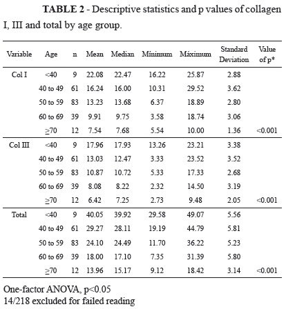
In a universe of 250 women evaluated during the study period, it was possible to select 218 patients who constituted the sample. The age ranged between 33 and 77 years, mean 54.9 ± 8.68 years (Table 1).
Microscopic analysis of histological sections stained with HE in the age groups showed no significant changes between them. The thickness of the epidermis measured in micrometers (µm) showed no significant differences between age groups (p = 0.152) and there were no significant differences in the thickness of the dermis (in µm) in relation to age (p = 0.506). There were no significant differences in the correlation between age and thickness of the epidermis (p = 0.546) and age and thickness of the dermis (p = 0.343).
At Ultrared Picrosirius staining, there was slight disruption and fragmentation of collagen fibers till the sixth decade, and became more evident from this age.
Histological sections of skin from the group of women aged below 50 years old showed large amounts of type I collagen fibers arranged neatly (Figure 1).
Histological sections of skin from the group of women aged 50 to 59 years showed mild fragmentation of collagen type I (Figure 2).
The histological sections of skin from the group of women aged 60 to 69 years showed marked reduction of type I collagen fibers and the presence of type III collagen fibers (Figure 3).
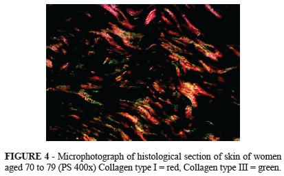
Histological sections of skin from the group of women aged 70 to 79 years showed marked disorganization and fragmentation of fibers of collagen type I and presence of type III collagen fibers (Figure 4).
Significant differences were found between the age groups and the percentage of collagens I, III and total (p < 0.001) (Table 2).
Comparing the age groups, two by two in relation to collagens I, III and total, differences were significant (p<0.001 for all comparisons except that between 60 to 69 x > 70 (p = 0.007 for collagen I; p = 0.076 for collagen III and p = 0.012 for total collagen).
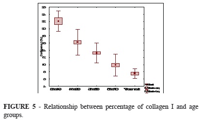
The comparison between age and percentage of collagen I, III and total showed a decrease of total collagen with increasing age (Figures 5, 6 and 7).
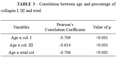
There were significant differences (p <0.001) between age and the percentage of collagens I, III and total (Table 3).
Discussion
Aging is a natural and unavoidable process for all organs of the body, and the skin is the ideal marker of chronological age. However, being an exposed organ, it is subject to environmental damage such as that caused by UVR. So, the skin aging can be classified into cutaneous intrinsic aging or chronological aging and cutaneous extrinsic aging or photo-aging, given the relevance of UVR3-5.
According to Landau, the most important aspect of aging would be the intrinsic flattening of the dermal-epidermal junction. However, while the thickness of the epidermis remains constant over the years, the thickness of the dermis reduces from the eighth decade6. In this sample there was no significant difference in the thickness of the epidermis and dermis among age groups assessed, going against the observations of this author.
Oriya et al.7 collected the abdominal skin of 18 cadavers between 20 and over 60 years, and found a significant reduction of the epidermis and dermis in individuals over 60 years of age, suggesting that these parameters are not fallen continuously, but it is more pronounced in advanced decades. The authors did not specify the causes of death and the general conditions of the research subjects. The patients who formed the present study were in perfect health, which could have contributed to a better quality of skin, especially among younger age groups.
Structural and functional alterations of dermo epidermal junction were cited by Le Varlet et al.8 after analysis of 14 samples of retro-auricular skin of women between nine to 79 years old. These changes were more homogeneous in young and older patients, and heterogeneous in intermediate group. The fact that retro-auricular skin has been examined is conflicting with the current study, because pre-auricular skin is more susceptible to UVR, being more exposed.
Rabe et al.9 noticed elongated and flattened fibroblasts in the photo aging skin, but still able to produce collagen, indicating that changes in collagen synthesis did not seem to be the cause of the decrease in collagen I and III.
The changes found in aged skin are: volume loss, reduction cell (fibroblasts and mast cells), reduction of blood vessels, shortening of capillary loops and abnormal nerve-endings10.
Moragas et al.11 showed changes of the dermal collagen in a study involving 86 patients from 3.5 months to 86 years, but they were more pronounced from the seventh decade. These authors obtained results similar to those of the present study, although the group of Moragas et al.11 presented extremes of age more pronounced and also had fewer cases.
In this study, the decrease in the percentage of collagen I, III and total across age groups was evident, and structural changes in organization of collagen fibers were more pronounced in the range 60 to 69 and > 70 unlike Varani et al.12, who concluded that the decrease in collagen, as well as the number of fibroblasts was more pronounced from 80 years.
In the study by El-Domyati et al.13 was evaluated exposed and unexposed skin of 38 donors between six to 84 years old. The changes started from the fifth decade, but they were only significant in the eighth and ninth decades, whith only traces of collagen were noticed. Perhaps the number of cases (218) in our sample may have contributed to the difference in results.
The changes that lead to decreased collagen with increasing age in this group and in the general population are not yet fully understood. Aging can vary from person to person according to lifestyle, current and previous diseases, genetic and environmental influences14.
In the elderly, the fibroblasts continue to produce collagen (even in vitro), which could suggest that the decrease of collagen would be subject to several factors, including increased degradation since the synthesis of matrix metalloproteinases (MMPs), enzymes responsible for degradation of collagen, increases with aging without an equivalent increase in their inhibitors. The beneficial effect of retinoids on skin aging due to the inhibitory effect on MMPs may be an indicator of this phenomenon15.
There are so many questions about skin aging and many studies are being made to clarify them. Only with the perfect understanding of this process will settle the ways to establish effective methods to prevent and to treat aging skin and perhaps to extend the benefits to other organs.
Conclusions
In white women, aging leads to: qualitative and degenerative changes in the dermis; decrease in total collagen and its fractions I and III; fragmentation and disorganization of collagen fibers especially above 60 years of age.
Acknowledgement
To Dr Gilberto Baroni for his valuable collaboration in all stages of this work.
Conflict of interest: none
Financial source: none
Received: May 10, 2012
Review: July 11, 2012
Accepted: August 14, 2012
-
1Available from www.ibge.gov.br/censo2010
» link - 2. Mine S, Fortunel NO, Pageon H, Asselineau D. Aging alters functionally human dermal papillary fibroblasts but not reticular fibroblasts: a new view of skin morphogenesis and aging. PloS One. 2008;3(12):e4066.
- 3. Faria JCM,Tuma PJ, Costa MP, Quagliano AP, Ferreira MC. Envelhecimento da pele e colágeno. Rev Hosp Clin Fac Med S. Paulo (Supl.). 1995;39-43.
- 4. Fisher GJ, Kang S, Varani J, Bata-Csorgo, Wan Y, Datta S, Voorhes JJ. Mechanisms of photoaging and chronological skin aging. Arch Dermatol. 2002;138:1462-70.
- 5. Yaar M, Gilchrest BA. Skin aging - postulated mechanisms and consequent changes in structure and function. Clin Geriatr Med. 2001;17:617-30.
- 6. Landau M. Exogenous factors in skin aging. Curr Probl Dermatol. 2007;35:1-13.
- 7. Oriá RB, Brito GAC, Ferreira FVA, Santana EM, Fernandes MR. Estudo das alterações relacionadas com a idade na pele humana utilizando métodos de histomorfometria e auto fluorescência. An Bras Dermatol. 2003;78(4):425-34.
- 8. Le Varlet B, Chaudagne C, Saunois A, Barré P, Sauvage C, Berthouloux B, Meybeck A, Dumas M, Bonté F. Age related functional and structural changes in human dermo-epidermal junction components. J Invest Dermatol Symp Proc. 1998;3:172-9.
- 9. Rabe JH, Mamelak AJ, McElgunn PJS, Morison WL, Sauder DN. Photoaging: mechanisms and repair. J Am Acad Dermatol. 2006;55:1-19.
- 10. Yaar M, Gilchrest BA. Photoageing: mechanism, prevention and therapy. Br J Dermatol. 2007;157:874-87.
- 11. Moragas A, Garcia-Bonafé M, Sans M, Torán N, Huguet P, Martin-Plata C. Image analysis of dermal collagen changes during skin aging. Analyt Quant Cytol Histol. 1998;20:493-9.
- 12. Varani J, Dame M, Rittie L, Voorhees J. Decreased collagen production in chronologically aged skin. Roles of age dependent alteration in fibroblasts functions and defective mechanical stimulation. Am J Pathol. 2006;168(6):1861-8.
- 13. El-Domyati M, Attia S, Saleh F, Brown D, Birk DE, Gasparro F, Ahmad H, Uitto J. Intrinsic aging VS photoaging: a comparative histopathological, immunohistochemical and ultrastructural study of skin. Exp Dermatol. 2002;11:398-405.
- 14. Yaar M, Eller MS, Gilchrest BA. Fifty years of skin aging. J Invest Dermatol. 2002;7:51-8.
- 15. Ortega YV, Tomey AV. Metaloproteinasas de la matriz y envejecimiento cutâneo. Rev Habanera Cienc Med. 2003;2(5). Available from http://www.ucmb.sld.cu/rhab/index.html
Publication Dates
-
Publication in this collection
27 Sept 2012 -
Date of issue
Oct 2012
History
-
Received
10 May 2012 -
Accepted
14 Aug 2012 -
Reviewed
11 July 2012