Abstracts
PURPOSE: To investigate the effect of cilostazol, in kidney and skeletal muscle of rats submitted to acute ischemia and reperfusion. METHODS: Fourty three animals were randomized and divided into two groups. Group I received a solution of cilostazol (10 mg/Kg) and group II received saline solution 0.9% (SS) by orogastric tube after ligature of the abdominal aorta. After four hours of ischemia the animals were divided into four subgroups: group IA (Cilostazol): two hours of reperfusion. Group IIA (SS): two hours of reperfusion. Group IB (Cilostazol): six hours of reperfusion. Group IIB (SS) six hours of reperfusion. After reperfusion, a left nephrectomy was performed and removal of the muscles of the hind limb. The histological parameters were studied. In kidney cylinders of myoglobin, vacuolar degeneration and acute tubular necrosis. In muscle interstitial edema, inflammatory infiltrate, hypereosinophilia fiber, cariopicnose and necrosis. Apoptosis was assessed by immunohistochemistry for cleaved caspase-3 and TUNEL. RESULTS: There was no statistically significant difference between groups. CONCLUSION: Cilostazol had no protective effect on the kidney and the skeletal striated muscle in rats submitted to acute ischemia and reperfusion in this model.
Ischemia; Reperfusion; Immunohistochemistry; Histology; Rats
OBJETIVO: Investigar o efeito do cilostazol no rim e na musculatura esquelética de ratos submetidos à isquemia aguda e reperfusão. MÉTODOS: Quarenta e três animais foram aleatoriamente distribuídos em dois grupos. Grupo I recebeu solução de cilostazol (10 mg/Kg) e Grupo II recebeu solução fisiológica a 0,9% (SF), após ligadura da aorta abdominal. Decorridas quatro horas de isquemia os animais foram distribuídos em quatro subgrupos: Grupo IA (Cilostazol): duas horas de reperfusão. Grupo IIA (SF): duas horas de reperfusão. Grupo IB (Cilostazol): seis horas de reperfusão. Grupo IIB (SF): seis horas de reperfusão. Após a reperfusão, realizou-se nefrectomia esquerda e a retirada da musculatura de membro posterior. Os parâmetros histológicos estudados em rim foram cilindros de mioglobina, degeneração vacuolar e necrose tubular. Em músculo foram edema, infiltrado inflamatório, hipereosinofilia de fibras, cariopicnose e necrose. A apoptose foi avaliada por imunohistoquímica, através da caspase-3 clivada e TUNEL. RESULTADOS: Não houve diferença estatisticamente significante entre os grupos estudados. CONCLUSÃO: O cilostazol não teve efeito protetor sobre o rim e sobre a musculatura estriada esquelética em ratos Wistar submetidos à isquemia aguda e reperfusão no modelo estudado.
Isquemia; Reperfusão; Imunoistoquímica; Histologia; Ratos
7 - ORIGINAL ARTICLE
ISCHEMIA/REPERFUSION
Effects of cilostazol in kidney and skeletal striated muscle of Wistar rats submitted to acute ischemia and reperfusion of hind limbs1 1 Research performed at Center for Technological Research (NPT), Mogi das Cruzes University (UMC), Sao Paulo-SP, Brazil.
Efeitos do cilostazol em rim e musculatura estriada esquelética de ratos Wistar submetidos à isquemia aguda e reperfusão de membros posteriores
Antonio Augusto Moreira NetoI; Sylvio Sebastião de Souza JúniorII; Vera Luíza CapelozziIII; Edwin Roger Parra-CuentasIV; Aurelino Fernandes Schmidt JúniorV; Acácio Francisco NetoVI; Olavo Ribeiro RodriguesVII
IPhD, Assistant Professor, Department of Surgery, Vascular Surgery Division, UMC, Mogi das Cruzes-SP, Brazil. Main author. Responsible for conception, design, intellectual and scientific content of the study
IIFellow PhD degree, Assistant Professor, Department of Surgery, Vascular Surgery Division, UMC, Mogi das Cruzes-SP, Brazil. Involved with technical procedures, acquisition of data and critical revision
IIIPhD, Assistant Professor, Department of Pathology, Head of Division of Pulmonary Pathology, University of Sao Paulo (USP), Brazil. Analysis of data
IVPhD, Assistant Professor, Department of Pathology, USP, Sao Paulo-SP, Brazil. Analysis and interpretation of data, revision
VPhD, Assistant Professor, Department of Surgery, Thoracic Surgery Division, UMC, Mogi das Cruzes-SP, Brazil. Manuscript writing, critical revision
VIPhD, Associate Professor, Department of Surgery, Head Division of Vascular Surgery, UMC, Mogi das Cruzes-SP, Brazil. Critical revision
VIIPhD, Associate Professor, Department of Surgery, Head Division of Thoracic Surgery, UMC, Mogi das Cruzes-SP, Brazil. Conception and design of the study, critical revision
Correspondence Correspondence: Antonio Augusto Moreira Neto Rua Alfredo Cardoso, 18 08710-280 Mogi das Cruzes SP Brasil Tel.: (55 11)4799-0440 femareis@yahoo.com.br
ABSTRACT
PURPOSE: To investigate the effect of cilostazol, in kidney and skeletal muscle of rats submitted to acute ischemia and reperfusion.
METHODS: Fourty three animals were randomized and divided into two groups. Group I received a solution of cilostazol (10 mg/Kg) and group II received saline solution 0.9% (SS) by orogastric tube after ligature of the abdominal aorta. After four hours of ischemia the animals were divided into four subgroups: group IA (Cilostazol): two hours of reperfusion. Group IIA (SS): two hours of reperfusion. Group IB (Cilostazol): six hours of reperfusion. Group IIB (SS) six hours of reperfusion. After reperfusion, a left nephrectomy was performed and removal of the muscles of the hind limb. The histological parameters were studied. In kidney cylinders of myoglobin, vacuolar degeneration and acute tubular necrosis. In muscle interstitial edema, inflammatory infiltrate, hypereosinophilia fiber, cariopicnose and necrosis. Apoptosis was assessed by immunohistochemistry for cleaved caspase-3 and TUNEL.
RESULTS: There was no statistically significant difference between groups.
CONCLUSION: Cilostazol had no protective effect on the kidney and the skeletal striated muscle in rats submitted to acute ischemia and reperfusion in this model.
Key words: Ischemia. Reperfusion. Immunohistochemistry. Histology. Rats.
RESUMO
OBJETIVO: Investigar o efeito do cilostazol no rim e na musculatura esquelética de ratos submetidos à isquemia aguda e reperfusão.
MÉTODOS: Quarenta e três animais foram aleatoriamente distribuídos em dois grupos. Grupo I recebeu solução de cilostazol (10 mg/Kg) e Grupo II recebeu solução fisiológica a 0,9% (SF), após ligadura da aorta abdominal. Decorridas quatro horas de isquemia os animais foram distribuídos em quatro subgrupos: Grupo IA (Cilostazol): duas horas de reperfusão. Grupo IIA (SF): duas horas de reperfusão. Grupo IB (Cilostazol): seis horas de reperfusão. Grupo IIB (SF): seis horas de reperfusão. Após a reperfusão, realizou-se nefrectomia esquerda e a retirada da musculatura de membro posterior. Os parâmetros histológicos estudados em rim foram cilindros de mioglobina, degeneração vacuolar e necrose tubular. Em músculo foram edema, infiltrado inflamatório, hipereosinofilia de fibras, cariopicnose e necrose. A apoptose foi avaliada por imunohistoquímica, através da caspase-3 clivada e TUNEL.
RESULTADOS: Não houve diferença estatisticamente significante entre os grupos estudados.
CONCLUSÃO: O cilostazol não teve efeito protetor sobre o rim e sobre a musculatura estriada esquelética em ratos Wistar submetidos à isquemia aguda e reperfusão no modelo estudado.
Descritores: Isquemia. Reperfusão. Imunoistoquímica. Histologia. Ratos.
Introduction
The reperfusion syndrome is characterized by metabolic acidosis, hyperkalemia by the loss of intracellular potassium, increased serum creatine kinase and myoglobin with myoglobinury1. May result in acute renal failure: coagulation disorders, accumulation of extracellular fluid and acute pulmonary distress2.
With the acute ischemia, initiating anaerobic metabolism, glycogen with transformation into lactate, leading to decreased production of adenosine triphosphate (ATP), which alters the permeability of the cell muscle, allowing the output of potassium and myoglobin and entry Sodium and calcium célula3.
The cell damage occurs only after an interval of thirty minutes of ischemia and irreversible changes in skeletal muscle occur after four to six hours of ischemia. Ischemia can lead to cell death by necrosis or apoptosis4.
The caspases are a group of protease essential for apoptosis, which makes irreversible cleavage of DNA. The cascade activation of caspases can be triggered by many factors, such as the presence of superoxide, which is a derivative of the mass muscular ischemia5.
After reperfusion muscle, is released into the circulation of acid metabolites and products of cell destruction that cause significant metabolic alterations, such as metabolic acidosis and hyperkalemia5. The more severe change is due to precipitation of myoglobin in the renal tubules in acid environment, causing acute tubular necrosis6.
Treatment of acute ischemia reperfusion is only the affected territory reperfusion of which may lead to ischemia and reperfusion syndrome.
Many times reperfusion cannot be performed immediately after the onset of ischemia, since there is a very variable period of time between the first symptoms of the disease and emergency medical care.
Cilostazol is an antiplatet drug and vasodilator with antimitogenics and cardiotonic actions intended to reduce the symptoms of peripheral vascular disease, intermittent claudication7 and prevention of recurrent cerebral stroke8.
We hypothesize that cilostazol inhibits platelet aggregation and promoting vasodilation could decrease the deleterious effects of ischemia and reperfusion syndrome. If administered at the onset of ischemia could reduce acute ischemic events, and consequently reduce renal injury after reperfusion.
The objective of this study is to assess the effect of cilostazol in the kidney and skeletal striated muscle of Wistar rats submitted to acute ischemia and reperfusion of hind limbs, since no experimental models in animals studies on the effectiveness of cilostazol in acute ischemia and reperfusion have been reported.
Methods
Forty three male Wistar rats aged ten months and average weight of 300g were used.
This study was approved by the Ethics Committee for Animal Experimentation and Manipulation (CEMEA) on 10.03.2008 and according to Federal Law No. 11.794, of October 8, 2008, and Decree No. 6689 of July 15, 2009 which regulated Law 11,794.
The experiment was developed at the Center for Technological Research (NPT) of Mogi das Cruzes University in the period from July/2008 to October /2010.
Surgical technique
The animals were anesthetized and placed an orogastric tube.
Laparotomy was performed in 4cm long. The abdominal aorta was ligated just below the renal artery with propylene 7.09.
Proceeded to divided into two randomly groups:
Group I (Cilostazol) - 21 animals, received by the tube solution of cilostazol (Cebralat®, Libbs) at a concentration of 1 mg/ml in 10mg/kg.
Group II (Sham) - 22 animals: received 10ml/kg saline solution 0.9%.
The solutions were administered immediately after aortic ligature.
The effectiveness of aortic ligature was confirmed by the appearance of pallor, cyanosis and decreased temperature on their hind legs for thermometry. The absence of pulse and flow in the aorta below the ligature was confirmed by intraoperative Dopplermetry.
Ischemic time
After aortic ligature, started to measure ischemia time and proceeded to the closure of the laparotomy.
After four hours of ischemia relaparotomy was performed in order to remove the aortic ligature and then, closed.
Reperfusion time
After removing aortic ligature, started to measure time of reperfusion.
Two animals were excluded from the previous steps of the experiment because they died before the final reperfusion time. Forty one animals underwent a second phase, remaining in the study.
Proceeded to the distribution of animals in four groups according to the time of reperfusion:
Group IA: 11 animals that received cilostazol with reperfusion time of two hours.
Group IIA: 11 animals receiving saline solution 0.9%, with time of two hours of reperfusion.
Group IB: nine animals receiving cilostazol with reperfusion time of six hours.
Group IIB: ten animals receiving saline solution 0.9%, with time of six hours of reperfusion.
Kidney and muscle harvesting and euthanasia
The animals were anesthetized, a left nephrectomy was performed, and the hind limb muscles harvested and proceded the euthanasia.
The kidney and muscles of the left hind limb were divided into two parts, fixed in peraformaldehyde 10%. One part was designed to histological analysis and the other for immunohistochemical study.
Histological and immunohistochemical analysis
The histological changes were examined in muscle: interstitial edema, inflammatory infiltrate, hypereosinophilia fiber, cariopicnose and necrosis (Figure 1). In kidney: presence of myoglobin cylinders, vacuolar degeneration of tubular cells and acute tubular necrosis (Figure 2)5.
For histological analysis were examined 10 microscopic fields non-coincident at 440x magnification of each sample.
The results of the histological analysis were expressed in a semi-quantitative percentage as the ratio of expression changes observed in the total:
0 = absent;
1 = less than 10%;
2 = 11% to 25%;
3 = 26% to 50%;
4 = more than 51%.
The presence of apoptosis was evaluated by immunohistochemical expression of cleaved caspase 3 and TUNEL (TdT mediated dUTP nick endlabeling).
A sample of each animal resulted in two slide sections for the immunohistochemical analysis.
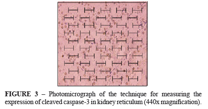
To determine the levels of immunohistochemical expression we used the technique of counting poins. We used a microscope equipped with an eyepiece and a grid of 50 rows and 100 points by 440x magnification. We examined 10 non-coincident microscopic fields on each piece to quantify all the points that fell on the stained portions, totaling 1000 points covering an area of 62.500 μm2 per field of study. Points falling on stained areas represent the proportion of positively stained cells in the tissue area10 (Figure 3).
The results of histological analysis and expression of immunohistochemical markers were tested using non-parametric Mann-Whitney (Wilcoxon Rank-Sum Test) for two independent samples, p ≤0.05 for significance. It was also made a graph of the 95% confidence interval of the mean for immunohistochemistry between groups with six hours of reperfusion.
Results
Histological evaluation
Histological changes observed in muscle and kidney that received cilostazol compared to control group showed no significant differences in two and six hours of reperfusion.
Immunohistochemical evaluation
The expression of cleaved caspase 3 in the kidney and TUNEL expression in the kidney and muscle are shown in Table 1 (next page).
The results showed that the total number of cells undergoing apoptosis quantified by cleaved caspase 3 and TUNEL was lower in the kidneys of animals that received cilostazol with six hours of reperfusion, however these differences were not statistically significant (Figures 4 and 5).
Discussion
The Cilostazol has been widely used in the treatment of chronic peripheral arterial disease and in treatment of ischemic coronary artery disease due to its antiplatelet and vasodilatation properties7.
The therapeutic use of cilostazol in acute ischemia and its role in prevention of reperfusion syndrome has not been recommended. The lack of randomized controlled studies, using cilostazol in ischemia and reperfusion in rat kidney and muscle, we have motivated the design of this research. In this study it was hypothesized that cilostazol might have efficacy in treating acute ischemia and would decrease the metabolic syndrome of reperfusion protects target organs such as muscle and kidney.
Experimental studies have demonstrated that cilostazol obtained a protective effect against ischemic injury in animal models when used in another organs than skeletal muscle and kidneys11-13.
Several experimental studies had model and design similar to our study to investigate the effectiveness of other drugs in ischemia and reperfusion6,15,16.
A recent experimental study demonstrated that cilostazol reduces the oxidative stress of ischemia and reperfusion in rats subjected to 45 minutes of spinal cord ischemia by clamping the aorta and a reperfusion period of 48 hours. The biochemical and histopathological analysis of the treated animals at a dose of cilostazol 20 mg/kg orally for three days before spinal cord ischemia, demonstrated a reduction in neurological damage and a reduction of oxidative stress16.
The tissue injury caused by ischemia and reperfusion is described as early onset, and studies have shown that histological changes are observable by microscopy after four hours of ischemia and 15 minutes of reperfusion14.
In this study, histological changes due to ischemia and reperfusion were observed in the kidney and striated muscle in similar intensity in the animals receiving cilostazol and those who received only saline solution. These changes were independent of reperfusion time, since it did not differ significantly in the two groups and underwent six hours of reperfusion.
The expression of cleaved caspase 318 and TUNEL19 for quantification of apoptosis currently have been used.
The expression of cleaved caspase 3 and TUNEL was lower in the cilostazol group with six hours of reperfusion, but not statistically significant. Therefore these results suggest that cilostazol could have a positive effect to reduce renal apoptosis in later periods of reperfusion.
Another hypothesis could be due to the number of subjects used, which the analysis of a few animals per group, had no power to find statistically significant differences.
Conclusion
Cilostazol had no protective effect on the kidney and the skeletal striated muscle in rats submitted to acute ischemia and reperfusion in this model.
Received: June 21, 2012
Review: August 23, 2012
Accepted: September 20, 2012
Conflict of interest: none
Financial source: none
- 1. Haimovici H. Peripheral arterial embolism. A study of 330 unselected cases of embolism to the extremities. Angiology. 1950;1:1-20.
- 2. Blaisdell FW. The reperfusion syndrome. Microcirc Endothelium Lymphatics. 1989;5(3-5):127-41.
- 3. Silveira M, Yoshida SB. Isquemia e reperfusão em músculo esquelético. J Vasc Br. 2004;3(4):367-78.
- 4. Badhwar A, Dungey AA, Harris KA. Limitations of ischemic tolerance in oxidative skeletal muscle: perfusion vs tissue protection. J Surg Res. 2003;109:62-7.
- 5. Lockshin RA. An attempt to understand the multiparametric control of the initiation of apoptosis. Apoptosis. 2008;13(1):1195-7.
- 6. Teruya R, Fagundes DJ, Oshima CTF, Brasileiro JL, Marks G, Ynouye CM, Simões MJ. The effects of pentoxifylline into the kidneys of rats in a model of unilateral hindlimb ischemia/reperfusion injury. Acta Cir Bras. 2008;23(1):29-35.
- 7. Akiyama H, Kudo S, Shimizu T. The absorption, distribution and excretion of a new antithrombotic and vasodilating agent, cilostazol, in rat, rabbit, dog and man. Arzneimittelforschung. 1985;35(7A):1124-32.
- 8. Rowlands TE, Donnelly R. Medical therapy for intermittent claudication. Eur J Vasc Endovasc Surg. 2007;34(3):314-21.
- 9. Huang Y, Cheng Y, Wu J, Li Y, Xu E, Hong Z. Cilostazol as an alternative to aspirin after ischaemic stroke: a randomised, double-blind, pilot study. Lancet Neurol. 2008;7(6):494-9.
- 10. Francisco Neto A, Silva JCCB, Fagundes DJ, Percário S, Novo NF, Juliano Y, Moreira Neto AA. Estudo das alterações oxidativas, da capacidade antioxidante total e do óxido nítrico, em ratos submetidos à isquemia e reperfusão de membros posteriores. Acta Cir Bras. 2005;20(2):134-9.
- 11. Gundersen HJ, Bendtsen TF, Korbo L, Marcussen N, Moller A, Nielsen K. Some new, simple and efficient stereological methods and their use in pathological research and diagnosis. APMIS. 1988;96(5):379-94.
- 12. Fukusawa M, Nishida H, Sato T, Miyazaki M, Nakaya H. [4-(1-Cyclohexyl-1H-tetrazol-5-yl)butoxy]-3,4-dihydro-2-(1H)quinolinone (cilostazol), a phosphodiesterase type 3 inhibitor, reduces infarct size via activation of mitochondrial Ca2+-activated K+ channels in rabbit hearts. J Pharmacol Exp Ther. 2008;326(1):100-4.
- 13. Lee JH, Park SY, Shin YW, Hong KW, Kim CD, Sung SM, Kim KY, Lee WS. Neuroprotection by cilostazol, a phosphodiesterase type 3 inhibitor, against apoptotic white matter changes in rat after chronic cerebral hypoperfusion. Brain Res. 2006;1082(1):182-91.
- 14. Iba T, Kidokoro A, Fukunaga M, Takuhiro K, Ouchi M, Ito Y. Comparison of the protective effects of type III phosphodiesterase (PDE3) inhibitor (cilostazol) and acetylsalicylic acid on intestinal microcirculation after ischemia reperfusion injury in mice. Shock. 2006;26(5):522-6.
- 15. Brasileiro JL, Fagundes DJ, Miiji LON, Oshima CTF, Teruya R, Marks G, Inouye CM, Santos MA. Isquemia e reperfusão de músculo sóleo de ratos sob ação da pentoxifilina. J Vasc Bras. 2007;6(1):50-63.
- 16. Francischetti I, Maffei FHA, Bitu-Moreno J, Fuhrmann Neto M, Coelho MPV, Kai FHT, Sequeira JL, Yoshida WB. Ação do ácido trissódio-cálcio-dietileno-triaminopentaacético (CaNa3DTPA) nas lesões de isquemia-reperfusão em membro posterior de rato. Acta Cir Bras. 2002;17(5):332-41.
- 17. Sahin MA, Onan B, Guler A, Oztas E, Uysal B, Arslan S, Demirkilic U, Tatar H. Cilostazol, a type III phosphodiesterase inhibitor, reduces ischemia/reperfusion-induced spinal cord injury. Heart Surg Forum. 2011;14(3):E171-7.
- 18. Lee YS, Kang YJ, Kim HJ, Park MK, Seo HG, Lee JH. Higenamine reduces apoptotic cell death by induction of heme oxygenase-1 in rat myocardial ischemia-reperfusion injury. Apoptosis. 2006;11(7):1091-100.
- 19. Gavrieli Y, Sherman Y, Bem-Sasson AS. Identification of programmed cell death in situ via specific labeling of nuclear DNA fragmentation. J Cell Biol. 1992;119(3):493-501.
Publication Dates
-
Publication in this collection
29 Oct 2012 -
Date of issue
Nov 2012
History
-
Received
21 June 2012 -
Accepted
20 Sept 2012 -
Reviewed
23 Aug 2012