Abstract
PURPOSE:
To evaluate KGF and human beta defensin-4 (HBD-4) levels produced by dermic fibroblasts and keratinocytes cultivated from burned patients' skin samples.
METHODS:
Keratinocytes and fibroblasts of 10 patients (four major burns, four minor burns and two controls) were primarily cultivated according to standard methods. HBD-4 and KGF genes were analyzed by quantitative PCR.
RESULTS:
In fibroblasts, KGF gene expression was 220±80 and 33.33±6.67 (M±SD; N=4), respectively for major and minor burn groups. In keratinocytes, KGF gene expression was 11.2±1.9 and 3.45±0.37 (M±SD; N=4), respectively for major and minor burn groups. In fibroblasts, HBD-4 gene expression was 15.0±4.0 and 11.5±0.5 (M±SD; N=4), respectively for major and minor burn. In keratinocyte, HBD-4 gene expression was 0.0±0.0 and 13.4±4.8 (M±SD; N=4), respectively for major and minor burn.
CONCLUSIONS:
KGF expression was increased in burn patient fibroblasts compared to control group. In keratinocytes culture, KGF suppression is inversely proportional to burn extension; it is active and increased in major burn but decreased in minor burn. HBD-4 expression was increased in fibroblasts and decreased in keratinocytes from all burned patients.
Burns; Gene expression; Fibroblasts; Keratinocytes; Fibroblast growth factor 7; Human beta defensin 4
Introduction
The high incidence of infection among burn patients is a major concern in its clinical evolution, which is directly related to morbidity and mortality rates of this group. Antimicrobial peptides, components of innate immune response, protect the organism by the activation of inflammatory cells or inactivating pathogen action directly. Therefore, the increased expression in these peptides by the epithelial cells improves hosts resistance against microbial infection11 Bals R, Weiner DJ, Moscioni AD, Meegalla RL, Wilson JM. Argumentation of innate host defense by expression of a cathelicidin antimicrobial peptide. Infect Immun. 1999 Nov;67(11):6084-9. , 2 2 Lee PHA, Ohtake T, Zaiou M, Murakami M, Rudisill JA, Lin KH. Expression of an additional cathelicidin antimicrobial peptide protects against bacterial skin infection. PNAS 2005 Mar 8;102(10):3750-5.. Among the great diversity of antimicrobial peptides involved in the inflammatory response, we highlight here the defensin family. Defensins are able to permeabilize bacterial membranes selectively, as has been proved in the study of peptides Crp-4, HBD-2, HBD-3, RTD-1 e BTD-733 Schmidt NW, Mishra A, Lai GH, Davis M, Sanders LK, Tran D. Criterion for amino acid composition of defensins and antimicrobial peptides based on geometry of membrane destabilization. J Am Chem Soc. 2011 May 4;133(17):6720-7..
HBD-4 presents a high antimicrobial activity against Pseudomonas aeruginosa when expressed by genetically modified primary epidermal keratinocytes44 Smiley AK, Gardner J, Klingenberg JM, Neely AN, Supp DM. Expression of human beta defensin 4 in genetically modified keratinocytes enhances antimicrobial activity. J Burn Care Res. 2007 Jan-Feb;28(1):127-32.. HBD-3 shows strong bactericidal activity against Staphylococcus aureus and MRSA when its expression is induced in human keratinocytes and fibroblasts55 Suzuki Y, Inokuchi S, Takazawa K, Umezawa K, Saito T, Kidokoro M, Tanaka M, Matsuzawa H, Inoue S, Tuchiya I, Ando K. Introduction of human beta-defensin-3 into cultured human keratinocytes and fibroblasts by infection of a recombinant adenovirus vector. Burns. 2011 Feb;37(1):109-16..
HBD-2 inhibits in vitro growth of the dermatophytes Trichophyton rubrum, T. mentagrophytes, Microsporum canis e Epidermophyton floccosum, despite not as much as psoriasin, RNAse 7 and fluconazol66 Fritz P, Beck-Jendroschek V, Brasch J. Inhibition of dermatophytes by the antimicrobial peptides human beta-defensin-2, ribonuclease 7 and psoriasin. Med Mycol. 2012 Aug;50(6):579-84..
Studies concerning keratinocyte growth factor (KGF) actions in skin lead to quantitative relations between KGF levels and immune response antimicrobial peptides. It was demonstrated that RNAm codifying HBD-2 and cathelecidin-37 quantity is increased proportionally to the KGF gene expression in cultured cells77 Werner S, Peters KG, Longaker MT, Fuller-Pace F, Banda MJ, Williams LT. Large induction of keratinocyte growth factor expression in the dermis during wound healing. Proc Natl Acad Sci U S A. 1992 Aug 1;89(15):6896-900. , 8 8 Heilborn JD, Nilsson MF, Kratz G, Weber G, Sørensen O, Borregaard N, Ståhle-Bäckdahl M. The cathelicidin anti-microbial peptide LL-37 is involved in re-epithelialization of human skin wounds and is lacking in chronic ulcer epithelium. J Invest Dermatol. 2003 Mar;120(3):379-89..
Keratinocytes cultures treated with interleukin-1-alpha presents high level of the antimicrobial peptides human beta-defensin 1 and 2, IL-37 and antileukoprotease. When infected with Escherichia coli, Pseudomonas aeruginosa and Staphylococcus aureus, its antibacterial activity is significantly increased99 Erdag G, Morgan JR. Interleukin-1alpha and interleukin-6 enhance the antibacterial properties of cultured composite keratinocyte grafts. Ann Surg. 2002 Jan;235(1):113-24..
In direct contact with bacteria as Pseudomonas aeruginosa, KGF alone is unable to prevent their proliferation. However, cultured human keratinocytes that produced KGF or other culture supplemented with this growth factor are able to exuberantly inhibit bacterial proliferation. Therefore, it is suggested that KGF is a skin immune response modulator, possibly through inducing defensins production by keratinocytes1010 Sobral CS, Gragnani A, Morgan J, Ferreira LM. Inhibition of proliferation of Pseudomonas aeruginosa by KGF in an experimental burn model using human cultured keratinocytes. Burns. 2007 Aug;33(5):613-620..
We infer that KGF acts indirectly, as an immune response modulator, as the substance itself does not present any effect in the microorganisms. The present study hypothesis is that KGF might be the trigger to some antimicrobial peptides production by keratinocytes, such as human beta defensins. Therefore, our objective is to perform a quantitative analysis of keratinocyte growth factor (KGF) gene expression and human beta-defensin-4 (HBD-4) produced by fibroblasts and keratinocytes, respectively, from cultured burned patients skin samples.
Methods
The project was approved by the Ethics Committee of Federal University of Sao Paulo (UNIFESP) in October 14th, 2011 (1454/11). The present study is experimental, in vitro, using discarded skin from burned patients. It is observational, analytic, controlled and conduced in a single center. Patients included in this study have read and signed the Free and Clarified Contentment Term.
The patients recruited to this study were burn victims admitted in the Burns Treatment Unit, Plastic Surgery Division, University Hospital (UNIFESP), and the control group with healthy patients, non-smoking, submitted to mammoplasty in the Plastic Surgery Division. The culture of cells was realized, and we have created a cell bank with the characteristics of these patients, which have been previously published1111 Gragnani A, Müller BR, da Silva ID, de Noronha SM, Ferreira LM. Keratinocyte growth factor, tumor necrosis factor-alpha and interleukin-1 beta gene expression in cultured fibroblasts and keratinocytes from burned patients. Acta Cir Bras. 2013 Aug;28(8):551-8..
Inclusion, exclusion and non-inclusion criteria
Inclusion criteria for the study were patients of both genders, over 18 years old, for the group with major burns: having deep partial thickness or full thickness burns affecting between 25% and 50% of total body area surface (TBSA) or which require partial skin graft in 10% TBSA. For the minor burns group, the criterion added was that the affected TBSA should be 5% or less, for deep partial thickness or full thickness burns and the need of partial skin graft. To the control group was included the criterion of not having previous diseases, not smoking, and performing aesthetic surgery.
Patients who had previous skin diseases or conditions that should interfere directly in the inflammatory process were not included. Exclusion criteria were the lost of the extracted material. The surgical procedure, fibroblasts and keratinocytes cultures, and total RNA extraction were reported earlier1111 Gragnani A, Müller BR, da Silva ID, de Noronha SM, Ferreira LM. Keratinocyte growth factor, tumor necrosis factor-alpha and interleukin-1 beta gene expression in cultured fibroblasts and keratinocytes from burned patients. Acta Cir Bras. 2013 Aug;28(8):551-8..
Commercially available TaqMan Gene Expression Assay kits (Applied Biosystems, Foster City, CA) were used to assess HBD-4 (Hs00823638_m1) and FGF7 (KGF) (Hs00384281_m1). B-actin gene (Hs99999903_m1) was used as housekeeping gene1111 Gragnani A, Müller BR, da Silva ID, de Noronha SM, Ferreira LM. Keratinocyte growth factor, tumor necrosis factor-alpha and interleukin-1 beta gene expression in cultured fibroblasts and keratinocytes from burned patients. Acta Cir Bras. 2013 Aug;28(8):551-8. , 1212 Greer S, Honeywell R, Geletu M, Arulanandam R, Raptis L.Housekeeping genes; expression levels may change with density of cultured cells. J Immunol Methods. 2010;355(1-2):76-9.. All qPCR reactions were performed on an ABI Prism 7000 Sequence Detection System (Applied Biosystems, Foster City, CA) using the fluorescent Taqman methodology (TaqMan One Step qPCR Master Mix Reagentes, Applied Biosytems, Foster City, CA). 100 ng of total RNA) was used for each qPCR reaction in a total volume of 50μL according to the manufacturer's protocol. Thermal cycling conditions were as follow: 30s at 48ºC, 10 min at 95ºC, 40 cycles of 15s, denaturation at 95ºC, and 60 s annealing at 60ºC. Quantification Cycle (Cq) values were used as endpoint defined as the PCR cycle number in which the fluorescence generated by the amplification crosses the threshold. Each gene was submitted to an absolute quantification assay by means of a standard curve, produced by dilutions to calculate the amount of a target sequence in a solution. The results were expressed in the same measure units as the standard curve. Three replicate reactions per sample were run to ensure statistical significance.
The assays were made in triplicate and the experimental values obtained in the study were presented by mean (M) and standard deviation (SD), because it is a cultured cells assay, where there is the homogenization of the cells lineage, with any stimulation differing from the culture medium. The calculation of the Cq were obtained as described in previous study1111 Gragnani A, Müller BR, da Silva ID, de Noronha SM, Ferreira LM. Keratinocyte growth factor, tumor necrosis factor-alpha and interleukin-1 beta gene expression in cultured fibroblasts and keratinocytes from burned patients. Acta Cir Bras. 2013 Aug;28(8):551-8.. The comparative Cq method (also known as the 2-DDqC method)1313 Schmittgen TD, Livak KJ. Analyzing real-time PCR data by the comparative C(T) method. Nat Protoc. 2008;3(6):1101-8. was used for data analysis in order to calculate relative quantities (RQs) of gene expression among the samples. DataAssistTM, software developed for quick analysis of TaqMan real-time PCR (Applied Biosystems, Foster City, CA), was used to confirm the data calculations performed in Microsoft Office Excel. Whenever receptor quantification exceeded 36 amplification rounds (qC), quantities were considered to be undetectable or very low, and measurements were taken as invalid. Statistical significance was assessed using the Student's t-test. Statistical significance was defined at p<0.05.
Results
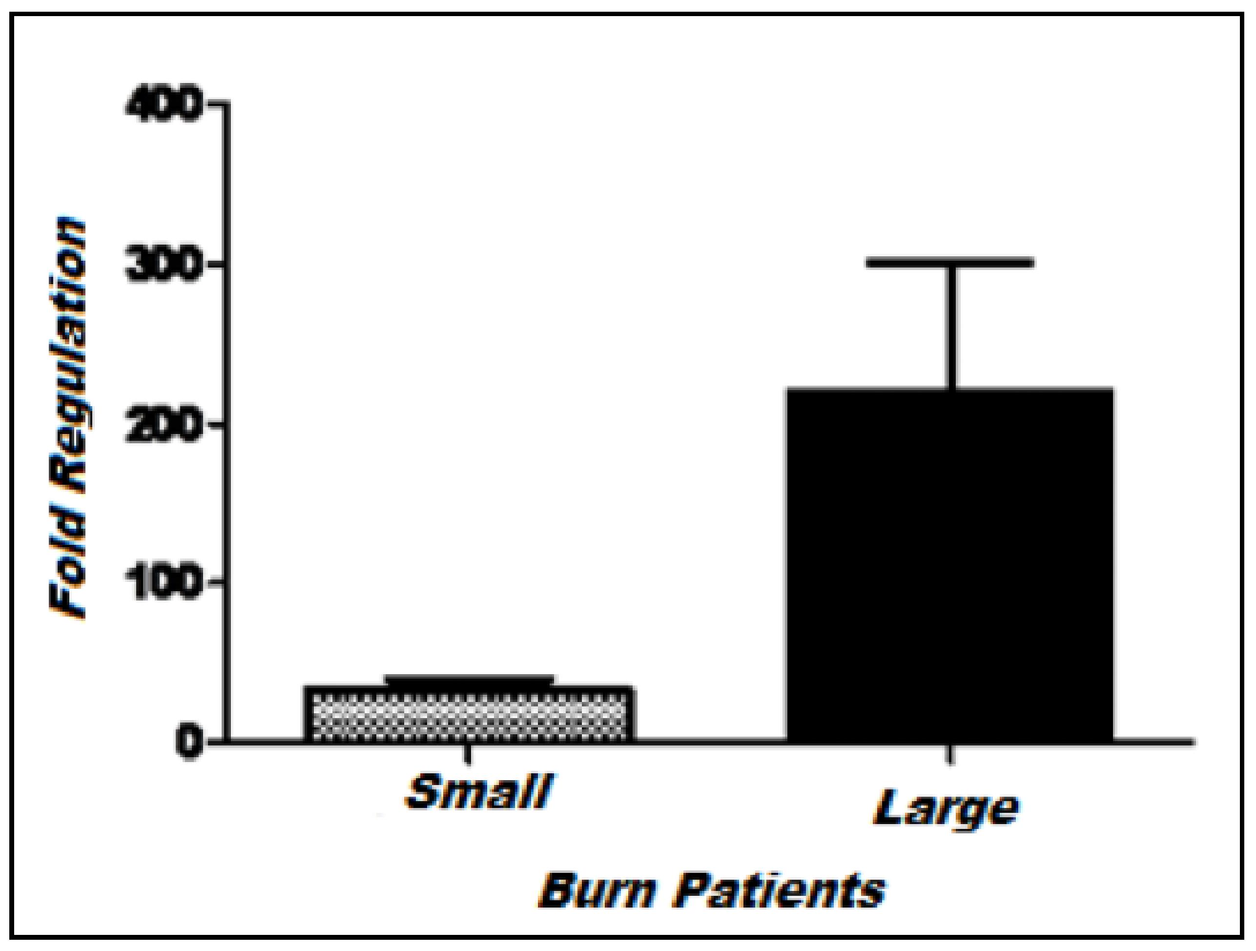
Keratinocyte growth factor (KGF) expression in fibroblast culture
The gene responsible for KGF synthesis is over expressed in burn patients' fibroblasts compared to the control group (Figure 1). Apparently there is no difference between expression levels among the minor and major burn groups and there is only a tendency to significance with p=0.081. The mean of the minor and major burn groups were (M ± SD): 33.33 ± 6.67 (n=4) and 220 ± 80 (n=4), respectively.
Keratinocyte growth factor (KGF) expression in keratinocyte culture
In keratinocyte culture, the expression of the KGF gene seems to be directly proportional to burn injury extension. It is active and over expressed in the major burn group and under expressed in the minor burn group (Figure 2). There was a difference (p=0.019) between both groups. The mean obtained for minor and major burn groups were (M ± SD): 3.45 ± 0.37 (n=4) and 11.2 ± 1.9 (n=4), respectively.
Human beta-defensin-4 (HBD-4) expression in fibroblast culture
HBD-4 is over expressed in burned patients' fibroblasts of both minor and major groups, compared to the control (Figure 3). No difference was shown between minor and major groups (p=0.43). The mean of minor and major burn were (M±SD): 11.5 ± 0.5 (n=4) and 15.0 ± 4.0 (n=4), respectively.
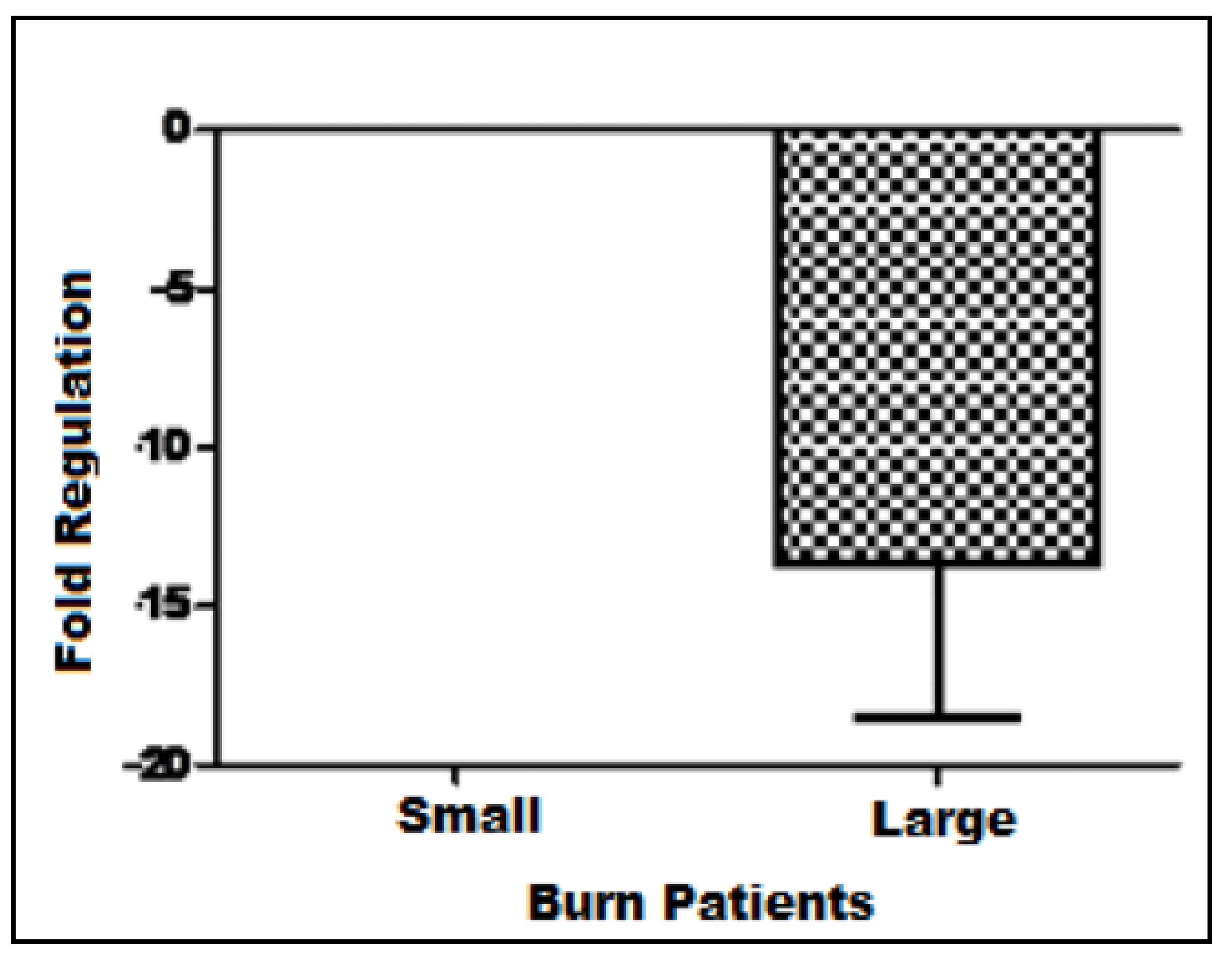
Human beta-defensin-4 (HBD-4) expression in keratinocyte culture
The keratinocytes cultured had the gene HBD-4 repressed in a directly proportional manner to the extension of the burn (Figure 4). In the major burn group, HDB-4 gene is active and over expressed compared to control, while it is no expressed in the minor burn group. The mean of minor and major burn were (M ± SD): 13.4 ± 4.8 (n=4) and 0 ± 0 (non-expressed), respectively.
Discussion
A large amount of studies shows KGF's importance as a paracrine mediator of epithelial growth. The induction of increased KGF expression in fibroblasts and keratinocytes grafted in burn models in vivo leads to significant acceleration of the reepithelialization process1414 Kopp J, Wang GY, Kulmburg P, Schultze-Mosgau S, Huan JN, Ying K, Seyhan H, Jeschke MD, Kneser U, Bach AD, Ge SD, Dooley S,Horch RE. Accelerated wound healing by in vivo application of keratinocytes overexpressing KGF. Mol Ther. 2004 Jul;10(1):86-96., while KGF ablation leads to decreased proliferation rates, angiogenesis and VEGF mRNA concentration in murine models submitted to burn injury1515 Peng C, He Q, Luo C. Lack of keratinocyte growth factor retards angiogenesis in cutaneous wounds. J Int Med Res. 2011;39(2):416-23..
Corroborating with the results obtained in the present study, assays utilizing RNAse has detected KGF expression in normal skin samples, without any injuries, while in situ hybridization has revealed an important increase in the expression of RNA codifying KGF only 1 day after skin damage. That increase is progressively proportional to the age of the damage infringed77 Werner S, Peters KG, Longaker MT, Fuller-Pace F, Banda MJ, Williams LT. Large induction of keratinocyte growth factor expression in the dermis during wound healing. Proc Natl Acad Sci U S A. 1992 Aug 1;89(15):6896-900..
Murine beta-defensin (mBD) has been detected in normal skin samples taken from burn murine models, while samples taken from areas which were submitted to thermal injury have shown a large decrease in mBD concentration. Gr-1(+)CD11b(+) cells, capable of suppress antimicrobial peptides production by keratinocytes, have been isolated from the circundating tissue around burned site in murine models. When those cells were inoculated in control rats, without burn injury, antimicrobial peptides production around inoculation site has decreased. Both groups have shown increase of intradermal infection incidence by Pseudomonas aeruginosa 1616 Kobayashi M, Yoshida T, Takeuchi D, Jones VC, Shigematsu K, Herndon DN, Suzuki F. Gr-1(+)CD11b(+) cells as an accelerator of sepsis stemming from Pseudomonas aeruginosa wound infection in thermally injured mice. J Leukoc Biol. 2008 Jun;83(6):1354-62. .
Similar to previously reported, our study has shown that occurred a significant increase in HBD-4 expression in area marginal to the burned skin. In detail, our results have shown that in the minor burn group the HBD-4 gene has its activity repressed, while in major burn group it is active, but hypo expressed.
The antibiotic use, although essential to the burned patient treatment, causes selection of multiresistant bacteria. Antibiotic resistant Acinetobacter baumannii, Staphylococcus aureus and Pseudomonas aeruginosa cultures were analyzed. All of them have shown sensibility to HBD-4 antimicrobial activity, without any relation between resistances to the most commonly used antibiotics and the sensibility to the human peptide1717 Supp DM, Gardner J, Klingenberg JM, Neely AN. Antibiotic resistance in clinical isolates of Acinetobacter baumannii, Pseudomonas aeruginosa, and Staphylococcus aureus does not impact sensitivity to human beta defensin 4. Burns. 2009 Nov;35(7):949-55..
These data, with are consistent with results shown in this study, indicates that the hypo expression of HBD-4 in burn patients must be related to the concerning high incidence of infection and sepsis, especially in major burn patients. The advance of knowledge about the altered genes and the possibility of using drugs and substances able to stimulate defensin production by keratinocytes open a possibility to the development of new efficient treatment against antibiotic-resistant pathogens.
Therefore, we have the perspective to ally the knowledge obtained regarding KGF expression and HBD-4 expression to a possible drug therapy to evaluate the control of these genes expression in major burn patients.
Conclusion
KGF gene expression is increased in burn patients fibroblasts, however, in keratinocyte culture, the expression of the KGF gene seems to be directly proportional to burn extension. HBD-4 is overexpressed in burn patients' fibroblasts and in keratinocyte culture this gene was repressed in a directly proportional manner to the extension of the burn.
Acknowledgement
To Sao Paulo Research Foundation (FAPESP) for Research Grants number 2011/12.945-4 and 2013/10.905-0.
References
-
1Bals R, Weiner DJ, Moscioni AD, Meegalla RL, Wilson JM. Argumentation of innate host defense by expression of a cathelicidin antimicrobial peptide. Infect Immun. 1999 Nov;67(11):6084-9.
-
2Lee PHA, Ohtake T, Zaiou M, Murakami M, Rudisill JA, Lin KH. Expression of an additional cathelicidin antimicrobial peptide protects against bacterial skin infection. PNAS 2005 Mar 8;102(10):3750-5.
-
3Schmidt NW, Mishra A, Lai GH, Davis M, Sanders LK, Tran D. Criterion for amino acid composition of defensins and antimicrobial peptides based on geometry of membrane destabilization. J Am Chem Soc. 2011 May 4;133(17):6720-7.
-
4Smiley AK, Gardner J, Klingenberg JM, Neely AN, Supp DM. Expression of human beta defensin 4 in genetically modified keratinocytes enhances antimicrobial activity. J Burn Care Res. 2007 Jan-Feb;28(1):127-32.
-
5Suzuki Y, Inokuchi S, Takazawa K, Umezawa K, Saito T, Kidokoro M, Tanaka M, Matsuzawa H, Inoue S, Tuchiya I, Ando K. Introduction of human beta-defensin-3 into cultured human keratinocytes and fibroblasts by infection of a recombinant adenovirus vector. Burns. 2011 Feb;37(1):109-16.
-
6Fritz P, Beck-Jendroschek V, Brasch J. Inhibition of dermatophytes by the antimicrobial peptides human beta-defensin-2, ribonuclease 7 and psoriasin. Med Mycol. 2012 Aug;50(6):579-84.
-
7Werner S, Peters KG, Longaker MT, Fuller-Pace F, Banda MJ, Williams LT. Large induction of keratinocyte growth factor expression in the dermis during wound healing. Proc Natl Acad Sci U S A. 1992 Aug 1;89(15):6896-900.
-
8Heilborn JD, Nilsson MF, Kratz G, Weber G, Sørensen O, Borregaard N, Ståhle-Bäckdahl M. The cathelicidin anti-microbial peptide LL-37 is involved in re-epithelialization of human skin wounds and is lacking in chronic ulcer epithelium. J Invest Dermatol. 2003 Mar;120(3):379-89.
-
9Erdag G, Morgan JR. Interleukin-1alpha and interleukin-6 enhance the antibacterial properties of cultured composite keratinocyte grafts. Ann Surg. 2002 Jan;235(1):113-24.
-
10Sobral CS, Gragnani A, Morgan J, Ferreira LM. Inhibition of proliferation of Pseudomonas aeruginosa by KGF in an experimental burn model using human cultured keratinocytes. Burns. 2007 Aug;33(5):613-620.
-
11Gragnani A, Müller BR, da Silva ID, de Noronha SM, Ferreira LM. Keratinocyte growth factor, tumor necrosis factor-alpha and interleukin-1 beta gene expression in cultured fibroblasts and keratinocytes from burned patients. Acta Cir Bras. 2013 Aug;28(8):551-8.
-
12Greer S, Honeywell R, Geletu M, Arulanandam R, Raptis L.Housekeeping genes; expression levels may change with density of cultured cells. J Immunol Methods. 2010;355(1-2):76-9.
-
13Schmittgen TD, Livak KJ. Analyzing real-time PCR data by the comparative C(T) method. Nat Protoc. 2008;3(6):1101-8.
-
14Kopp J, Wang GY, Kulmburg P, Schultze-Mosgau S, Huan JN, Ying K, Seyhan H, Jeschke MD, Kneser U, Bach AD, Ge SD, Dooley S,Horch RE. Accelerated wound healing by in vivo application of keratinocytes overexpressing KGF. Mol Ther. 2004 Jul;10(1):86-96.
-
15Peng C, He Q, Luo C. Lack of keratinocyte growth factor retards angiogenesis in cutaneous wounds. J Int Med Res. 2011;39(2):416-23.
-
16Kobayashi M, Yoshida T, Takeuchi D, Jones VC, Shigematsu K, Herndon DN, Suzuki F. Gr-1(+)CD11b(+) cells as an accelerator of sepsis stemming from Pseudomonas aeruginosa wound infection in thermally injured mice. J Leukoc Biol. 2008 Jun;83(6):1354-62.
-
17Supp DM, Gardner J, Klingenberg JM, Neely AN. Antibiotic resistance in clinical isolates of Acinetobacter baumannii, Pseudomonas aeruginosa, and Staphylococcus aureus does not impact sensitivity to human beta defensin 4. Burns. 2009 Nov;35(7):949-55.
-
1
Research performed at Division of Plastic Surgery, Department of Surgery, Paulista School of Medicine (EPM), Federal University of Sao Paulo (UNIFESP), Brazil.
Publication Dates
-
Publication in this collection
2014