Abstract
PURPOSE:
To investigate the deposition of collagen in the colon wall of patients with sigmoid diverticulitis.
METHODS:
Samples of sigmoid tissue from 15 patients (disease group), seven men and eight women aged 37-77 years who underwent surgery for the treatment of diverticulitis, were selected. For the control group, specimens from five patients, three men and two women aged 19-58 years undergoing emergency surgery for sigmoid trauma were selected. These subjects had no associated diseases. The histological study of the surgical specimens was performed by staining with hematoxylin-eosin and picrosirius and using a histochemical method for collagen quantification.
RESULTS:
Collagen deposition in the colon wall in terms of area (F), glandular epithelium (E) and total area was significantly higher in the disease group compared to control (p=0.003, p=0.026 and p=0.010, respectively). The collagen volume fraction (F fraction) and muscle tissue (M fraction) were also significantly higher compared to control (p=0.044 and p=0.026, respectively). The muscle (M area) and volume fraction of glandular epithelium (E fraction) did not differ significantly between the two groups, (p=0.074 and p=1.000, respectively).
CONCLUSION:
In this study, collagen deposition in the colon wall of the patients operated for sigmoid diverticulitis was higher compared to patients without the disease.
Colon; Collagen; Diverticulitis; Colon, Sigmoid
Introduction
Diverticulosis is an acquired disease involving herniations of the colonic mucosa at sites of low resistance of the muscle layer of the colon wall. A considerable increase in the prevalence of diverticular disease has been observed in western populations, which is related to the diet of industrialized nations11. Painter NS, Burkitt DP. Diverticular disease of the colon: a 20th century problem. Clin Gastroenterol. 1975;4(1):3-21. PMID: 1109818.. Diverticula occur in parallel to the mesenteric and antimesenteric taeniae22. Hobson GK, Roberts LP. Etiology and pathophysiology of diverticular disease. Clin Colon Rectal Surg. 2004;17(3):147-53. doi: 10.55/s-2004-832695.
https://doi.org/10.55/s-2004-832695...
and the etiology of diverticular disease includes changes in the colon wall related to intestinal motility disorders and disturbances in cholinergic activity, accompanied by thickening of the muscle layer33. Jeyarajah S, Papagrigoriadis S. Review article: the pathogenesis of diverticular disease - current perspectives on motility and neurotransmitters. Aliment Pharmacol Ther. 2011;33:789-800. doi: 10.1111/j.1365-2036.2011.04586.X.
https://doi.org/10.1111/j.1365-2036.2011...
. An increase of 26% in hospital admissions due to diverticulitis has been observed in the United States between 1998 and 200544. Etzioni DA, Mack TM, Beart RW Jr, Kaiser AM. Diverticulitis in the United States: 1998- 2005: changing patterns of disease and treatment. Ann Surg. 2009;249(2):210-7. doi: 10.1097/SLA.0b013e3181952888.
https://doi.org/10.1097/SLA.0b013e318195...
. In the United Kingdom, the incidence of diverticulosis has been shown to increase with age, with approximately 5% of individuals being affected by the age of 5055. Humes D, Smith JK, Spiller R. Colonic diverticular disease. Am Fam Physician. 2011;84(10):1163-4. PMID: 22085672..
Golder et al .66. Golder M, Burleigh DE, Belai A, Ghali L, Ashby D, Lunniss PJ, Navsaria HA, Williams NS. Smooth muscle cholinergic denervation hypersensitivity in diverticular disease. Lancet. 2003;361(9373):1945-51. doi: http://dx.doi.org/10.1016/S0140-6736(03)13583-0.
https://doi.org/10.1016/S0140-6736(03)13...
observed lower activity of choline acetyltransferase in patients with diverticular disease compared to controls and suggested that cholinergic deficits play an important role in the pathogenesis of the disease. The submucosal layer, which mainly consists of collagen fibers, plays an important role in the maintenance, strength, shape, and integrity of the colon wall77. Wess L, Eastwood MA, Wess TJ, Busuttil A, Miller A. Cross linking of collagen is increased in colonic diverticulosis. Gut. 1995;37(1):91-4. PMCID: PMC1382775.. With increasing age, collagen fibers become smaller and more compact and these changes are more marked in left-sided diverticular disease88. Thomson HJ, Busuttil A, Eastwood MA, Smith AN, Elton RA. Submucosal collagen changes in the normal colon and in diverticular disease. Int J Colorect Dis. 1987;2(4):208-13. PMID: 3694019.. No hyperplasia or hypertrophy of muscle cells is observed in diverticular disease, only an increase in elastin in the taeniae coli99. Whiteway J, Morson BC. Elastosis in diverticular diseases of the sigmoid colon. Gut. 1985;26(3):258-66. PMID: 3972272..
An increase in type I collagen is observed in malignant tumors of the colon, but not in diverticular disease1010. Bode MK, Karttunen TJ, Mäkelä J, Risteli L, Risteli J. Type I and III collagens in human colon cancer and diverticulosis. Scand J Gastroenterol. 2000;35(7):747-52. PMID: 10972180.. In the same line of research, Mimura et al .1111. Mimura T, Bateman AC, Lee RL, Johnson PA, McDonald PJ, Talbot IC, Kamm MA, MacDonald TT, Pender SL. Up-regulation of collagen and tissue inhibitors of matrix metalloproteinase in colonic diverticular disease. Dis Colon Rectum. 2004;47(3):371-9. PMID: 14991500. demonstrated an increase of collagen content in the mucosal and submucosal layers in complicated diverticular disease and Stumpf et al .1212. Stumpf M, Cao W, Klinge U, Klosterhalfen B, Kasperk R, Schumpelink V. Incresead distribution of collagen type lll and reduced expression of matrix metalloproteinase 1 in patients with diverticular disease. Int J Colorectal Dis. 2001;16(5):271-5. PMID: 11686522. observed alterations in the collagen metabolism of patients with diverticular disease. Another study demonstrated a reduction in total collagen content in the colon wall of patients with Crohn's disease1313. Stumpf M, Cao W, Klinge U, Klosterhalfen B, Junge K, Krones CJ, Schumpelick V. Reduced expression of collagen type I and increased expression of matrix metalloproteinases 1 in patients with Crohn's Disease. J Invest Surg. 2005;18(1):33-8. PMID: 15804950..
Although the morphology and alterations found in diseases of the colon wall have been extensively studied, little is known about the distribution of collagen fibers in diverticular disease and its complications. Thus, it seems reasonable to investigate the quantitative alterations that occur in these fibers in diverticular disease of the colon. On this basis, the objective of the present study was to evaluate alterations in the deposition of collagen in the colon wall of patients with diverticular disease of the sigmoid colon and controls without diverticular disease, by quantification of collagen, muscle tissue and glandular epithelium.
Methods
The study protocol was approved by the Ethics Committee of UNIFESP - Escola Paulista de Medicina, Protocol No. 304.795. The study was conducted during the period from 2009 to 2012.
Samples of surgical specimens (sigmoid) from 15 patients operated for diverticular disease and its complications, median age 58.5 (range 37-77) years (disease group), were selected. The control group consisted of three men and two women, median age 44.8 (range 19-58) years, who were operated due to traumatism of the sigmoid.
Patients with ulcerative colitis and Crohn's disease, colon neoplasia, diabetes mellitus, aneurysm of the aorta, chronic hepatitis, and diseases that affect the liver, patients using hepatotoxic drugs and steroids, and HIV-positive patients were excluded, since these conditions can reduce the capacity of collagen synthesis by fibroblasts.
In the disease group, the samples were taken from defined regions macroscopically free of abscesses, fistulas and necrotic tissue along the sigmoid. The sigmoid specimens resected from both groups measured 30 to 50 mm in length by approximately 20 mm in width and included the mucosa, submucosa, muscle and serous layers.
Collagen study
A Sirius red test was performed (EasyPath code EP-11-20011) using a kit purchased from Erviegas Surgical Ltda (Brazil). For the test, initially the slides were deparaffinized through three successive baths lasting five minutes (each batch) in xylene. After dehydration, the slides were submerged in absolute alcohol at 95%, 80% and 50%, and washed for five minutes in running water and then in distilled water, then 10 drops of picrosirius reagent was added and maintained for one hour.
The total collagen content of the colonic wall was measured by the Sirius red test, as shown in Figures 1 and 2. Specimens were considered to be positive for collagen when stained red in the areas and were used for quantitative fiber analysis. Specimens showing other colors (that did not display collagen) were identified and quantified on the slides as muscle tissue (M) and gland epithelium (E). The collagen fibers, muscle tissue and gland tissue were quantified using an image capture system (Qcapture Pro Image Capture and Analysis software, QImaging) which consists of a Qcolor 50 pixels microcamera coupled to an Olympus Bx 53 light microscope with a 40x zoom.
The captured images were transferred to a CellSens version 1.5 program processor (Olympus software platform) in which the highlighted images displaying collagen, muscle tissue and gland tissue were separated on each slide.
Computerized quantification was performed by counting the picrosirius-stained red areas (F), the grey areas for the colon wall muscle (M) and the blue areas for the gland epithelium (E). The collagen fibers (F) were compared in terms of area proportionality and the relationship between wall muscles (M) and gland epithelium (E) areas. The F fraction, M fraction and E fraction were determined based on the ratio of each area in each region (collagen, muscle tissue and gland epithelium) to the total area.
All measurements and the sum of all stained areas were determined and a measure was generated for each patient. The morphometric analysis was performed in all areas of each tissue fragment (not in fields).
Statistical analysis
Data were analyzed statistically by the Wilcoxon test to determine possible differences between independent groups for a given measure, with the level of significance set at 0.05. The analyses were carried out with the aid of the SAS Software (Statistical Analysis System).
Initially, a pilot project was carried out in order to estimate the variability of the main measure, i.e., collagen concentration. The variability thus estimated was used in the design of the sample investigated in the present study.
Results
Figures 1 and 2 below illustrate the Sirius red test in the colonic wall.
- Photomicrograph of the disease group showing red-stained collagen tissue and tissue areas: A - submucosa; B - glandular tissue; C - muscle tissue.
- Photomicrograph of the control group showing red-stained collagen tissue and tissue areas: A - submucosa; B - glandular tissue; C - muscle tissue.
In the samples analyzed, an increase in collagen deposition was present on the colonic wall of the disease group compared to control, whereas no difference was observed between groups in the analysis of the muscle layer (Table 1).
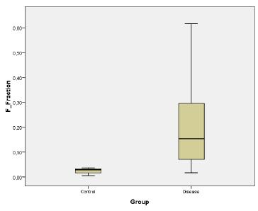
Comparison of the colonic wall (F fraction) and muscle (M fraction) areas of the two groups showed a statistically significant difference (p=0.044 and p=0.026, respectively). Gland tissue areas also differed significantly between groups (Figure 3).
- Box plot showing the collagen fraction in the colonic wall (F fraction) of the two groups.
Comparative analysis of the colonic wall (F area) showed a statistically significant difference (p=0.003) between the disease group and the control group.
Discussion
Diverticular disease of the sigmoid colon predominates in western populations and is a result of increased intraluminal pressure1414. Fong SS, Tan EY, Foo A, Sim R, Cheong DMO. The changing trend of diverticular disease in a developing Nation. Colorectal Dis. 2011;13(3):312-6. doi: 10.1111/j.1463-1318.2009.02121.x.
https://doi.org/10.1111/j.1463-1318.2009...
. These mechanical loads on the tissues, in addition to chemical factors, stimulate the proliferation of fibroblasts1515. Junqueira LC, Montes GS, Sanchez EM. The influence of tissue section thickness on the study of collagen by the picrosirius-polarization method. Histochemistry. 1982;74(1):153-6. PMID: 7085347.. Most studies within this line of research have compared the concentration of collagen between patients with diverticular disease and patients with colon cancer. Other authors have used sigmoid colon specimens obtained from cadavers77. Wess L, Eastwood MA, Wess TJ, Busuttil A, Miller A. Cross linking of collagen is increased in colonic diverticulosis. Gut. 1995;37(1):91-4. PMCID: PMC1382775.,88. Thomson HJ, Busuttil A, Eastwood MA, Smith AN, Elton RA. Submucosal collagen changes in the normal colon and in diverticular disease. Int J Colorect Dis. 1987;2(4):208-13. PMID: 3694019.,1010. Bode MK, Karttunen TJ, Mäkelä J, Risteli L, Risteli J. Type I and III collagens in human colon cancer and diverticulosis. Scand J Gastroenterol. 2000;35(7):747-52. PMID: 10972180.,1616. Stumpf M, Krones CJ, Klinge U, Rosch R, Junge K, Schumpelink V. Collagen in colon disease. Hernia. 2006;10(6): 498-501. PMID: 17080262..
The extracellular matrix exerts structural and biochemical functions and plays a role in cell metabolism and in the storage and release of growth factors. Collagen is the main protein component of the extracellular matrix1717. Gelsea K, Poschlb E, Aignera T. Collagens - structure, function, and biosynthesis. Adv Drug Deliv Rev. 2003;55:1531-46. PMID: 14623400.. Most references involve a traditional model, in which the main cause of diverticulosis is the association between low dietary fiber intake, small feces volume and increased intraluminal pressure. Specific analysis of diverticulosis shows the presence of acquired changes and chronic evolution. On the other hand, diverticulitis is an acute disease associated with severe complications, which affects individuals of different ages.
The main doubt about the alterations that occur in the colon wall encouraged us to determine whether the concentration of collagen is altered in the colon wall of patients with diverticular disease. The coexistence of colorectal cancer may influence the synthesis of collagen. Malignant tumors often induce a fibroproliferative response in the stroma adjacent to the tumor, which is characterized by an increase in the deposition of type I and type III procollagen. The same authors reported the presence of defective cross-linking of collagens and disorganized arrangement of collagen fibers in ovarian carcinoma1818. Kauppila S, Bode MK, Stenbäck F, Risteli L, Risteli J. Cross-linked telopeptides of type I and III collagens in malignant ovarian tumors in vivo. Br J Cancer. 1999;81:654-61. PMID: 2362884.. In a retrospective study, Krones et al .1919. Krones CJ, Klinge U, Butz N, Junge K, Stumpf M, Rosch R, Hermanns B, Heussen N, Schumpelick V. The rare epidemiologic coincidence of diverticular disease and advanced colonic neoplasia. Int J Colorectal Dis. 2006;21(1):18-24. PMID: 15889263., observed that the simultaneous occurrence of diverticular disease and cancer seems to be less frequent than individual epidemiological estimates of either disease.
Rodrigues Jr et al .2020. Rodrigues AJ Jr, Rodrigues CJ, Cunha ACP. Quantitative analysis of collagen and elastic fibers in the transversalis fascia in direct and indirect inguinal hernia. Rev Hosp Clin Fac Med Univ São Paulo. 2002;57(6):265-70. PMID: 12612758. evaluated collagen concentration in direct and indirect inguinal hernias by picrosirius staining and observed a lower collagen concentration in the transversalis fascia of patients with direct hernias compared to those with indirect hernias. Picrosirius staining combined with polarized light microscopy is specific for the identification of collagen2121. Junqueira LC, Bignolas G, Brentani RR. Picrosirius staining plus polarization microscopy: specific method for collagen detection in tissue sections. Histochem J. 1979;11(4):447-55. PMID: 91593.. Fibrous tissue is characterized by an increase in type I and III collagen and fibrosis can have the aspect of tissue repair or of a response to a pathological stimulus. Excessive collagen deposition can damage the function of the affected organ2222. Fritz D, Cai L, Stefanovic L Stefanovic B. Progress towards discovery of antifibrotic drugs targeting synthesis of type I collagen. Curr Med Chem. 2011;18(22):3410-6. PMID: 21728959..
Steroids used for the treatment of chronic diseases induce the activation, recruitment and proliferation of defense cells, including neutrophils and macrophages. The prolonged use of corticosteroids exerts a protective effect on the survival of neutrophils and delays apoptosis (death of neutrophils). These cells may be involved in the pathogenesis of severe inflammation in diverticulitis2323. Burkhard HA, Rahden BH, Kircher S, Thiery S, Landmann D, Jurowich CF, Germer CT, Grimm M. Association of steroid use with complicated sigmoid diverticulitis: potential role of activated CD68+/CD163+ macrophages. Langenbecks Arch Surg. 2011;396(6):759-68. PMID: 21553154..
The present study demonstrated an increasing trend in the amount of collagen in diverticulitis. Collagen deposition in the F area was significantly higher in the disease group than in the control group (p=0.003). The submucosal layer, which mainly consists of collagen and elastic fibers, plays an important role in the early healing stage of anastomoses and confers tensile strength and resistance to sutures in the gastrointestinal tract2424. Thornton FJ, Barbul A. Healing in the gastrointestinal tract. Surg Clin North Am. 1997;77(3):546-73. PMID: 9194880.. Analysis of the deposition of collagen in the glandular epithelium (E area) showed significantly higher deposition in the disease group compared to control (p=0.02). Comparison of the M fraction between the disease group and control revealed a significant difference (p=0.02). In the present investigation, the lower fraction represented by the musculature of the disease group compared to the control group could be explained by the higher collagen deposition on the colon wall of the disease group.
According to previous theories, the formation of diverticula is a slow process that can take several years and involves the remodeling of connective tissue. Furthermore, the abnormalities observed in the wall of the sigmoid colon may be related to motility disorders and diet.
Conclusion
Collagen deposition in the colon wall was increased in patients with diverticulitis of the sigmoid colon compared to patients without the disease.
Acknowledgements
To Eliani Guelli Mosquim for the statistical analysis, and Kerstin Markendorf for English language.
References
-
1Painter NS, Burkitt DP. Diverticular disease of the colon: a 20th century problem. Clin Gastroenterol. 1975;4(1):3-21. PMID: 1109818.
-
2Hobson GK, Roberts LP. Etiology and pathophysiology of diverticular disease. Clin Colon Rectal Surg. 2004;17(3):147-53. doi: 10.55/s-2004-832695.
» https://doi.org/10.55/s-2004-832695 -
3Jeyarajah S, Papagrigoriadis S. Review article: the pathogenesis of diverticular disease - current perspectives on motility and neurotransmitters. Aliment Pharmacol Ther. 2011;33:789-800. doi: 10.1111/j.1365-2036.2011.04586.X.
» https://doi.org/10.1111/j.1365-2036.2011.04586.X -
4Etzioni DA, Mack TM, Beart RW Jr, Kaiser AM. Diverticulitis in the United States: 1998- 2005: changing patterns of disease and treatment. Ann Surg. 2009;249(2):210-7. doi: 10.1097/SLA.0b013e3181952888.
» https://doi.org/10.1097/SLA.0b013e3181952888 -
5Humes D, Smith JK, Spiller R. Colonic diverticular disease. Am Fam Physician. 2011;84(10):1163-4. PMID: 22085672.
-
6Golder M, Burleigh DE, Belai A, Ghali L, Ashby D, Lunniss PJ, Navsaria HA, Williams NS. Smooth muscle cholinergic denervation hypersensitivity in diverticular disease. Lancet. 2003;361(9373):1945-51. doi: http://dx.doi.org/10.1016/S0140-6736(03)13583-0.
» https://doi.org/10.1016/S0140-6736(03)13583-0 -
7Wess L, Eastwood MA, Wess TJ, Busuttil A, Miller A. Cross linking of collagen is increased in colonic diverticulosis. Gut. 1995;37(1):91-4. PMCID: PMC1382775.
-
8Thomson HJ, Busuttil A, Eastwood MA, Smith AN, Elton RA. Submucosal collagen changes in the normal colon and in diverticular disease. Int J Colorect Dis. 1987;2(4):208-13. PMID: 3694019.
-
9Whiteway J, Morson BC. Elastosis in diverticular diseases of the sigmoid colon. Gut. 1985;26(3):258-66. PMID: 3972272.
-
10Bode MK, Karttunen TJ, Mäkelä J, Risteli L, Risteli J. Type I and III collagens in human colon cancer and diverticulosis. Scand J Gastroenterol. 2000;35(7):747-52. PMID: 10972180.
-
11Mimura T, Bateman AC, Lee RL, Johnson PA, McDonald PJ, Talbot IC, Kamm MA, MacDonald TT, Pender SL. Up-regulation of collagen and tissue inhibitors of matrix metalloproteinase in colonic diverticular disease. Dis Colon Rectum. 2004;47(3):371-9. PMID: 14991500.
-
12Stumpf M, Cao W, Klinge U, Klosterhalfen B, Kasperk R, Schumpelink V. Incresead distribution of collagen type lll and reduced expression of matrix metalloproteinase 1 in patients with diverticular disease. Int J Colorectal Dis. 2001;16(5):271-5. PMID: 11686522.
-
13Stumpf M, Cao W, Klinge U, Klosterhalfen B, Junge K, Krones CJ, Schumpelick V. Reduced expression of collagen type I and increased expression of matrix metalloproteinases 1 in patients with Crohn's Disease. J Invest Surg. 2005;18(1):33-8. PMID: 15804950.
-
14Fong SS, Tan EY, Foo A, Sim R, Cheong DMO. The changing trend of diverticular disease in a developing Nation. Colorectal Dis. 2011;13(3):312-6. doi: 10.1111/j.1463-1318.2009.02121.x.
» https://doi.org/10.1111/j.1463-1318.2009.02121.x -
15Junqueira LC, Montes GS, Sanchez EM. The influence of tissue section thickness on the study of collagen by the picrosirius-polarization method. Histochemistry. 1982;74(1):153-6. PMID: 7085347.
-
16Stumpf M, Krones CJ, Klinge U, Rosch R, Junge K, Schumpelink V. Collagen in colon disease. Hernia. 2006;10(6): 498-501. PMID: 17080262.
-
17Gelsea K, Poschlb E, Aignera T. Collagens - structure, function, and biosynthesis. Adv Drug Deliv Rev. 2003;55:1531-46. PMID: 14623400.
-
18Kauppila S, Bode MK, Stenbäck F, Risteli L, Risteli J. Cross-linked telopeptides of type I and III collagens in malignant ovarian tumors in vivo. Br J Cancer. 1999;81:654-61. PMID: 2362884.
-
19Krones CJ, Klinge U, Butz N, Junge K, Stumpf M, Rosch R, Hermanns B, Heussen N, Schumpelick V. The rare epidemiologic coincidence of diverticular disease and advanced colonic neoplasia. Int J Colorectal Dis. 2006;21(1):18-24. PMID: 15889263.
-
20Rodrigues AJ Jr, Rodrigues CJ, Cunha ACP. Quantitative analysis of collagen and elastic fibers in the transversalis fascia in direct and indirect inguinal hernia. Rev Hosp Clin Fac Med Univ São Paulo. 2002;57(6):265-70. PMID: 12612758.
-
21Junqueira LC, Bignolas G, Brentani RR. Picrosirius staining plus polarization microscopy: specific method for collagen detection in tissue sections. Histochem J. 1979;11(4):447-55. PMID: 91593.
-
22Fritz D, Cai L, Stefanovic L Stefanovic B. Progress towards discovery of antifibrotic drugs targeting synthesis of type I collagen. Curr Med Chem. 2011;18(22):3410-6. PMID: 21728959.
-
23Burkhard HA, Rahden BH, Kircher S, Thiery S, Landmann D, Jurowich CF, Germer CT, Grimm M. Association of steroid use with complicated sigmoid diverticulitis: potential role of activated CD68+/CD163+ macrophages. Langenbecks Arch Surg. 2011;396(6):759-68. PMID: 21553154.
-
24Thornton FJ, Barbul A. Healing in the gastrointestinal tract. Surg Clin North Am. 1997;77(3):546-73. PMID: 9194880.
-
Financial source: CAPES
-
1
Research performed at Department of Pathology, Medical School of Jundiai, Jundiai-SP, Brazil. Part of Master degree thesis, Postgraduate Program in Interdisciplinary Surgery Science, UNIFESP. Tutor: Prof. Dr. Gaspar de Jesus Lopes Filho.
Publication Dates
-
Publication in this collection
Oct 2015
History
-
Received
11 June 2015 -
Reviewed
12 Aug 2015 -
Accepted
15 Sept 2015