Abstract
Purpose:
To evaluate the influence of nandrolone decanoate on fracture healing and bone quality in normal rats.
Methods:
Male rats were assigned to four groups (n=28/group): Control group consisting of animals without any intervention, Nandrolone decanoate (DN) group consisting of animals that received intramuscular injection of nandrolone decanoate, Fracture group consisting of animals with a fracture at the mid-diaphysis of the femur, and Fracture and nandrolone decanoate group consisting of animals with a femur fracture and treatment with nandrolone decanoate. Fractures were created at the mid-diaphysis of the right femur by a blunt trauma and internally fixed using an intramedullary steel wire. The DN was injected intramuscularly twice per week (10 mg/kg of body mass). The femurs were measured and evaluated by densitometry and mechanical resistance after animal euthanasia. The newly formed bone and collagen type I levels were quantified in the callus.
Results:
The treated animals had longer femurs after 28 days. The quality of the intact bone was not significantly different between groups. The bone callus did show a larger mass in the treated rats.
Conclusion:
The administration of nandrolone decanoate did not affect the quality of the intact bone, but might have enhanced the bone callus formation.
Key words:
Anabolic Agents; Femoral Fractures; Bony Callus; Rats.
Introduction
The androgenic anabolic steroids (AAS) are a group of testosterone derived substances classically indicated for hypogonadism and protein deficiency11 Cunha TS, Cunha NS, Moura MJCS, Marcondes FK. Esteróides anabólicos androgênicos e sua relação com a prática desportiva. Rev Bras de Cienc Farm. 2004:169-79.. The compounds are also used as adjuvant therapy in chronic diseases, multiple injuries, extensive burns, and following radiotherapy treatments22 Kanayama G, Brower KJ, Wood RI, Hudson JI, Pope HG, Jr. Treatment of anabolic-androgenic steroid dependence: Emerging evidence and its implications. Drug Alcohol Depend. 2010;109(1-3):6-13. doi: 10.1016/j.drugalcdep.2010.01.011.
https://doi.org/10.1016/j.drugalcdep.201...
. Nandrolone decanoate is an AAS used in the musculoskeletal system to improve bone mass, resistance to fracture, and muscular tropism33 Sun L, Pan J, Peng Y, Wu Y, Li J, Liu X, Qin Y, Bauman WA, Cardozo C, Zaidi M, Qin W. Anabolic steroids reduce spinal cord injury-related bone loss in rats associated with increased Wnt signaling. J Spinal Cord Med. 2013;36(6):616-22. doi: 10.1179/2045772312Y.0000000020.
https://doi.org/10.1179/2045772312Y.0000...
4 Ahmad F, Yunus SM, Asghar A, Faruqi NA. Influence of anabolic steroid on tibial fracture healing in rabbits - a study on experimental model. J Clin Diagn Res. 2013;7(1):93-6. doi: 10.7860/JCDR/2012/4863.2679.
https://doi.org/10.7860/JCDR/2012/4863.2...
5 Cardozo CP, Qin WP, Peng YZ, Liu X, Wu Y, Pan JP, Bauman WA, Sun L. Nandrolone slows hindlimb bone loss in a rat model of bone loss due to denervation. An N Y Acad Sci. 2010;1192:303-6. doi: 10.1111/j.1749-6632.2009.05313.x.
https://doi.org/10.1111/j.1749-6632.2009...
6 Gadeleta SJ, Boskey AL, Paschalis E, Carlson C, Menschik F, Baldini T, Peterson M, Rimnac CM. A physical, chemical, and mechanical study of lumbar vertebrae from normal, ovariectomized, and nandrolone decanoate-treated cynomolgus monkeys (Macaca fascicularis). Bone. 2000;27(4):541-50. PMID: 11033450.
7 Aerssens J, Vanaudekercke R, Geusens P, Schot LPC, Osman AAH, Dequeker J. Mechanical-properties, bone-mineral content, and bone-composition (Collagen, Osteocalcin, Igf-I) of the rat femur - influence of ovariectomy and nandrolone decanoate (Anabolic-Steroid) treatment. Calcif Tissue Int. 1993;53(4):269-77. PMID: 8275356.
8 Hassager C, Jensen LT, Johansen JS, Riis BJ, Melkko J, Podenphant J, Risteli L, Christiansen C, Risteli J. The carboxy-terminal propeptide of type I procollagen in serum as a marker of bone formation: the effect of nandrolone decanoate and female sex hormones. Metabolism. 1991;40(2):205-8. PMID: 1988778.-99 Johansen JS, Hassager C, Podenphant J, Riis BJ, Hartwell D, Thomsen K, Christiansen C. Treatment of postmenopausal osteoporosis - Is the anabolic-steroid nandrolone decanoate a candidate. Bone Miner. 1989;6(1):77-86. PMID: 2665884.. There are several clinical and laboratory investigations that support its therapeutic benefits for conditions of aging bone and muscular diseases such as osteoporosis and sarcopenia1010 Reginster JY, Beaudart C, Buckinx F, Bruyere O. Osteoporosis and sarcopenia: two diseases or one? Curr Opin Clin Nutr Metab Care. 2016;19(1):31-6. doi: 10.1097/MCO.0000000000000230.
https://doi.org/10.1097/MCO.000000000000...
.
In modern society bone fractures affects young people (sports, work and traffic accidents) and older people (fall and fragile bones). In the United States of America it was estimated that in 2009 there were 7.9 million fractures, and these injuries have an important impact on society and the health system1111 Victoria G, Petrisor B, Drew B, Dick D. Bone stimulation for fracture healing: What's all the fuss? Indian J Orthop. 2009;43(2):117-20. PMID: 2762251.. Additionally, 10% of the fractures may have improper healing or fail to heal1212 Einhorn TA. Enhancement of fracture-healing. J Bone Joint Surg Am. 1995;77a(6):940-56. PMID: 7782368.. Complications affecting older patients can be devastating and may lead to death. As a result, many studies have been conducted to combat the fracture epidemic and its complications. There are many local orthopedic approaches that enhance fracture healing including electric electromagnetic stimulation1313 Bassett CAL, Becker RO. Generation of electric potentials by bone in response to mechanical stress. Science. 1962;137(3535):1063-4. PMID: 13865637.,1414 Korenstein R, Somjen D, Fischler H, Binderman I. Capacitative pulsed electric stimulation of bone cells. Induction of cyclic-AMP changes and DNA synthesis. Biochim Biophys Acta. 1984;803(4):302-7. PMID: 6322860., low intensity ultrasound1515 O'Herlihy L, Elkins MR. Ultrasound may promote fracture healing but this does not necessarily accelerate return of function. Br J Sports Med. 2013;47(6):397-8. doi: 10.1136/bjsports-2013-092229.
https://doi.org/10.1136/bjsports-2013-09...
, vibration therapy1616 Komrakova M, Sehmisch S, Tezval M, Ammon J, Lieberwirth P, Sauerhoff C, Trautmann L, Wicke M, Dullin C, Stuermer KM, Stuermer EK. Identification of a vibration regime favorable for bone healing and muscle in estrogen-deficient rats. Calcif Tissue Int. 2013;92(6):509-20. doi: 10.1007/s00223-013-9706-x.
https://doi.org/10.1007/s00223-013-9706-...
, and the injection of growth factors1717 Kawaguchi H, Oka H, Jingushi S, Izumi T, Fukunaga M, Sato K, Matsushita T, Nakamura K, TESK Group. A local application of recombinant human fibroblast growth factor 2 for tibial shaft fractures: a randomized, placebo-controlled trial. J Bone Miner Res. 2010;25(12):2459-67. doi: 10.1002/jbmr.146.
https://doi.org/10.1002/jbmr.146...
. The systemic agents used include the administration of parathyroid hormone1818 Peichl P, Holzer LA, Maier R, Holzer G. Parathyroid hormone 1-84 accelerates fracture-healing in pubic bones of elderly osteoporotic women. J Bone Joint Surg Am. Volume. 2011;93(17):1583-7. doi: 10.2106/JBJS.J.01379.
https://doi.org/10.2106/JBJS.J.01379...
, bisphosphonates1919 Kates SL, Ackert-Bicknell CL. How do bisphosphonates affect fracture healing? Injury. 2016;47 Suppl 1:S65-8. doi: 10.1016/S0020-1383(16)30015-8.
https://doi.org/10.1016/S0020-1383(16)30...
, strontium ranelate2020 Komrakova M, Weidemann A, Dullin C, Ebert J, Tezval M, Stuermer KM, Sehmisch S. The impact of strontium ranelate on metaphyseal bone healing in ovariectomized rats. Calcif Tissue Int. 2015;97(4):391-401. doi: 10.1007/s00223-015-0019-0.
https://doi.org/10.1007/s00223-015-0019-...
, and raloxifene2121 Stuermer EK, Sehmisch S, Rack T, Wenda E, Seidlova-Wuttke D, Tezval M, Wuttke W, Frosch KH, Stuermer KM. Estrogen and raloxifene improve metaphyseal fracture healing in the early phase of osteoporosis. A new fracture-healing model at the tibia in rat. Langenbecks Arch Surg. 2010;395(2):163-72. doi: 10.1007/s00423-008-0436-x.
https://doi.org/10.1007/s00423-008-0436-...
. However, there are several publications that report conflicting results. There are currently a limited number of studies assessing the role of nandrolone decanoate on fracture healing despite its well-recognized positive effect on bone quality and muscular tropism33 Sun L, Pan J, Peng Y, Wu Y, Li J, Liu X, Qin Y, Bauman WA, Cardozo C, Zaidi M, Qin W. Anabolic steroids reduce spinal cord injury-related bone loss in rats associated with increased Wnt signaling. J Spinal Cord Med. 2013;36(6):616-22. doi: 10.1179/2045772312Y.0000000020.
https://doi.org/10.1179/2045772312Y.0000...
,44 Ahmad F, Yunus SM, Asghar A, Faruqi NA. Influence of anabolic steroid on tibial fracture healing in rabbits - a study on experimental model. J Clin Diagn Res. 2013;7(1):93-6. doi: 10.7860/JCDR/2012/4863.2679.
https://doi.org/10.7860/JCDR/2012/4863.2...
. Thus, we conducted an animal study to explore the possible positive effects of the decanoate of nandrolone on fracture healing and bone quality.
Methods
The experimental procedure was approved by the Committee on Ethics and Animal Experimentation of the School of Medicine, Universidade de São Paulo (protocol 115/2014). The procedures were performed based on the guidelines of Brazilian Law No. 11.7942222 FIOCRUZ. Diretrizes da prática de eutanásia do CONCEA. Ministério da ciência, tecnologia e inovação: CONCEA; 2013, p.1-54. Availaable at http://www.unifesp.br/reitoria/ceua/images/Diretrizes%20Eutanasia%20CONCEA.pdf.
The rats were provided by the University Campus vivarium. One hundred twelve young adult (280-300 g of body mass) male Wistar rats were randomly assigned to the following four experimental groups (n=28/group): the Control (CON) group consisting of animals without any intervention, the nandrolone decanoate (DECA) group consisting of animals treated with intramuscular injection of nandrolone decanoate; the Fracture (FRAT) group consisting of animals with a fracture at the mid-diaphysis of the right femur, and the Fracture and nandrolone decanoate (FRAT + DECA) group consisting of animals with a fracture at the mid-diaphysis of the femur treated with nandrolone decanoate. Each group was divided into two subgroups according to the follow-up periods of 14 or 28 days. The animals were housed in cages with transparent walls in a calm isolated environment with controlled temperature (22±2°C). The air relative humidity was 55±10% and the room had 12 h dark-light cycles. There was no restriction to access of chow and water.
Fracture production
The anesthesia was administered using equal volumes of xylazine 2% and ketamine 10% injected intramuscularly at the dose of 0.1 mg /100 g of body mass. The right thigh of each animal was placed on two metal blocks located under a second class lever with a blunt blade attached at its center (guillotine). The lever arm was lowered manually with the blade aimed at the mid part of the thigh and force was applied until there was no resistance. The fracture was confirmed by the abnormal mobility at the center of the femur under manipulation.
The animals were placed on the operating table and kept on a warm mattress. The pelvic limb was shaved and antisepsis was performed with povidone-iodine. The procedure was performed under routine surgical isolation. The skin above the fracture site was incised from the center of the lateral aspect of the thigh and a 1.0 cm long straight skin incision was made. The septum muscularis between the vastus lateralis and the biceps was identified and used to deepen the incision until reaching the bone (conventional approach to the diaphysis of the femur). The fracture was minimally exposed and inspected. When the fracture presented more than four fragments the rat was euthanized and properly discarded. The adequate fractures were stabilized with a 1.0 mm steel Kirschner wire (K-wire) that was first introduced into the medullary canal of the proximal fragment until protruding through the skin at the lateral aspect of the pelvis. The fracture was then reduced, and the wire was reversed through the medullary canal of the distal bone fragment until the tip reached the condylar region of the knee. The excess length of the K-wire protruding through the pelvic region was cut off and the wire tip was buried under the abductor muscles. The incision was closed in layers with 4-0 absorbable separated stitches. A lateral digital X-ray image view was obtained of the entire pelvic limb to assess the adequacy of fracture reduction and K-wire positioning. Each rat was allowed to recover in a protected environment and housed in an individual cage for one week. The animals were then housed two or three animals per cage. The animals received 80 mL of dipyrone diluted 1:5 in saline, which was injected subcutaneously every 12 hours for the first three days postoperatively.
Nandrolone decanoate
The nandrolone decanoate (Deca-Durabolin 50 mg, Schering-Plough 1.0 mL, São Paulo) was injected intramuscularly twice per week (10 mg/kg of body mass)2323 Saito M, Shiraishi A, Ito M, Sakai S, Hayakawa N, Mihara M, Marumo K. Comparison of effects of alfacalcidol and alendronate on mechanical properties and bone collagen cross-links of callus in the fracture repair rat model. Bone. 2010;46(4):1170-9. doi: 10.1016/j.bone.2009.12.008.
https://doi.org/10.1016/j.bone.2009.12.0...
for either two or four weeks for the DECA and FRAT+DECA subgroups. The fracture control and non-fractured groups received the same volume of vehicle.
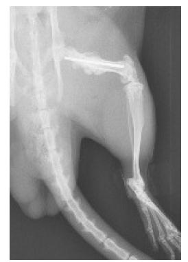
The rats were euthanized with an excessive dose of xylazine and ketamine2222 FIOCRUZ. Diretrizes da prática de eutanásia do CONCEA. Ministério da ciência, tecnologia e inovação: CONCEA; 2013, p.1-54. Availaable at http://www.unifesp.br/reitoria/ceua/images/Diretrizes%20Eutanasia%20CONCEA.pdf. A lateral X-ray of the hind limb was obtained to evaluate bone healing (Figure 1).
Lateral X-ray of the pelvic limb of a control rat, 28 days after fracture. It is possible to note the adequate alignment of the fragments, the intramedullary fixation with the Kirschner wire, and the external bone callus formed (arrow).
The right femur was removed and cleaned of soft tissue prior to measurement and weighing. The bones were examined with the DEXA technique (Lunar DPX-IQ, GE Healthcare, United Kingdom). The region of interest (ROI) encompassed the whole fracture callus and the distal metaphysis for groups without fracture. The bone mineral density (BMD) and bone mineral content (BMC) were obtained for both areas with software for small specimens (Lunar®; software version 4.7e).
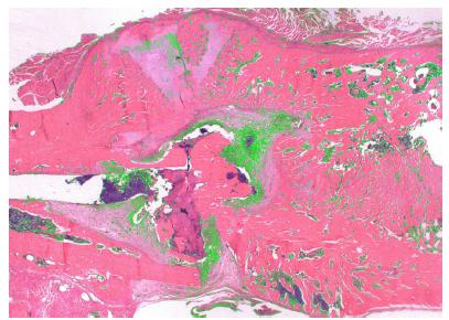
The fractured specimens with the bone callus were routinely processed for histological analysis and 48 coronal semi-serial sections 5 µm thick were obtained. From each specimen, after collecting eight sections, the following 10 sections were discarded and this procedure was repeated for the whole callus. The sections were stained with H&E and picrosirius red. The analysis of the central area of the callus was examined under 1.25x at AxioImager Z2 microscope (Zeiss, Germany) and the newly formed bone was quantified with Axionvision® software (Zeiss, Germany) (Figure 2).
Photomicrograph of histological section for identification of neoformed bone. The green region corresponds to other tissues of no interest that were erased so that only the neoformed bone was left for histomorphometric analysis. Hematoxylin and eosin, original magnification x1.25.
The picrosirius red stained sections of the callus were examined under polarized light, which permits identification of collagen type I as red-orange color (Figure 3). The same software was used to quantify the collagen.
Photomicrograph using polarized light from the bone callus area with type I collagen fibers selected in light green color (Picrosirus red, original magnification x50).
The bones not reserved for the histological studies were evaluated in a two-point bending mechanical test using a universal test machine (EMIC® DL10.000, PR, Brazil) with a load cell of 50 kgf. The distal condylar region of the femur was embedded in a spherical block of methyl methacrylate to facilitate horizontal positioning of the bone. A vertical force was applied at the intertrochanteric region of the femur at a speed of 0.5 mm/min until breaking the bone callus (Figure 4). The Tesc software (EMIC®, PR, Brazil) was used to calculate the maximum force and stiffness.
Illustration of the mechanical test at two points on a femur from an animal on day 28 after the fracture. The callus is prominent in the central region of the bone. The condylar end of the femur was set in a spherical block of acrylic resin, which was attached to a table vise. The force was applied at the proximal end of the femur (A) until fracture occurred (B).
Statistical analysis
The SPSS 20.0 software package was used for statistical analysis. The Shapiro-Wilk test was used to determine normal distribution. The parameter comparisons were tested with linear models, and the Bonferroni test was used with the significance level set at 5%. The graphs were generated using GraphPad Prism5® (GraphPad Software, Inc, USA).
Results
We used 112 rats in this study. However, there were 20 animals discarded (36%) due to comminuted fractures (27%) or external migration of the K-wire (9%). Therefore, 28 animals remained in Con and DECA, 17 in CON + FRAT and 19 in DECA + FRAT.
The body mass of the animals increased in all groups from day 0 to day 28. However, at day 7 both of the nandrolone decanoate groups rats showed a difference between the non-steroid groups (p=0.001). However, this difference did not maintain at 14, 21 and 28 days (p=0.683) (Figure 5). The length of the non-fractured femurs was longer in animals that received steroid (p<0.001).
The bone mineral density at the distal metaphysis was higher at 28 days (p<0.001) than at day 14 in the control and steroid-treated animals. There was no difference for BMD at 28 days between the nandrolone decanoate treated and non-treated groups (p=0.261). The bone mineral content (BMC) was higher at 28 days than at 14 days (p<0.001). However, the inter-group comparison between the two 14 day groups revealed no difference (p=0.611). There was also no difference between the two 28 day groups (p=0.307). The callus area was measured by DXA and the results did not show any difference between CON+FRAt-14 and DECA+FRAT-14 (p=0.990) or between the groups CON+FRAT-28 and DECA+FRAT-28.
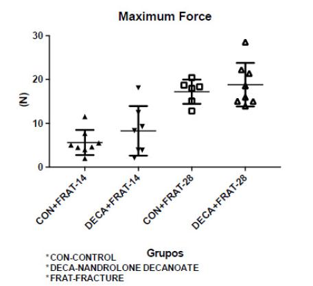
The mechanical tests did not show any significant difference between the non-fractured femurs with or without nandrolone decanoate administration for the maximum force (p=0.323) or stiffness (p=0.211) (Figure 6).
Maximum force of the intact femurs in the Control and Nandrolone decanoate groups after 14 and 28 days.
Our data indicate there were no differences in maximum force between the fractured bones at 14 days and 28 days (p=0.542) (Figure 7). The stiffness was significantly higher for the 28-day groups than the 14-day groups (p=0.0380).
Maximum force of fractured femurs in the Control and Nandrolone decanoate groups after 14 and 28 days.
Histological results
The volume of newly formed bone in the callus was significantly higher at 28 days for the nandrolone decanoate treated animals (p<0.001, Figure 8).
Mean of the volume of formed bone in the callus of different groups treated with and without nandrolone decanoate at 14 and 28 days. Animals with fracture and nandrolone decanoate had significantly higher callus bone volume at 28 days (p=0.001).
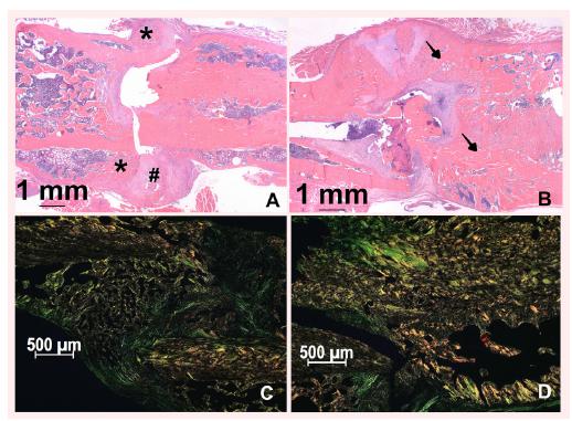
Furthermore, the hematoxylin-eosin slides evidenced an improved bone callus quality in the nandrolone deacanoate treated rats (Figure 9B). At 28 days post-fracture these bone callus were mainly formed by thick and dense trabeculae bone. On the other hand, the control rats exhibited bone callus mainly formed by cartilaginous tissue, with sparse and thin trabecular bone (Figure 9A).
Histologic section of the bone callus 28 days after fracture. A - The control bone callus shows immature neoformed bone (*) and a large amount of cartilaginous tissue (#). B - Animal from the nandrolone decanoate group with a fracture showing a substantial amount of new bone and more mature trabeculae (arrows), evidencing a better and larger bone callus formation. (Hematoxylin and eosin, x1.25). Microphotographs under polarized light also illustrate the region of the bone callus 28 days after the fracture and confirm the HE findings. D - The bone callus of animals treated with nandrolone decanoate shows higher deposition of type I collagen in red-orange color than in control animals (C) (Picrosirus red, x50).
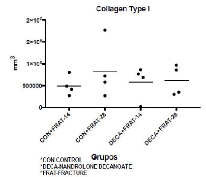
Mean area of type I collagen in the various groups at 14 and 28 days. There was no significant difference in collagen content.
Discussion
Human life expectancy has increased, and the aging population has dramatically changed disease epidemiology. Within the locomotor system there are concerns regarding impaired bone quality and the different types of osteoporosis that facilitate fractures and muscular deterioration. The syndrome of fragility is characterized by sarcopenia (muscular mass decrease) and functional impairment that compromises the quality of life of older people. This condition leads to old age fracture syndrome that results from health deterioration and falls2424 Farooqi V, van den Berg ME, Cameron ID, Crotty M. Anabolic steroids for rehabilitation after hip fracture in older people. Cochrane Database Syst Rev. 2014(10):CD008887. doi: 10.1002/14651858.CD008887.pub2.
https://doi.org/10.1002/14651858.CD00888...
. These injuries can favor complications such as skin breakage, pneumonia, urinary tract infection, and malnutrition that can lead to death.
The use of drugs to improve bone quality either by stimulating its formation or by decreasing its resorption is invaluable in minimizing the effects of skeletal decay. The most used and studied antiresorptive drugs are bisphosphonates (alendronate, pamidronate, risedronate, zoledronate, etc.) and estrogen. There are also anabolic drugs such as parathormone, strontium ranelate, antisclerostin, and anabolizing steroids. Current data supports the continuation of such therapies even in the event of fractures2525 Vannucci L, Brandi ML. Healing of the bone with anti-fracture drugs. Expert Opin Pharmacother. 2016;17(17):2267-72. doi: 10.1080/14656566.2016.1241765.
https://doi.org/10.1080/14656566.2016.12...
.
Attempts to change the abnormal occurrence of fracture mostly in at-risk patients focus on local interventions, and the systemic drugs have been overlooked. However, the interest in such drugs has increased due to promising results for bone healing2626 Brandi ML. Drugs for bone healing. Expert Opin Investig Drugs. 2012;21(8):1169-76. doi: 10.1517/13543784.2012.696610.
https://doi.org/10.1517/13543784.2012.69...
. Several preclinical studies have demonstrated the positive impact of both anti-catabolic and anabolic drugs on fracture healing in osteoporotic bone models2525 Vannucci L, Brandi ML. Healing of the bone with anti-fracture drugs. Expert Opin Pharmacother. 2016;17(17):2267-72. doi: 10.1080/14656566.2016.1241765.
https://doi.org/10.1080/14656566.2016.12...
. Despite the long existence and use of nandrolone decanoate to treat both intact bone and muscular deteriorations, no studies have specifically investigated bone callus formation. The study by Demling2727 Demling RH. Oxandrolone, an anabolic steroid, enhances the healing of a cutaneous wound in the rat. Wound Repair Regen. 2000;8(2):97-102. PMID: 10810035. reported that anabolic steroids are capable of releasing beta growth factors that stimulate bone formation. Interestingly, such drugs were not described by Vanucci and Brandi2525 Vannucci L, Brandi ML. Healing of the bone with anti-fracture drugs. Expert Opin Pharmacother. 2016;17(17):2267-72. doi: 10.1080/14656566.2016.1241765.
https://doi.org/10.1080/14656566.2016.12...
in a recent literature update on anti-fracture drugs. There are currently no preclinical or clinical trials examining the effects of nandrolone decanoate on fracture healing44 Ahmad F, Yunus SM, Asghar A, Faruqi NA. Influence of anabolic steroid on tibial fracture healing in rabbits - a study on experimental model. J Clin Diagn Res. 2013;7(1):93-6. doi: 10.7860/JCDR/2012/4863.2679.
https://doi.org/10.7860/JCDR/2012/4863.2...
. This study was designed to test the effects of nandrolone decanoate on fracture healing. We first studied the effects of nandrolone on normal intact and normally fractured bone. However, we recognize that more helpful studies should focus on osteoporotic bone fracture. The closed and minimal exposition of this fracture model with stabilization is an adequate experimental method because it allows healing by callus formation (secondary healing) without interference of fracture misalignment that might make it difficult to interpret the results2828 Ogasawara A, Nakajima A, Nakajima F, Goto K, Yamazaki M. Molecular basis for affected cartilage formation and bone union in fracture healing of the streptozotocin-induced diabetic rat. Bone. 2008;43(5):832-9. doi: 10.1016/j.bone.2008.07.246.
https://doi.org/10.1016/j.bone.2008.07.2...
,2929 Urabe K, Kim HJ, Sarkar G, Bronk JT, Bolander ME. Determination of the complete cDNA sequence of rat type II collagen and evaluation of distinct expression patterns of types IIA and IIB procollagen mRNAs during fracture repair in rats. J Orthop Sci. 2003;8(4):585-90. doi:n10.1007/s00776-003-0658-2.
https://doi.org/n10.1007/s00776-003-0658...
. The parameters of bone and callus quality that we used are currently employed in experimental practice3030 Falcai MJ, Zamarioli A, Leoni GB, de Sousa Neto MD, Volpon JB. Swimming activity prevents the unloading induced loss of bone mass, architecture, and strength in rats. Biomed Res Int. 2015;2015:507848. doi: 10.1155/2015/507848.
https://doi.org/10.1155/2015/507848...
31 Zamarioli A, Maranho DA, Butezloff MM, Moura PA, Volpon JB, Shimano AC. Anatomic changes in the macroscopic morphology and microarchitecture of denervated long bone tissue after spinal cord injury in rats. Biomed Res Int. 2014;2014:853159. doi: 10.1155/2014/853159.
https://doi.org/10.1155/2014/853159...
-3232 Santiago HA, Zamarioli A, Sousa Neto MD, Volpon JB. Exposure to secondhand smoke impairs fracture healing in rats. Clin Orthop Relat Res. 2017;475(3):894-902. doi: 10.1007/s11999-016-5184-6.
https://doi.org/10.1007/s11999-016-5184-...
. We chose the post-fracture periods of 14 and 28 days because these time intervals represent important landmarks for the femoral fracture healing in the rat when there is conversion of the soft callus into hard callus3333 Fazzalari NL. Bone fracture and bone fracture repair. Osteoporos Int. 2011;22(6):2003-6. doi: 10.1007/s00198-011-1611-4.
https://doi.org/10.1007/s00198-011-1611-...
.
The overall results of our study did not show any significant contribution of nandrolone decanoate on callus improvement or on the intact bone quality in this experimental model. Therefore, our initial hypothesis of a positive drug effect could not be confirmed. There are several publications with controversial opinions on the favorable effects of nandrolone decanoate on fracture healing. However, all of the studies involve osteoporotic conditions3434 Geusens P, Dequeker J. Long-term effect of nandrolone decanoate, 1 alpha-hydroxyvitamin D3 or intermittent calcium infusion therapy on bone mineral content, bone remodeling and fracture rate in symptomatic osteoporosis: a double-blind controlled study. Bone Miner. 1986;1(4):347-57. PMID: 3333018.,3535 Craig SJ, Youssef PP, Vaile JH, Sullivan L, Bleasel JF. Intravenous zoledronic acid and oral alendronate in patients with a low trauma fracture: experience from an osteoporosis clinic. Intern Med J. 2011;41(2):186-90. doi: 10.1111/j.1445-5994.2010.02198.x.
https://doi.org/10.1111/j.1445-5994.2010...
. The present study showed more significant results for increased femoral growth in the 28 day treated animals. This result is consistent with the expected effects of an increased metabolism caused by the nandrolone and suggests that our rats were still growing and immature. The results could be different with fully mature animals. Another positive outcome was the increased bone callus mass observed in the 28 day treated animals. However, as an isolated result it is difficult to interpret this finding as it is expected that a more developed and mature callus might be stronger and denser and contain more collagen type I. However, these results were not obtained.
There are several methodological limitations in this investigation, and they should be avoided in future studies. One concern is the dosage of nandrolone decanoate. The dose used was based on the study by Saito et al.2323 Saito M, Shiraishi A, Ito M, Sakai S, Hayakawa N, Mihara M, Marumo K. Comparison of effects of alfacalcidol and alendronate on mechanical properties and bone collagen cross-links of callus in the fracture repair rat model. Bone. 2010;46(4):1170-9. doi: 10.1016/j.bone.2009.12.008.
https://doi.org/10.1016/j.bone.2009.12.0...
s. Several other authors used higher dosages because they were interested in studying the side effects of overdosing3636 Jannatifar R, Shokri S, Farrokhi A, Nejatbakhsh R. Effect of supraphysiological dose of Nandrolone Decanoate on the testis and testosterone concentration in mature and immature male rats: A time course study. Int J Reprod Biomed (Yazd). 2015;13(12):779-86. PMID: 27141538.,3737 Breuer ME, McGinnis MY, Lumia AR, Possidente BP. Aggression in male rats receiving anabolic androgenic steroids: effects of social and environmental provocation. Horm Behav. 2001;40(3):409-18. doi: 10.1006/hbeh.2001.1706.
https://doi.org/10.1006/hbeh.2001.1706...
. We used young adult rats in this study and it is possible fully mature animals could show a better response.
Future research studies should test the nandrolone decanoate in osteoporotic fractures. It is difficult to obtain an osteoporotic condition in male rats so a fracture could be provoked in a model of regional osteoporosis could be used by causing sciatic nerve injury to affect one pelvic limb. Additionally, a longer administration period for the nandrolone decanoate could be valuable. Other methods could also be used to investigate bone formation using molecular biology and micro-CT studies of the bone structure.
Conclusion
The administration of nandrolone decanoate did not affect the quality of the intact bone but might have enhanced the bone callus formation.
References
-
1Cunha TS, Cunha NS, Moura MJCS, Marcondes FK. Esteróides anabólicos androgênicos e sua relação com a prática desportiva. Rev Bras de Cienc Farm. 2004:169-79.
-
2Kanayama G, Brower KJ, Wood RI, Hudson JI, Pope HG, Jr. Treatment of anabolic-androgenic steroid dependence: Emerging evidence and its implications. Drug Alcohol Depend. 2010;109(1-3):6-13. doi: 10.1016/j.drugalcdep.2010.01.011.
» https://doi.org/10.1016/j.drugalcdep.2010.01.011 -
3Sun L, Pan J, Peng Y, Wu Y, Li J, Liu X, Qin Y, Bauman WA, Cardozo C, Zaidi M, Qin W. Anabolic steroids reduce spinal cord injury-related bone loss in rats associated with increased Wnt signaling. J Spinal Cord Med. 2013;36(6):616-22. doi: 10.1179/2045772312Y.0000000020.
» https://doi.org/10.1179/2045772312Y.0000000020 -
4Ahmad F, Yunus SM, Asghar A, Faruqi NA. Influence of anabolic steroid on tibial fracture healing in rabbits - a study on experimental model. J Clin Diagn Res. 2013;7(1):93-6. doi: 10.7860/JCDR/2012/4863.2679.
» https://doi.org/10.7860/JCDR/2012/4863.2679 -
5Cardozo CP, Qin WP, Peng YZ, Liu X, Wu Y, Pan JP, Bauman WA, Sun L. Nandrolone slows hindlimb bone loss in a rat model of bone loss due to denervation. An N Y Acad Sci. 2010;1192:303-6. doi: 10.1111/j.1749-6632.2009.05313.x.
» https://doi.org/10.1111/j.1749-6632.2009.05313.x -
6Gadeleta SJ, Boskey AL, Paschalis E, Carlson C, Menschik F, Baldini T, Peterson M, Rimnac CM. A physical, chemical, and mechanical study of lumbar vertebrae from normal, ovariectomized, and nandrolone decanoate-treated cynomolgus monkeys (Macaca fascicularis). Bone. 2000;27(4):541-50. PMID: 11033450.
-
7Aerssens J, Vanaudekercke R, Geusens P, Schot LPC, Osman AAH, Dequeker J. Mechanical-properties, bone-mineral content, and bone-composition (Collagen, Osteocalcin, Igf-I) of the rat femur - influence of ovariectomy and nandrolone decanoate (Anabolic-Steroid) treatment. Calcif Tissue Int. 1993;53(4):269-77. PMID: 8275356.
-
8Hassager C, Jensen LT, Johansen JS, Riis BJ, Melkko J, Podenphant J, Risteli L, Christiansen C, Risteli J. The carboxy-terminal propeptide of type I procollagen in serum as a marker of bone formation: the effect of nandrolone decanoate and female sex hormones. Metabolism. 1991;40(2):205-8. PMID: 1988778.
-
9Johansen JS, Hassager C, Podenphant J, Riis BJ, Hartwell D, Thomsen K, Christiansen C. Treatment of postmenopausal osteoporosis - Is the anabolic-steroid nandrolone decanoate a candidate. Bone Miner. 1989;6(1):77-86. PMID: 2665884.
-
10Reginster JY, Beaudart C, Buckinx F, Bruyere O. Osteoporosis and sarcopenia: two diseases or one? Curr Opin Clin Nutr Metab Care. 2016;19(1):31-6. doi: 10.1097/MCO.0000000000000230.
» https://doi.org/10.1097/MCO.0000000000000230 -
11Victoria G, Petrisor B, Drew B, Dick D. Bone stimulation for fracture healing: What's all the fuss? Indian J Orthop. 2009;43(2):117-20. PMID: 2762251.
-
12Einhorn TA. Enhancement of fracture-healing. J Bone Joint Surg Am. 1995;77a(6):940-56. PMID: 7782368.
-
13Bassett CAL, Becker RO. Generation of electric potentials by bone in response to mechanical stress. Science. 1962;137(3535):1063-4. PMID: 13865637.
-
14Korenstein R, Somjen D, Fischler H, Binderman I. Capacitative pulsed electric stimulation of bone cells. Induction of cyclic-AMP changes and DNA synthesis. Biochim Biophys Acta. 1984;803(4):302-7. PMID: 6322860.
-
15O'Herlihy L, Elkins MR. Ultrasound may promote fracture healing but this does not necessarily accelerate return of function. Br J Sports Med. 2013;47(6):397-8. doi: 10.1136/bjsports-2013-092229.
» https://doi.org/10.1136/bjsports-2013-092229 -
16Komrakova M, Sehmisch S, Tezval M, Ammon J, Lieberwirth P, Sauerhoff C, Trautmann L, Wicke M, Dullin C, Stuermer KM, Stuermer EK. Identification of a vibration regime favorable for bone healing and muscle in estrogen-deficient rats. Calcif Tissue Int. 2013;92(6):509-20. doi: 10.1007/s00223-013-9706-x.
» https://doi.org/10.1007/s00223-013-9706-x -
17Kawaguchi H, Oka H, Jingushi S, Izumi T, Fukunaga M, Sato K, Matsushita T, Nakamura K, TESK Group. A local application of recombinant human fibroblast growth factor 2 for tibial shaft fractures: a randomized, placebo-controlled trial. J Bone Miner Res. 2010;25(12):2459-67. doi: 10.1002/jbmr.146.
» https://doi.org/10.1002/jbmr.146 -
18Peichl P, Holzer LA, Maier R, Holzer G. Parathyroid hormone 1-84 accelerates fracture-healing in pubic bones of elderly osteoporotic women. J Bone Joint Surg Am. Volume. 2011;93(17):1583-7. doi: 10.2106/JBJS.J.01379.
» https://doi.org/10.2106/JBJS.J.01379 -
19Kates SL, Ackert-Bicknell CL. How do bisphosphonates affect fracture healing? Injury. 2016;47 Suppl 1:S65-8. doi: 10.1016/S0020-1383(16)30015-8.
» https://doi.org/10.1016/S0020-1383(16)30015-8 -
20Komrakova M, Weidemann A, Dullin C, Ebert J, Tezval M, Stuermer KM, Sehmisch S. The impact of strontium ranelate on metaphyseal bone healing in ovariectomized rats. Calcif Tissue Int. 2015;97(4):391-401. doi: 10.1007/s00223-015-0019-0.
» https://doi.org/10.1007/s00223-015-0019-0 -
21Stuermer EK, Sehmisch S, Rack T, Wenda E, Seidlova-Wuttke D, Tezval M, Wuttke W, Frosch KH, Stuermer KM. Estrogen and raloxifene improve metaphyseal fracture healing in the early phase of osteoporosis. A new fracture-healing model at the tibia in rat. Langenbecks Arch Surg. 2010;395(2):163-72. doi: 10.1007/s00423-008-0436-x.
» https://doi.org/10.1007/s00423-008-0436-x -
22FIOCRUZ. Diretrizes da prática de eutanásia do CONCEA. Ministério da ciência, tecnologia e inovação: CONCEA; 2013, p.1-54. Availaable at http://www.unifesp.br/reitoria/ceua/images/Diretrizes%20Eutanasia%20CONCEA.pdf
-
23Saito M, Shiraishi A, Ito M, Sakai S, Hayakawa N, Mihara M, Marumo K. Comparison of effects of alfacalcidol and alendronate on mechanical properties and bone collagen cross-links of callus in the fracture repair rat model. Bone. 2010;46(4):1170-9. doi: 10.1016/j.bone.2009.12.008.
» https://doi.org/10.1016/j.bone.2009.12.008 -
24Farooqi V, van den Berg ME, Cameron ID, Crotty M. Anabolic steroids for rehabilitation after hip fracture in older people. Cochrane Database Syst Rev. 2014(10):CD008887. doi: 10.1002/14651858.CD008887.pub2.
» https://doi.org/10.1002/14651858.CD008887.pub2 -
25Vannucci L, Brandi ML. Healing of the bone with anti-fracture drugs. Expert Opin Pharmacother. 2016;17(17):2267-72. doi: 10.1080/14656566.2016.1241765.
» https://doi.org/10.1080/14656566.2016.1241765 -
26Brandi ML. Drugs for bone healing. Expert Opin Investig Drugs. 2012;21(8):1169-76. doi: 10.1517/13543784.2012.696610.
» https://doi.org/10.1517/13543784.2012.696610 -
27Demling RH. Oxandrolone, an anabolic steroid, enhances the healing of a cutaneous wound in the rat. Wound Repair Regen. 2000;8(2):97-102. PMID: 10810035.
-
28Ogasawara A, Nakajima A, Nakajima F, Goto K, Yamazaki M. Molecular basis for affected cartilage formation and bone union in fracture healing of the streptozotocin-induced diabetic rat. Bone. 2008;43(5):832-9. doi: 10.1016/j.bone.2008.07.246.
» https://doi.org/10.1016/j.bone.2008.07.246 -
29Urabe K, Kim HJ, Sarkar G, Bronk JT, Bolander ME. Determination of the complete cDNA sequence of rat type II collagen and evaluation of distinct expression patterns of types IIA and IIB procollagen mRNAs during fracture repair in rats. J Orthop Sci. 2003;8(4):585-90. doi:n10.1007/s00776-003-0658-2.
» https://doi.org/n10.1007/s00776-003-0658-2 -
30Falcai MJ, Zamarioli A, Leoni GB, de Sousa Neto MD, Volpon JB. Swimming activity prevents the unloading induced loss of bone mass, architecture, and strength in rats. Biomed Res Int. 2015;2015:507848. doi: 10.1155/2015/507848.
» https://doi.org/10.1155/2015/507848 -
31Zamarioli A, Maranho DA, Butezloff MM, Moura PA, Volpon JB, Shimano AC. Anatomic changes in the macroscopic morphology and microarchitecture of denervated long bone tissue after spinal cord injury in rats. Biomed Res Int. 2014;2014:853159. doi: 10.1155/2014/853159.
» https://doi.org/10.1155/2014/853159 -
32Santiago HA, Zamarioli A, Sousa Neto MD, Volpon JB. Exposure to secondhand smoke impairs fracture healing in rats. Clin Orthop Relat Res. 2017;475(3):894-902. doi: 10.1007/s11999-016-5184-6.
» https://doi.org/10.1007/s11999-016-5184-6 -
33Fazzalari NL. Bone fracture and bone fracture repair. Osteoporos Int. 2011;22(6):2003-6. doi: 10.1007/s00198-011-1611-4.
» https://doi.org/10.1007/s00198-011-1611-4 -
34Geusens P, Dequeker J. Long-term effect of nandrolone decanoate, 1 alpha-hydroxyvitamin D3 or intermittent calcium infusion therapy on bone mineral content, bone remodeling and fracture rate in symptomatic osteoporosis: a double-blind controlled study. Bone Miner. 1986;1(4):347-57. PMID: 3333018.
-
35Craig SJ, Youssef PP, Vaile JH, Sullivan L, Bleasel JF. Intravenous zoledronic acid and oral alendronate in patients with a low trauma fracture: experience from an osteoporosis clinic. Intern Med J. 2011;41(2):186-90. doi: 10.1111/j.1445-5994.2010.02198.x.
» https://doi.org/10.1111/j.1445-5994.2010.02198.x -
36Jannatifar R, Shokri S, Farrokhi A, Nejatbakhsh R. Effect of supraphysiological dose of Nandrolone Decanoate on the testis and testosterone concentration in mature and immature male rats: A time course study. Int J Reprod Biomed (Yazd). 2015;13(12):779-86. PMID: 27141538.
-
37Breuer ME, McGinnis MY, Lumia AR, Possidente BP. Aggression in male rats receiving anabolic androgenic steroids: effects of social and environmental provocation. Horm Behav. 2001;40(3):409-18. doi: 10.1006/hbeh.2001.1706.
» https://doi.org/10.1006/hbeh.2001.1706
-
Financial source:
CAPES
-
1
Research performed at Laboratory of Bioengineering, Department of Biomechanics, Medicine and Rehabilitation of the Locomotor System, School of Medicine, Universidade de São Paulo (USP), Ribeirao Preto-SP, Brazil.
Publication Dates
-
Publication in this collection
Nov 2017
History
-
Received
10 July 2017 -
Reviewed
15 Sept 2017 -
Accepted
12 Oct 2017