Abstracts
Two-dimensional gel electrophoresis analysis was performed on adventitious and storage root in cassava (Manihot esculenta Crantz). Adventitious root lacking swelling formation and swelled storage root were obtained from the accession WU104 grown in the green house of the Department of Biology in Washington University in St. Louis (MO-USA). Saline buffer-soluble proteins were extracted, separated in a high-resolution 2-D electrophoresis system, visualized with silver staining gel procedure, and digital image generate for further analysis. Quantitative and qualitative protein spots analysis was performed with a computer assisted image software system. Results revealed large variation in the complexity of the gel protein profile between the two root systems. About 90% of the protein spots appeared in the pI range value of 4.0 to 6.5 and between 14 to 80 Kda of molecular mass. Detailed computer assisted analysis of this gel allowed us to establish 5 distinct classes of protein based on spot quantification that could be associated with swelling and non-swelling roots. Variation in the complexity of protein pattern was related with different type of root. Whereas the adventitious root showed a more simple profile related to primary growth, the storage root showed to be a more complex profile related to secondary growth and starch accumulation.
cassava; Manihot esculenta; storage root; 2-D gel electrophoresis; proteins
2-DE géis foram usados para separar proteínas de raízes de reserva de mandioca (Manihot esculenta Crantz). Raízes adventícias desprovidas de engrossamento e raízes adventícias de reservas foram obtidas com o crescimento da cultivar WU104 em vasos em condições de casa-de-vegetação durante o verão de 1995 no Departamento de Biologia da Universidade de Washington em St. Louis (MO, USA). Proteínas solúveis em tampão fosfatado salino foram extraídas, separadas em um sistema de 2-DE de alta resolução, visualizadas com coloração em prata e utilizadas para gerar imagens digitais para análises computacionais posteriores. Análises quantitativos e qualitativos das proteínas foram proces-sadas em programas de computadores específicos. Os resultados revelaram uma grande variabilidade e complexidade nos perfis protéicos dos dois sistemas de raízes. Cerca de 90% das proteínas estavam distribuídas entre os valores de pI entre 4.0 a 6.5 e de peso molecular entre 14 a 80Kda de massa, formando padrões específicos para cada tipo de raiz. Com as analises computacionais detalhadas desses perfis protéicos diferenciados, foi possível estabelecer cinco classes de proteínas baseadas na quantificação das mesmas. A variação na complexidade do padrão dos perfis protéicos indicaram que raízes adventícias apresentam perfis simples de composição protéica relacionadas com padrões de crescimento primário de raízes, enquanto os perfis protéicos das raízes de reservas tinham perfis protéicos de complexidade superior e relacionados com crescimento secundário e acúmulo de amido.
mandioca; Manihot esculenta; raízes de reserva; 2-DE géis; proteínas
ANALYSIS OF PROTEINS ASSOCIATED WITH STORAGE ROOT FORMATION IN CASSAVA USING TWO-DIMENSIONAL GEL ELECTROPHORESIS
GLAUCIA B. CABRAL1 1 Received: 16/12/2000 Accepetd: 2/5/2001 . EMBRAPA-Genetic Resources and Biotechnology, Brasilia-DF, Brazil. * Corresponding author: carvalho@cenargen.embrapa.br , LUIZ J.C.B. CARVALHO1* 1 Received: 16/12/2000 Accepetd: 2/5/2001 . EMBRAPA-Genetic Resources and Biotechnology, Brasilia-DF, Brazil. * Corresponding author: carvalho@cenargen.embrapa.br
EMBRAPA-Genetic Resources and Biotechnology, Brasilia-DF, Brazil.
ABASTRACT - Two-dimensional gel electrophoresis analysis was performed on adventitious and storage root in cassava (Manihot esculenta Crantz). Adventitious root lacking swelling formation and swelled storage root were obtained from the accession WU104 grown in the green house of the Department of Biology in Washington University in St. Louis (MO-USA). Saline buffer-soluble proteins were extracted, separated in a high-resolution 2-D electrophoresis system, visualized with silver staining gel procedure, and digital image generate for further analysis. Quantitative and qualitative protein spots analysis was performed with a computer assisted image software system. Results revealed large variation in the complexity of the gel protein profile between the two root systems. About 90% of the protein spots appeared in the pI range value of 4.0 to 6.5 and between 14 to 80 Kda of molecular mass. Detailed computer assisted analysis of this gel allowed us to establish 5 distinct classes of protein based on spot quantification that could be associated with swelling and non-swelling roots. Variation in the complexity of protein pattern was related with different type of root. Whereas the adventitious root showed a more simple profile related to primary growth, the storage root showed to be a more complex profile related to secondary growth and starch accumulation.
ADDITIONAL INDEX TERMS: cassava, Manihot esculenta, storage root, 2-D gel electrophoresis, proteins.
ABBREVIATIONS: SR - Storage root, 2-DE - Two-dimensional Gel electrophoresis.
ANÁLISES DE PROTEÍNAS ASSOCIADAS À FORMAÇÃO DE RAÍZES DE RESERVA DE MANDIOCA UTILIZANDO-SE 2-DE GEL
RESUMO - 2-DE géis foram usados para separar proteínas de raízes de reserva de mandioca (Manihot esculenta Crantz). Raízes adventícias desprovidas de engrossamento e raízes adventícias de reservas foram obtidas com o crescimento da cultivar WU104 em vasos em condições de casa-de-vegetação durante o verão de 1995 no Departamento de Biologia da Universidade de Washington em St. Louis (MO, USA). Proteínas solúveis em tampão fosfatado salino foram extraídas, separadas em um sistema de 2-DE de alta resolução, visualizadas com coloração em prata e utilizadas para gerar imagens digitais para análises computacionais posteriores. Análises quantitativos e qualitativos das proteínas foram proces-sadas em programas de computadores específicos. Os resultados revelaram uma grande variabilidade e complexidade nos perfis protéicos dos dois sistemas de raízes. Cerca de 90% das proteínas estavam distribuídas entre os valores de pI entre 4.0 a 6.5 e de peso molecular entre 14 a 80Kda de massa, formando padrões específicos para cada tipo de raiz. Com as analises computacionais detalhadas desses perfis protéicos diferenciados, foi possível estabelecer cinco classes de proteínas baseadas na quantificação das mesmas. A variação na complexidade do padrão dos perfis protéicos indicaram que raízes adventícias apresentam perfis simples de composição protéica relacionadas com padrões de crescimento primário de raízes, enquanto os perfis protéicos das raízes de reservas tinham perfis protéicos de complexidade superior e relacionados com crescimento secundário e acúmulo de amido.
TERMOS ADICIONAIS PARA INDEXAÇÃO: mandioca, Manihot esculenta, raízes de reserva, 2-DE géis, proteínas.
INTRODUCTION
Cassava (Manihot esculenta Crantz) crop has multiple and diversified uses in tropical world. It has been utilized as basic food for mankind and animals as well as in the starch industry. Diversified because up to 600 derived products from the root of this crop can be processed by the starch industry. The edible part of this plant is the storage root, which is an organ of unique and peculiar characteristics, because of its vegetative character rather than reproductive and/or propagation, like the majority of the storage organs in other crops. Systematic information on the biology, physiology and biochemistry of this organ is not available, what make it lags behind knowledge in relation to other fields such as molecular genetics and plant transformation in this plant. There is, therefore, an urgent need to understand the biology of storage root formation.
Mastering the formation of storage root in cassava implies a better understanding of the biochemical mechanisms involved on its formation. The storage root of cassava results from the increasing in diameter of an adventitious root, given rise to secondary growth and differentiation of parenchyma cells of the secondary xylem (Indira, 1977; Ramanujam and Indira, 1984) to accumulate exclusively starch (OHair, 1989, OHair et al., 1983; Cabral et al., 2000; Sarmento et al., 2000). Unlike others storage organs (i.e. seeds and tubers), little is know about gene regulation and protein patterns during the secondary growth of storage root of any other plant, specially cassava storage root (Carvalho et al., 2000; Cabral et al., 2000). However it has been shown that presence of specific proteins can be correlated with secondary growth.
Two-dimensional polyacrilamide gel electrophoresis (2-DE), which separates polypeptides by their charge (pI) and relative molecular mass (Mr.), has been shown to be a powerful technique for analyzing complex mixture of denatured proteins (OFarrell, 1975). This technique has recently received large number of technological improvements on reproducibility, advanced image analysis techniques and computer assisted analysis (Gorg et al., 1988; Gorg et al., 1998; Rabilloud et al., 1997; Molloy et al., 1998; Costa et al., 1999). It nowadays has been proved to be useful for many applications including assignment of function to gene products (Peltier, et al., 2000) as well as providing physiological and biological explanation for differential protein expression (Jeknie and Chen, 1999; Sallandrouze, 1999) given the establishment of the PROTEOME concept (Williams, 1999).
Here we are reporting our setting up study toward the application of this technique in attempting to understand the molecular basis of cassava storage root formation to attend the following step on the isolation of storage root specific gene promoter.
Our objective in this study is three fold. First, to set our knowledge upon the innovations on 2-DE protein analysis. Second to examine quantitative and qualitative changes of saline buffer-soluble proteins in adventitious and storage root of cassava. Third to establish classes of proteins associated with adventitious and storage root of cassava.
MATERIALS AND METHODS
Plant Material
Cassava (cv. WU104) stem cuttings from the lower part of the plants having 3-4 buds were planted in pots containing commercial soil mixture, in the greenhouse of the Biology Department-WU, during spring and summer 1995. Watering and fertilizer applications were performed as required.
Root Protein Extraction
Adventitious roots (AR) and storage roots (SR) were washed and the SR peeled, sliced, and homogenized (1:3) in ice-cold saline buffer (25 mM NaPO4, pH 7.0, 250 mM NaCl, 10 mM EDTA, 10 mM Thiourea, 10 mM DTT, 1 mM PMSF and 1.5% Polyvinylpolypyrolydone) in a polytron. The samples were under constant agitation (150 RPM) in a rotary shaker overnight at 4°C. Extracts were filtered through 4 layers of cheesecloth and centrifuged twice (2 hours at 10000g) to eliminate debris and starch. The crude extracts were collected and proteins precipitated by addition of 2 volumes of cold ethanol. Samples were centrifuged (1 hour at 10000g), and the pellets were dissolved in suspension buffer (20 mM NaPO4 pH 7.0, 100 mM NaCl, 20 mM EDTA, 1 mM PMSF, 5 mM DTT). Proteins were concentrated by lyophilization, and resuspended in H2O. Insoluble material was removed by microcentrifugation (12000g/20 min.). Protein concentration was estimated by standard Bradfords method (Bradford, 1976; Bio-Rad Protein Assay kit).
Protein Separation in 2-D Electrophoresis
Saline buffer soluble protein samples were exhaustively dialyzed against nanopure water for 24 hours at 4°C with changes of water every 6 hours. After lyophilization, the samples were resuspended in Sample Buffer I (0.3% w/v SDS, 200 mM DTT, 28 mM Tris-HCl, 22 mM Tris base). About 40 µg of protein from each sample was boiled for 5 min. and chilled on ice. Two volumes of Sample Buffer III (9.9 M urea, 4% NP-40, 2.2% pre-blended ampholites [pH 3-10, 5-7], 100mM DTT) were added to the samples, just before loading on the first dimension gel.
Two-dimensional electrophoresis was performed by the method of OFarrell (1975) with some modifications. Millipore 2-D Investigator apparatus as well as reagents from Millipore Corp., Bedford, (MA) were used. Protein samples were loaded onto 4.1% acrylamide tube gels for isoelectric focusing (IEF) in the first dimension. The ampholites used were a pre-blended mixture of pH 3-10, reinforced in the pH 5-7 range. IEF was carried out for 18,000 Volt-hours. The second dimension consisted of a large format (22x22 cm.), 11.5% polyacrylamide slab gel (Duracryl), which was run for 6 h at a maximum voltage of 500V. Gels were fixed in a solution of methanol:acetic acid:water (50:10:40) overnight. Silver staining was done by a modified Morrissey (1981) technique with silver nitrate (Sigma, St. Louis, MO).
Gel Image Acquisition and Analysis
Two-dimensional gel images were generated using the method of Burns (Personal communication). Gels were scanned into a Sun workstation using a high resolution (1024 x 1024 pixel) Kodak Microvision¨ CCD camera. Images were analyzed with the BioImage Visage 2-D and automatic Gel Matcher (AGM) software. Protein spots were automatically quantified using 36 inflection points per spot and a local background to ensure accuracy and reproducibility among gel data sets. The software uses actual spot boundaries instead of assuming elliptical or Gaussian models of fitting. Quantification includes spot height, width, location, size (mm2), optical density (OD), integrated intensity (OD/mm2), and percent-integrated intensity. Protein patterns among gels are compared using the AGM software, which employs pattern recognition algorithms and constellation matching instead of X and Y coordinate matching. The standards software assigns molecular weight and pI as defined by the actual pattern observed. Images were enhanced to improve clarity. RATIO and NORMALIZE programs were also applied from the BioImage Comparator software. RATIO calculates the antitative ratio of the integrated intensities of the spots contained in two compared images. The ratio is computed by first determining which spots in the image match and then calculating the central tendency of the distribution of the log ratios of the integrated intensities for the matched spots. RATIO was automatically corrected for the unavoidable quantitative differences resulting from sample preparation and loading and normalizes the images in the third (z) dimension. The NORMALIZE program was then applied with the multiplicative factor obtained from RATIO program to the quantifying numbers in the list of all the spots for each gel. The same procedure was applied uniformly for both gels generated with adventitious roots and storage root.
RESULTS AND DISCUTIONS
2-D gel quality and reproducibility
Protein solubility as well as reproducibility of the gel proved to be critically dependent on the buffer composition. Buffer with high concentration of urea (6M) followed by boiling the samples was showed to ensure maximum protein solubility and gel reproducibility. Gel pattern reproducibility was also dependent on the amount of protein loaded in the first dimension. While, by loading 60mg protein resulted in a breakage of the first dimension gel due to proteins get stacked in a particular pH range, the loading of less than 20mg resulted in faint undefined spot. Gel pattern reproducibility and stain uniformity across each gel run was considered to be very critical because of our image analysis used system as well as the kind of comparison we aimed. The Investigator 2-D Electrophoresis System from MILLIPORE allowed us to run six gels at the same time, and to gain great improvement in our overall performance in obtaining reproducibility and stain uniformity across gel runs. Figure 1 shows a sample gel image revealed in one of the three replicated gel runs. Replicate gels for the same sample and samples from different extractions showed good reproducibility and resolution, as indicated by the symmetric shape of the spots. Uniform protein solubility across repeated procedure of extraction and gel run was also obtained. Although the isoelectric focusing for the carrier ampholites extended from pH 3.0 to 10, the zone of pI between 4.0 and 6.5 revealed more than 90% of the detectable proteins. This zone corresponded to the part of the gel where the pH gradient was well stabilized as indicated by the expected position of the three proteins standard. Therefore all gels analyzed were restricted to the pI range of 4.0 to 6.5 and to molecular mass between 14 to 80 Kda. Figure 1 also shows spots identification in regarding to their putative association with the kind of root sample analyzed.
Analysis of protein pattern
The gel zone of pI and molecular mass described above was further analyzed for spot identification, spot quantification, and protein characterization in a computer analysis system. In our analysis we considered only spots with optical density of percent intensity greater than 0.1, to avoid introducing mistakes across gel comparison. Under our conditions, a total of 382 reproducible spots were observed in adventitious root versus 302 for a storage root (Table 1).
These values are comparable to reported values for roots under similar experimental conditions (Jernstedt and Mansfield, 1985). Up to 60 spots are common to both kind of roots, however, the number of unique to each particular root type were different. Up to 322 spots are unique to adventitious root, while 242 spots are unique to storage root. This comparison would be expected to be contrary, if one consider a storage root of differential physiology and metabolic active root. However since we loaded the same amount of protein onto each IEF gel and considered in our analysis only spots of percent OD intensity over 01 % this lower amount o spots in storage root could be attributed to the relative abundance of all peptides in the different mixture.
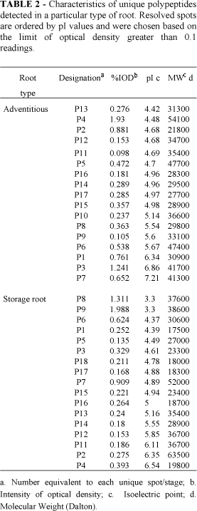
The seventeen more abundant unique protein spots to each root type marked in the gel image (Figure 1) were further characterized. Protein spot quantification, their pI value and their relative molecular mass are showed in Table 2.
A great variation on protein characteristics is observed. While the pI values ranged from 4.42 to 7.21 in AR, in the SR it ranged from 3.3 to 6.54. The estimated molecular mass for AR ranges between 21.8 to 54.1 Kda and for SR between 17.5 to 63.5 Kda.
Characterization of specific protein markers associated with a particular root type
A computer-assisted analysis of the 2-D gel profile was performed to map and establish classes of proteins related to the formation of cassava storage root. The number of unique proteins spots to each root types were classified based on their intensity of optical density (%), in 5 protein classes (Table 3): class 1 (persisting spots), spots originally present in AR, which do not appear to undergo major change from in SR; class 2 (decreasing spots), spots originally present in AR, but decreasing in SR; class 3 (increasing spots), spots originally present in AR which increase the intensity in SR; class 4 (appearing spots), spots not originally present in AR which appear with the SR;class 5 (disappearing spots), spots originally present in AR and disappeared in SR. It is observed that most of proteins appear to be present in both root types varying only in the intensity from one root type to the other.
However, spots 6913, 6884, and 6850 are of high intensity in AR but absent in SR. It indicates it may be associated only with adventitious root. On the other hand spots 6901, 6849, and 6917 increase or appeared only in the SR. In all case the proteins qualitative characteristics showed abundant variation. The high intensity (1.919 %OD) spot number 6917 appearing only in SR showed pI value of 7.44 and molecular mass of 37.6 Kda. While the highest intensity (6.72 %OD) spot number 6850 is present only in adventitious root. Those proteins markers associated to each root may be differentially related with the primary growth in AR and secondary growth and starch accumulation in SR.
In conclusion, we have shown that, (i) variation in the complexity of protein pattern was connected with different type of root, whereas a simplification and a specialization type of the profile characterized the storage root type. This observation is in concern with the fact that SR does not accumulate storage protein, (ii) identification of protein markers for each type of root is possible to be gained by using this technique. Further studies are in progress to incorporate detail characterization of the AR and SR unique proteins by using mass spectrometry analysis (MALD-TOF) and amino acid sequence. Identification and cellular localization of these polypeptides are also considered as a mean to provide a better understanding of the involvement in the storage root formation of cassava.
- BRADFORD, M. M. A rapid and sensitive method for the quantification of microgram quantities of protein utilizing the principle of protein-dye binding. Anal. Biochem, 72:248-254, 1976.
- CABRAL, G.B.; CARVALHO, L.J.C.B.; & SCHAAL, B.A The formation of storage roots in cassava: An Anatomical and a 2-D protein analysis. In: Carvalho, LJCB, AM Thro, & AD Vilarinhos (Ed.), Cassava Biotechnology IV International Scientific Meeting, CBN EMBRAPA-Genetic Resources and Biotechnology. Brasilia-DF, Brazil. 2000, p 345-356.
- CARVALHO, L.J.C.B.; CABRAL, G.B.; & CAMPOS, L. Raiz de reserva de mandioca: Um sistema biológico de múltiplas utilidade. EMBRAPA-Recursos Genéticos e Biotecnologia. Brasilia,DF. Brazil. Serie Documentos # 44: 2000, 16p.
- GORG, A., BOGUTH, G., OBERMAIER, C., & WEISS, W. Two-dimensional electrophoresis of proteins in an immobilized pH 4-12 gradient. Electrophoresis 19:1516-1519, 1998.
- GORG, A.; POSTEL W.; & GUBTHER, S. The current state of two-dimensional electrophoresis with immobilized pH gradients. Electrophoresis, 9:531-546, 1988.
- INDIRA, P.; & KURIAN, T. 1977. A study on the comparative anatomy changes undergoing tuberrization in roots of cassava and sweet potato. J. Root Crops 3:29-32, 1997.
- JEKNIE, Z.; & CHEN, T.H.H. Changes in protein profiles of poplar tissues during the induction of bud dormancy by short-day photoperiods. Plant Cell Physiology, 40, 25-35, 1999.
- MOLLOY, J.H. Silver stain for proteins in polyacrylamide gels: A modified procedure with enhanced uniform sensitivity. Anal. Biochem, 117:307-310, 1981.
- MOLLOY, M.P.; HERBERT, B.R.; WALSH, B.J.; TYLER, M.I.; TRAINI, M.; SANCHEZ, J.C.; HOCHSTRASSER, D.F.; WILLIAMS, K.L.; & GOOLEY, A. A. Extraction of membrane proteins by differential solubilization for separation using two-dimensional gel electrophoresis. Electrophoresis,19:837-844, 1998.
- O’FARRELL, P.H. High resolution two-dimentional electrophoresis for proteins. J. Biol. Chem 250:4007-4021, 1975.
- O’HAIR, S.K. Cassava root starch content and distribution varies with tissue age. HortScience 24:505-506, 1989.
- O’HAIR, S.K.; FORBES, R.B.; LOCASCIO, S.J.; RICH, J.R.; & STANLEY, R.L. HortScience, 18:735-737, 1983.
- PELTIER, J.B.; FRISO, G.; KALUME, D.E.; ROEPSTORFF, P.; NILSSON, F.; ADAMSKA, I.; & VAN WIJK, K.J. Proteomics of the chloroplast: Systematic identification and targeting analysis of lumenal and peripheral thylakoid proteins. Plant Cell, 12, 319-341, 2000.
- PLOMION, C. Separation and characterization of needle and xylem maritime pine proteins. Electrophoresis, 20, 1098-1108, 1999.
- RABILLOUD, T.; ADESSI, C.; GIRAUDEL, A.; & LUNARDI, J. Improvement of the solubilization of proteins in two-dimensional electrophoresis with immobilized pH gradients. Electrophoresis, 18: 307-316, 1997.
- RAMANUJAM, T.; & INDIRA, P. Indian J. Plant Physiol, 27:355-360, 1984.
- SALLANDROUZE, A.; FAUROBERT, M.; EL MAATAOUI, M.; & ESPAGNAC, H. Two-dimensional electrophoresis analysis of proteins associated with somatic embryogenesis development in Cupressus sempervirens L. Electrophoresis, 20, 1109-1119, 1999.
- SARMENTO, S.B.S.; CARVALHO, L.J.C.B.; CEREDA, M.P.; PENTEADO, M.V.C.; & PIEDADE, S.M.S. Growth rate, starch and fiber content in cassava store root as an indication of harvesting time. In: Carvalho, LJCB, AM Thro, & AD Vilarinhos (Ed.), Cassava Biotechnology IV International Scientific Meeting, CBN. EMBRAPA-Genetic Resources and Biotechnology. Brasilia-DF, Brazil. 2000, p 357-366.
- WILLIAMS, K.L. 1999 Genomes and proteomes: Toward a multidimensional view of biology. Electrophoresis, 20:678-688, 1999.
Publication Dates
-
Publication in this collection
20 Nov 2002 -
Date of issue
2001
History
-
Accepted
02 May 2001 -
Received
16 Dec 2000