
Abstract
The aim of this study was to evaluate the μTBS in different dentin substrates and water-storage periods. Twenty-four dentin blocks obtained from sound third molars were randomly divided into 3 groups: Sound dentin (Sd), Caries-affected dentin (Ca) and Caries-infected dentin (Ci). Dentin blocks from Ca and Ci groups were subjected to artificial caries development (S. mutans biofilm). The softest carious tissue was removed using spherical drills under visual inspection with Caries Detector solution (Ca group). It was considered as Ci (softer and deeply red stained dentin) and Ca (harder and slightly red stained dentin). The Adper Single Bond 2 adhesive system was applied and Z350 composite blocks were built in all groups. Teeth were stored in deionized water for 24 h at 37 ºC and sectioned into beams (1.0 mm2 section area). The beams from each tooth were randomly divided into three storages periods: 24 h, 6 months or 1 year. Specimens were submitted to µTBS using EZ test machine at a crosshead speed of 1.0 mm/min. Failure mode was examined by SEM. Data from µTBS were submitted to split plot two-way ANOVA and Tukey’s HSD tests (a=0.05). The µTBS (MPa) of Sd (41.2) was significantly higher than Ca (32.4) and Ci (27.2), regardless of storage. Ca and Ci after 6 months and 1 year, presented similar µTBS. Mixed and adhesive failures predominated in all groups. The highest µTBS values (48.1±9.1) were found for Sd at 24 h storage. Storage of specimens decreased the µTBS values for all conditions.
Key Words:
dentin; artificial caries; S. mutans; storage time; microtensile bond strength.
Resumo
O objetivo neste estudo foi avaliar a resistência de união à microtração (RUµT) de um sistema adesivo convencional (Adper Single Bond 2 - SB) em diferentes substratos dentinários e períodos de armazenagem. Vinte e quatro blocos de dentina foram obtidos de terceiros molares hígidos e separados aleatoriamente em 3 grupos (n=8): dentina sadia (Ds), dentina afetada (Da) e dentina infectada (Di). A Da e a Di foram submetidas ao desenvolvimento biológico artificial de cárie (S. mutans). O tecido cariado amolecido foi removido usando broca esférica sob inspeção visual com a solução Caries Detector (grupo Da). Considerou-se como Di a dentina amolecida e fortemente pigmentada de vermelho e como Da, a dentina hígida e levemente pigmentada de vermelho. O sistema adesivo SB foi aplicado de acordo com as recomendações do fabricante e blocos da resina composta Z350 foram construídos (6 mm de altura). O conjunto (dente/bloco de resina) foi armazenado em água deionizada por 24 horas a 37 °C. Estes foram seccionados em palitos (1,0 mm2 de área), que foram separados aleatoriamente em 3 períodos de armazenagem: 24 horas, 6 meses e 1 ano. Os palitos foram submetidos ao ensaio de resistência de união à microtração na máquina EZ teste a uma velocidade de 1,0 mm/min. Dados de RUµT foram submetidos à Análise de Variância 2 fatores em esquema de parcela subdividida e ao teste de Tukey (a=0,05). Os valores de resistência (MPa) da Ds (41,2) foram significativamente maiores do que os da Da (32,4) e Di (27,2), independente do tempo de armazenagem. Di e Da, 6 meses e 1ano, apresentaram valores similares de resistência de união. As falhas adesivas e mistas foram predominantes para todos os grupos. Em conclusão, os maiores valores de RUµT (48,1±9,1) foram verificados para a Ds e 24 h de armazenagem. A armazenagem diminuiu os valores de RUµT para todas as condições.
Introduction
The minimally invasive operative management of carious dentin has arisen a discussion concerning the supposed requirement for complete removal of carious tissues when cleaning and restoring lesions in a deeply decayed tooth 11 Banerjee, A. Minimal intervention dentistry: part 7. Minimally invasive operative caries management: rationale and techniques. Br Dent J 2013;214:107-111.,22 Almeida Neves, A; Coutinho, E; Cardoso, MV; Lambrechts, P; Van Meerbeek, B. Current concepts and techniques for caries excavation and adhesion to residual dentin. J Adhes Dent 2011;13:7-22.. Several studies have been performed on bonding to sound (non-carious) dentin specimens, but it is well known that the caries-affected dentin is clinically the most frequent substrate after cavity preparation 33 Huang, X; Li, L; Huang, C; Du, X. Effect of ethanol-wet bonding with hydrophobic adhesive on caries-affected dentine. Eur J Oral Sci 2011;119:310-315.,44 Mobarak, EH; El-Badrawy, WH. Microshear bond strength of self-etching adhesives to caries-affected dentin identified using the dye permeability test. J Adhes Dent 2012;14:245-250.,55 Macedo, GV; Yamauchi, M; Bedran-Russo, AK. Effects of chemical cross-linkers on caries-affected dentin bonding. J Dent Res 2009;88:1096-1100.,66 Yoshiyama, M; Tay, FR; Doi, J; Nishitani, Y; Yamada, T; Itou, K, et al.. Bonding of self-etch and total-etch adhesives to carious dentin. J Dent Res 2002;81:556-560..
Caries-infected and caries-affected dentins are distinct substrates that have different chemical composition and morphological structures 11 Banerjee, A. Minimal intervention dentistry: part 7. Minimally invasive operative caries management: rationale and techniques. Br Dent J 2013;214:107-111.,22 Almeida Neves, A; Coutinho, E; Cardoso, MV; Lambrechts, P; Van Meerbeek, B. Current concepts and techniques for caries excavation and adhesion to residual dentin. J Adhes Dent 2011;13:7-22.. The caries-infected dentin is a superficial necrotic zone of vastly demineralized substrate 11 Banerjee, A. Minimal intervention dentistry: part 7. Minimally invasive operative caries management: rationale and techniques. Br Dent J 2013;214:107-111.,22 Almeida Neves, A; Coutinho, E; Cardoso, MV; Lambrechts, P; Van Meerbeek, B. Current concepts and techniques for caries excavation and adhesion to residual dentin. J Adhes Dent 2011;13:7-22., with degenerated collagen fibrils that lost their cross-linking 55 Macedo, GV; Yamauchi, M; Bedran-Russo, AK. Effects of chemical cross-linkers on caries-affected dentin bonding. J Dent Res 2009;88:1096-1100., it may also be seen a bacterial biomass 77 Banerjee, A; Kellow, S; Mannocci, F; Cook, RJ; Watson, TF. An in vitro evaluation of microtensile bond strengths of two adhesive bonding agents to residual dentine after caries removal using three excavation techniques. J Dent 2010;38:480-489.. Conversely, caries-affected dentin is considered a variation of reactionary dentin, formed in reaction to bland stimuli like caries, presenting small alterations in the cross-linking of its collagen fibrils 11 Banerjee, A. Minimal intervention dentistry: part 7. Minimally invasive operative caries management: rationale and techniques. Br Dent J 2013;214:107-111.,22 Almeida Neves, A; Coutinho, E; Cardoso, MV; Lambrechts, P; Van Meerbeek, B. Current concepts and techniques for caries excavation and adhesion to residual dentin. J Adhes Dent 2011;13:7-22.. Additionally, it contrasts with sound dentin by the mineralized precipitates within the tubules 44 Mobarak, EH; El-Badrawy, WH. Microshear bond strength of self-etching adhesives to caries-affected dentin identified using the dye permeability test. J Adhes Dent 2012;14:245-250.. Investigations established that the caries-affected dentin may be remineralized 88 ten Cate, JM. Remineralization of caries lesions extending into dentin. J Dent Res 2001;80:1407-1411., making a more reliable adhesion to this tissue. Therefore, it has been recommended to remove only the caries-infected dentin before the bonding procedures involving dentin carious lesions 77 Banerjee, A; Kellow, S; Mannocci, F; Cook, RJ; Watson, TF. An in vitro evaluation of microtensile bond strengths of two adhesive bonding agents to residual dentine after caries removal using three excavation techniques. J Dent 2010;38:480-489.. Altered dentin characteristics can affect the resin/dentin bonding interface, since caries-affected dentin leads to greater variability and a highly irregular composition along the interface 99 Wang ,Y; Spencer, P; Walker, MP. Chemical profile of adhesive/caries-affected dentin interfaces using Raman microspectroscopy. J Biomed Mater Res A 2007;81:279-286.. Although, clinically caries affected dentin shows mineralized precipitates within the tubules (66 Yoshiyama, M; Tay, FR; Doi, J; Nishitani, Y; Yamada, T; Itou, K, et al.. Bonding of self-etch and total-etch adhesives to carious dentin. J Dent Res 2002;81:556-560., Pacheco et al. 1010 Pacheco, LF; Banzi, ECF; Rodrigues, E; Soares, LES; Pascon, FM; Correr-Sobrinho, LC, et al.. Molecular and structural evaluation of dentin caries-like lesions produced by different artificial models. Braz Dent J 2013;24:238-244. showed that similar mineral content concerning Ca, CO3 and PO4 and organic content like CH bonds, may be found also in vitro when S mutans biofilm provided dentin carious lesions compared with the natural ones.
The short and long-term bond strengths to caries-infected and caries-affected dentins commonly present lower values for both substrates, though some of these bond strength values were found to be clinically acceptable, since there is some evidence that macrotensile and microtensile bond strength tests correlates with microtensile and tensilen bond strength tests 55 Macedo, GV; Yamauchi, M; Bedran-Russo, AK. Effects of chemical cross-linkers on caries-affected dentin bonding. J Dent Res 2009;88:1096-1100.,66 Yoshiyama, M; Tay, FR; Doi, J; Nishitani, Y; Yamada, T; Itou, K, et al.. Bonding of self-etch and total-etch adhesives to carious dentin. J Dent Res 2002;81:556-560.,1112,1313 Heintze, SD. Clinical relevance of tests on bond strength, microleakage and marginal adaptation. Dent Mater 2013;29:59-84.?. There is a challenge on bonding to caries-affected dentin, since the deposition of acid-resistant minerals at the intratubular regions (33 Huang, X; Li, L; Huang, C; Du, X. Effect of ethanol-wet bonding with hydrophobic adhesive on caries-affected dentine. Eur J Oral Sci 2011;119:310-315.,55 Macedo, GV; Yamauchi, M; Bedran-Russo, AK. Effects of chemical cross-linkers on caries-affected dentin bonding. J Dent Res 2009;88:1096-1100.,66 Yoshiyama, M; Tay, FR; Doi, J; Nishitani, Y; Yamada, T; Itou, K, et al.. Bonding of self-etch and total-etch adhesives to carious dentin. J Dent Res 2002;81:556-560.,1414 Nakajima, M; Kitasako, Y; Okuda, M; Foxton, RM; Tagami, J. Elemental distributions and microtensile bond strength of the adhesive interface to normal and caries-affected dentin. J Biomed Mater Res B App Biomater 2005;72:268-275. makes this substrate almost impermeable to water 66 Yoshiyama, M; Tay, FR; Doi, J; Nishitani, Y; Yamada, T; Itou, K, et al.. Bonding of self-etch and total-etch adhesives to carious dentin. J Dent Res 2002;81:556-560.. Additionally, a more porous intertubular dentin may be found because of the partial demineralization (33 Huang, X; Li, L; Huang, C; Du, X. Effect of ethanol-wet bonding with hydrophobic adhesive on caries-affected dentine. Eur J Oral Sci 2011;119:310-315.. While these mineral deposits reduce dentin permeability from harmful agents, able to injure the pulp, they may impair an adequate bonding to dentin (1515 Nakajima, M; Sano, H; Burrow, MF; Tagami, J; Yoshiyama, M; Ebisu, S, et al.. Tensile bond strength and SEM evaluation of caries-affected dentin using dentin adhesives. J Dent Res 1995;74:1679-1688.. This barrier to bonding procedures is clinically also true for the caries-infected dentin, due to the large alterations affecting this substrate. Therefore, there are still some concerns regarding the long-term bonding to these altered dentin substrates. Assessments in literature comparing the immediate, short-term (up to 6 months) and long-term (1 year or more) bond strengths on sound, caries-infected and caries-affected dentins are inconclusive 44 Mobarak, EH; El-Badrawy, WH. Microshear bond strength of self-etching adhesives to caries-affected dentin identified using the dye permeability test. J Adhes Dent 2012;14:245-250.,99 Wang ,Y; Spencer, P; Walker, MP. Chemical profile of adhesive/caries-affected dentin interfaces using Raman microspectroscopy. J Biomed Mater Res A 2007;81:279-286.. High bond strength values can be verified immediately after the bonding procedures. However, these findings are not completely related to the long-term bond stability, since the resin-dentin bonding degradation may start in earlier stages (6 months or less) 1616 Costa, AR; Correr-Sobrinho, L; Ambrosano, GMB; Sinhoreti, MAC; Borges, GA; Platt, JA, et al.. Dentin bond strength of a fluoride-releasing adhesive system submitted to pH-cycling. Braz Dent J 2014;25:472-478.. This may be worsened for bonding to carious substrates after long-term water exposure 1313 Heintze, SD. Clinical relevance of tests on bond strength, microleakage and marginal adaptation. Dent Mater 2013;29:59-84.?. The bond strength and durability seem to be more associated to the hybrid layer quality than to other factors 77 Banerjee, A; Kellow, S; Mannocci, F; Cook, RJ; Watson, TF. An in vitro evaluation of microtensile bond strengths of two adhesive bonding agents to residual dentine after caries removal using three excavation techniques. J Dent 2010;38:480-489.. Overall, some studies on bonding strength to carious-affected dentin showed a thicker hybrid layer and lower bond strength than those from bonding to sound dentin and more prone to hydrolytic degradation than the sound ones. In addition, compared with caries-affected dentin, the sound dentin interface showed remarkable differences in demineralization depth, adhesive infiltration and interfacial bond strength 1111 Erhardt, MC; Toledano, M; Osorio, R; Pimenta, LA. Histomorphologic characterization and bond strength evaluation of caries-affected dentin/resin interfaces: effects of long-term water exposure. Dent Mater 2008;24:786-798..
Procedures and materials that improve the quality of bonding, reducing water infiltration and collagen degradation at the hybrid layer, are beneficial for making the resin-dentin interface more stable, even in altered substrates. The removal of caries-infected dentin layer, leaving the caries-affected dentin lining the cavity with sound enamel margins has been proposed in order to enable a good marginal seal with the dentin adhesive systems and otherwise avoid pulp exposition 66 Yoshiyama, M; Tay, FR; Doi, J; Nishitani, Y; Yamada, T; Itou, K, et al.. Bonding of self-etch and total-etch adhesives to carious dentin. J Dent Res 2002;81:556-560.,77 Banerjee, A; Kellow, S; Mannocci, F; Cook, RJ; Watson, TF. An in vitro evaluation of microtensile bond strengths of two adhesive bonding agents to residual dentine after caries removal using three excavation techniques. J Dent 2010;38:480-489.. In addition, a demineralized dentin substrate (caries affected-dentin) can be wetter than the sound one, since it shows great number of spaces between fibrils and the hydrophilicity of the demineralized substrate. Thus, the association of caries-affected dentin (S. mutans biofilm) and the application of a water wet-bonding strategy with available adhesive systems on the bond strength to different dentin substrates subjected to storage, seem an important topic to be evaluated. Some features, which can be found at real caries-affected dentin substrate 1414 Nakajima, M; Kitasako, Y; Okuda, M; Foxton, RM; Tagami, J. Elemental distributions and microtensile bond strength of the adhesive interface to normal and caries-affected dentin. J Biomed Mater Res B App Biomater 2005;72:268-275., certainly, cannot be observed in vitro, like the pulp tissue reaction to caries injuries. However, the slight demineralization of the dentin substrate can be found in vitro caries procedure models when caries-affected dentin is observed.
Therefore, the purpose of the present study was to assess the in vitro bond strength of an etch-and-rinse 2-step adhesive system to different dentin conditions (sound, caries-affected and caries-infected dentin) according to storage periods (24 h, 6 and 1 year). The hypotheses investigated were: 1) the bond strength of the adhesive system will be influenced by the different dentin conditions; 2) the bond strength of the adhesive system to the different dentin conditions will be affected by the storage periods.
Material and Methods
Artificial Caries Development
Twenty-four sound human third molars were selected, frozen and used within 3 months following extraction (gathered following informed consent approved by the Committee for Ethics in Research, #041/2010). The remaining soft tissues on teeth were hand removed using dental curette and polishing was performed with rubber cup and pumice slurry. The teeth were fixed in acrylic plates and occlusal enamel was cut by a water-cooled low-speed diamond saw (Isomet 1000; Buehler, Lake Bluff, IL, USA) followed by polishing with 600-grit SiC papers to expose a uniform enamel-free occlusal dentin surface. All surfaces of the teeth submitted to the microbiologic challenge were protected with an acid-resistant varnish layer (Colorama CEIL Ltda., Sao Paulo, SP, Brazil), except for the occlusal dentin surface.
Orthodontic looped wires and silicon hot glue were used to individually fix the teeth to the lid of glass vials immersing them in 40 mL sterile distilled water in order to be sterilized using gamma irradiation (GC-220E; MDS Nordion, Ottawa, Canada) for 32 h at 27 ºC with a 14.5 kGy total radiation dose 1717 Carvalho, FG; Fucio, SB; Pascon, FM; Kantovitz, KR; Correr-Sobrinho, L; Puppin-Rontani, RM. Effect of gamma irradiation on fluoride release and antibacterial activity of resin dental materials. Braz Dent J 2009;20:122-126.. The teeth from groups Ca and Ci were relocated in a sterile second glass vial immersed in 40 mL sterile brain-heart infusion broth (BHI; Difco Laboratories, Detroit, MI, USA) supplemented with yeast extract (Himedia Laboratories, PVT Ltd., Mumbai, India), 0.5% glucose (Synth; LabSynth, São Paulo, SP, Brazil), 1.0% sucrose (Synth; LabSynth) and 2.0% S. mutans strain (UA159) for development of the artificial carious lesions. The concentration of the bacterial suspension was determined by the absorption at 600 nm (A660). The adjustment of viable bacteria at A660 (colony formation unit - CFU per mL of bacterial suspension) was determined by the optical density of the suspension. The inoculation occurred only in the first day of the experiment, since the culture medium was changed every 48 h, during 14 days 1818 Carvalho, FG; de Fucio, SB; Sinhoreti, MA; Correr-Sobrinho, L; Puppin-Rontani, RM. Confocal laser scanning microscopic analysis of the depth of dentin caries-like lesions in primary and permanent teeth. Braz Dent J 2008;19:139-144.. The teeth were randomly divided by simple draw, into three groups (n=8): Sd - sound dentin; Ca - caries affected dentin and Ci - caries infected dentin.
Dentin Caries Differentiation
After the artificial caries development (broth culture with S. mutans), the specimens were washed with distilled water and a dye solution (Caries Detector; Kuraray Medical Inc., Okayama, Japan) was used for visual identification of the infected and affected dentin in association to inspection using a sharp explorer, according to Momoi et al. guidelines 1919 Momoi ,Y; Hayashi, M; Fujitani, M; Fukushima, M; Imazato, S; Kubo, S et al.. Clinical guidelines for treating caries in adults following a minimal intervention policy - Evidence and consensus based report. J Dent 2012;40:95-105.. The solution was applied for 10 s over the carious dentin surface, followed by water rinsing for 10 s. The dentin was identified as: Sd - sound dentin (hard substrate without dyeing coloration); Ca - caries affected dentin (soft dentin with light-pink coloration); Ci - caries infected dentin (soft dentin with dark-pink coloration). The groups submitted to the microbiologic challenge were assigned to two different procedures according to the dentin characteristics: Caries infected dentin (Ci) - soft deeply pigmented dentin remained after microbiology challenge. Caries affected dentin (Ca) - soft pigmented carious tissue was partially removed using spherical carbide burs (n. 8; KG Sorensen, Barueri, SP, Brazil) till hard and slightly pigmented dentin was reached.
Previous to bonding procedures, roots were sectioned 1.0 mm bellow the cement-enamel junction using double-sided diamond disc (#7020, KG Sörensen, Barueri, SP, Brazil) under copious water irrigation. The remaining pulp tissue was removed using excavators and the coronal chamber was etched with 35% phosphoric-acid (Scotchbond Etchant; 3M ESPE, St. Paul, MN, USA) for 30 s, rinsed with water and gently dried with air-spray for 2 s. Adper Single Bond 2 (3M ESPE) adhesive system was applied according to the manufacturer’s instruction and photo activated for 10 s using a light-curing unit XL2500 (3M ESPE) with 700 mW/cm2. The light power output was checked with digital radiometer Hilux Light Meter (Firt Medica, Greensboro, NC, USA). The coronal chamber was filled with composite resin Filtek Z350 (3M ESPE) and photo activated for 40 s using a light-curing unit XL2500 (3M ESPE).
Bonding Procedures
For all groups, the dentin surface was etched with 35% phosphoric acid for 15 s (Scotchbond Etchant, 3M ESPE), rinsed with water for 30 s and the water excess was removed with absorbent papers. After that, the dentin surface was rehydrated with deionized water using microbrush disposable applicators for 60 s, and the excess water was removed with absorbent papers. The Adper Single Bond 2 (3M ESPE) adhesive system was applied according to the manufacturer’s instruction and photo activated for 10 s by a light-curing unit XL2500 (3M ESPE). The adhesive bonded surfaces received composite resin Filtek Z350 (3M ESPE) applied in 2 mm increments and photo activated using a curing unit XL2500 (3M ESPE) for 40 s for each increment until building a 6 mm thick block. The restored teeth were stored in distilled water at 37 °C for 24 h.
Specimen Forming and Storage
The restored teeth were sectioned perpendicular to the bonded area to obtain beams with a 1 mm2 transversal area, by a water-cooled diamond blade (EXTEC Corporation, Enfield, CT, USA) in a low speed saw machine (Isomet 1000, Buehler). The restored teeth were sectioned before storage 2020 De Munck, J; Van Landuyt, K; Peumans, M; Poitevin, A; Lambrechts, P; Braem, M, et al.. A critical review of the durability of adhesion to tooth tissue: methods and results. J Dent Res 2005;84:118-132.. On average, 13 beams were obtained from each restored tooth, in a total of 117 beams per group. Thirty-nine beams per group were randomly (simple draw) divided into three storage times in deionized water at 37 °C for 24 h, 6 months and 1 year (Table 3). The deionized water was changed every week.
Microtensile Bonding Test
After the storage times, each beam was fixed to the grips of a microtensile device using a cyanoacrylate adhesive (Zapit; Dental Ventures of America Inc., Corona, CA, USA) and the test was conducted in a testing machine (EZ Test; EZS, Shimadzu, Tokyo, Japan) at a crosshead speed of 1.0 mm/min until failure. Bond strength values were calculated in MPa. Data were subjected to split-plot two-way ANOVA; the factors were: substrate (3 levels) and water-storage periods (3 levels), followed by the Tukey’s post hoc test (SAS Institute Inc., Cary, NC, USA, version 9.3) (p<0.05). The beams unable to be tested due to debonding on the pre-test were discarded and were not accounted for in the statistical analysis (Table 3).
Failure Mode Analysis
The specimen failure mode was determined using scanning electron microscopy (LEO 435 VP; LEO Electron Microscopy Ltd., Cambridge, UK) at 100x to 3000x magnifications. The fractured surfaces were classified according to the prevailing remaining structure as: Mode 1- adhesive; Mode 2 - cohesive in dentin; Mode 3 - cohesive in composite and Mode 4 - mixed, involving bonding agent, composite and/or tooth structure and part as an adhesive failure.
Analysis of the Bonding Interfaces
Two additional teeth were restored according to the method described above were used to this analysis. They were vertically sectioned in 2 mm slices obtaining 6 slices per tooth. They were randomly divided according to the storage time. After each storage period, four slices per group were randomly selected and evaluated (n=4). Then, they were embedded in epoxy resin and wet-polished using 600, 1200 and 2000-grit SiC papers and with decreasingly fine diamond compounds (3 µm, 1 µm, ½ µm, ¼ µm - Metadi II; Buehler). After each polishing step, the specimens were ultrasonically cleaned for 10 min. The specimens were demineralized with 50% H3PO4 solution during 5 s, rinsed in distilled water, deproteinized with 2.5% NaOCl for 10 min. Then, the specimens were fixed in 2.5% glutaraldehyde in 0.1 M cacodylate buffered solution titrated to pH 7.2 for 72 h and rinsed several times with 0.1 M sodium cacodylate buffered solution. They were dehydrated in ascending ethanol concentrations (50%, 60%, 70%, 80% and 90%) for 2 h in each solution and in 100% ethanol for 24 h. The final chemical drying was conducted by immersion in hexamethyldisilazane for 10 min on filter paper inside a covered glass vial, air-dried at room temperature. The specimens were gold-sputtered under high-vacuum environment (MED 010, Balzers Union, Liechtenstein) and analyzed by SEM (LEO 435 VP; LEO Electron Microscopy Ltd., Cambridge, UK) operating on secondary electron mode at 20 kV under 1000-5000x magnifications. The specimens were examined focusing on the depth of etching, micromechanical entanglement, integrity, homogeneity and continuity along the bonded interface.
Results
Bond Strength
The two-way ANOVA in split plot design showed significant difference for the factors dentin conditions (p<0.001) and storage time (p<0.001). There was no significant interaction between the studied factors (p=0.875). The µTBS averages and standard deviations for the three dentin conditions and storage time are in Table 1.
Regardless of the storage time, the results showed that µTBS values (MPa) of Sd were significantly higher than for Ca (p=0.01) and Ci (p<0.001). No difference was found for Ca and Ci (p=0.187). Regardless the dentin conditions, the 24 h storage time produced significantly higher values than those obtained at 6 months (p=0.004) and 1 year (p=0.002). No difference was found between 6 months and 1 year (p=0.926). The highest uTBS was found on sound dentin after 24 h storage.
The percentage of failed beams on pre-test was very low. The highest frequency of failure among pre-test beams occurred in caries infected dentin and for 6 and 12 month storage times. Twelve-month storage time increased pre-test failure for all substrates. (Table 2)
Failure Mode Analysis
The failure mode distribution is in Table 3.
There was a predominance of mixed and adhesive failures for all groups. The most representative micrographs of failure modes are in Figure 1.

A- Adhesive failure; B: Magnification showing the adhesive detachment from dentin (d); C: Magnification showing the detached resin tags; D: Cohesive failure in dentin; E and F: Higher magnifications showing the dentin’s fractured surfaces (arrow in F); G: Cohesive failure at the resin-based composite; H: aspect of an internal flaw located at the fractured composite surface; I: aspect of composite fractured surface; J: Mixed failures comprehending the materials of the bonded interfaces showing resin-based composite I, adhesive (a) and dentin (d); K: mixed failure showing the three materials; L: magnification of a mixed failure showing the materials of the bonded interface, resin-based composite I, adhesive (a) and dentin (d) (Magnifications ranged from 100× to 3000×).
Bonding Interfaces
The SEM characterization of the bonding interfaces for the dentin substrates and storage periods is in Figure 2. The water wet-bonding protocol applied for all experimental groups resulted in hybrid layers presenting continuous and homogenous interfaces for the sound dentin (Sd) after 24 h storage, with cone shaped resin tags (Fig. 2A). After 6 months and 1 year storage there was tag formation and conventional hybrid layer (hl) conformation. A high decrease of microtag branches suggesting slight degradation of the hl was detected (Figs. 2B, 2C). The caries-affected dentin group (Ca) hybrid layer presented low resin tags formation after 24 h storage, with cylindrical shaped resing tags (Fig. 2D). After 6 months storage hl presenting shorter penetration of tags into dentin were verified (Fig. 2E) and extensive changes were seen after 1 year of storage, suggesting increased hl degradation (Fig. 2F). There were irregular spaced resin tags (Fig. 2F). The caries-infected dentin group (Ci) exhibited hl conformation with abnormal tags and gaps at the interface (Fig. 2G). After 6 months storage, the hl showed disrupted interface and unusual dentin aspect (Fig. 2H), and after 1 year storage, increased hl degradation showing altered dentin aspect, with some failures at bottom and top of hl and some small and rare tags were identified (Fig. 2I).
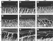
A: (Sd 24 h) Conventional hybrid layer (hl) conformation, presenting a continuous and homogenous bonding interface; lateral extensions of micro tags branching off at angles from the main resin tags (rt) are visible (pointer); B: (Sd 6 months) tag formation and conventional hl conformation; high decrease of microtag branches suggesting slight degradation of the hl over time; C: (Sd 1 year) tag formation and conventional hl conformation; high decrease of microtag branches suggesting slight degradation of the hl over time; D: (Ca 24h) hl (pointer) formation and low resin tags formation - unfilled spaces at the bottom of hl ; E: (Ca 6 months) hl formation with tags presenting shorter penetration into dentin; F: (Ca 1 year) increased disruptive areas at the interface (pointer) and an abnormal dentin aspect (arrow); G: (Ci 24 h) gaps at the interface suggesting dentin degradation by microorganism activity during caries production (arrow) and abnormal tag aspect (pointer); H: (Ci 6 months) hl showing disrupted interface (pointer) and unusual dentin aspect (arrow); I: (Ci 1 year) increased degradation of the hl with some failures at bottom (arrow) and top of hl (pointer) and presence of some small and sparse tags. Arrows also show altered dentin aspect, suggesting dentin degradation by microorganism activity during caries production. c: composite; ad: adhesive; hl: hybrid layer; rt: resin tag and d: dentin.
Discussion
The first hypothesis that different dentin conditions affect the bond strength to adhesive system was accepted, since the sound dentin group (Sd) presented the highest bond strengths. The bond strengths obtained with the same bonding protocol for the caries-affected (Ca) and caries-infected (Ci) groups were statistically similar (Table 1).
The mineral and organic changes that occur in caries-affected dentin make the adhesion more complex than to sound dentin 99 Wang ,Y; Spencer, P; Walker, MP. Chemical profile of adhesive/caries-affected dentin interfaces using Raman microspectroscopy. J Biomed Mater Res A 2007;81:279-286., associated to lower bond strengths for the contemporary adhesive systems. The present findings agree with other investigations indicating that bonding to caries-affected dentin results in lower bond strengths as related to sound dentin 55 Macedo, GV; Yamauchi, M; Bedran-Russo, AK. Effects of chemical cross-linkers on caries-affected dentin bonding. J Dent Res 2009;88:1096-1100.,2121 Doi, J; Itota, T; Torii, Y; Nakabo, S; Yoshiyama, M. Micro-tensile bond strength of self-etching primer adhesive systems to human coronal carious dentin. J Oral Rehabil 2004;31:1023-1028., even using etch-and-rinse systems 66 Yoshiyama, M; Tay, FR; Doi, J; Nishitani, Y; Yamada, T; Itou, K, et al.. Bonding of self-etch and total-etch adhesives to carious dentin. J Dent Res 2002;81:556-560.,1111 Erhardt, MC; Toledano, M; Osorio, R; Pimenta, LA. Histomorphologic characterization and bond strength evaluation of caries-affected dentin/resin interfaces: effects of long-term water exposure. Dent Mater 2008;24:786-798.,2222 Zanchi, CH; Lund, RG; Perrone, LR; Ribeiro,GA; del Pino, FA; Pinto, MB, et al.. Microtensile bond strength of two-step etch-and-rinse adhesive systems on sound and artificial caries-affected dentin. Am J Dent 2010;23:152-156.. These results are attributed to the weaker structure of the demineralized caries-affected dentin that limits adhesive infiltration because of the tubules filled with acid-resistant mineral deposits 33 Huang, X; Li, L; Huang, C; Du, X. Effect of ethanol-wet bonding with hydrophobic adhesive on caries-affected dentine. Eur J Oral Sci 2011;119:310-315.,55 Macedo, GV; Yamauchi, M; Bedran-Russo, AK. Effects of chemical cross-linkers on caries-affected dentin bonding. J Dent Res 2009;88:1096-1100.,66 Yoshiyama, M; Tay, FR; Doi, J; Nishitani, Y; Yamada, T; Itou, K, et al.. Bonding of self-etch and total-etch adhesives to carious dentin. J Dent Res 2002;81:556-560.,1414 Nakajima, M; Kitasako, Y; Okuda, M; Foxton, RM; Tagami, J. Elemental distributions and microtensile bond strength of the adhesive interface to normal and caries-affected dentin. J Biomed Mater Res B App Biomater 2005;72:268-275.) and to the unusual conformation of the hybrid layer, which is commonly thicker 1515 Nakajima, M; Sano, H; Burrow, MF; Tagami, J; Yoshiyama, M; Ebisu, S, et al.. Tensile bond strength and SEM evaluation of caries-affected dentin using dentin adhesives. J Dent Res 1995;74:1679-1688.. In addition, the present study showed low resin tags formation with short penetration into demineralized dentin regions (Figs. 2D-2F). Moreover, the increasing water content and the more permeable condition of caries-affected dentin are concerns that may compromise bonding quality/stability over time when using hydrophilic adhesives systems 1111 Erhardt, MC; Toledano, M; Osorio, R; Pimenta, LA. Histomorphologic characterization and bond strength evaluation of caries-affected dentin/resin interfaces: effects of long-term water exposure. Dent Mater 2008;24:786-798..
Regardless of the adhesive approach, lower bond strengths were already expected for caries-infected dentin 2121 Doi, J; Itota, T; Torii, Y; Nakabo, S; Yoshiyama, M. Micro-tensile bond strength of self-etching primer adhesive systems to human coronal carious dentin. J Oral Rehabil 2004;31:1023-1028., since the hypomineralized dentin is even more porous, with lower mechanical properties and high water content 11 Banerjee, A. Minimal intervention dentistry: part 7. Minimally invasive operative caries management: rationale and techniques. Br Dent J 2013;214:107-111.,22 Almeida Neves, A; Coutinho, E; Cardoso, MV; Lambrechts, P; Van Meerbeek, B. Current concepts and techniques for caries excavation and adhesion to residual dentin. J Adhes Dent 2011;13:7-22.,55 Macedo, GV; Yamauchi, M; Bedran-Russo, AK. Effects of chemical cross-linkers on caries-affected dentin bonding. J Dent Res 2009;88:1096-1100., besides the bacterial biomass 77 Banerjee, A; Kellow, S; Mannocci, F; Cook, RJ; Watson, TF. An in vitro evaluation of microtensile bond strengths of two adhesive bonding agents to residual dentine after caries removal using three excavation techniques. J Dent 2010;38:480-489.. Caries-infected dentin is also presumed to present almost complete loss of the mineral phase of dentin with a small number of larger apatite crystals and denaturation of the collagen matrix 66 Yoshiyama, M; Tay, FR; Doi, J; Nishitani, Y; Yamada, T; Itou, K, et al.. Bonding of self-etch and total-etch adhesives to carious dentin. J Dent Res 2002;81:556-560.. On this way, bonding can be hampered on this dentin, because fewer large crystals provide surface area for interacting with the adhesive system. Additionally, it was assumed that the chemical link between the adhesive systems and dentin collagen may contribute for bonding to sound and caries-affected dentin if the last has normal collagen, but it cannot take place at the denaturated matrix of caries-infected dentin 66 Yoshiyama, M; Tay, FR; Doi, J; Nishitani, Y; Yamada, T; Itou, K, et al.. Bonding of self-etch and total-etch adhesives to carious dentin. J Dent Res 2002;81:556-560.. The adhesive infiltration into dentin tubules may be also complicated by the presence of acid-resistant minerals within dentin tubules. Despite all these facts, there were no significant differences between the bond strength to the caries-affected or caries-infected dentin. This can be explained by the demineralization in Ca and the partially denatured collagen acted in a similar way which totally disorganized and demineralized the Ci dentin, also in long-term storage.
The second hypothesis that the bond strength of the adhesive system to the different dentin conditions would be affected by the storage periods was also fully accepted. Similar bond strengths were verified for the specimens stored for 6 or 12 months, irrespective of the dentin substrate. However, lower bond strength values were observed for both storage periods compared to those obtained with the specimens tested immediately (24 h), for all dentin conditions. There were a large number of adhesive failures among the Sd specimens after 1 year storage period, and for the specimens from Ca and Ci groups after 6 months. An increased number of cohesive failures in dentin were observed for the groups Ca after 1 year and for Ci after 6 months. This result may be related to the association of long-term storage and the lower mechanical properties of the dentin altered by caries (Ca and Ci), which probably fractured when the bonding strength exceeded the cohesive strength of dentin 66 Yoshiyama, M; Tay, FR; Doi, J; Nishitani, Y; Yamada, T; Itou, K, et al.. Bonding of self-etch and total-etch adhesives to carious dentin. J Dent Res 2002;81:556-560.. The adhesive failures observed for the Ca group may also be related to issues with the collagen partially denatured fibrils 66 Yoshiyama, M; Tay, FR; Doi, J; Nishitani, Y; Yamada, T; Itou, K, et al.. Bonding of self-etch and total-etch adhesives to carious dentin. J Dent Res 2002;81:556-560., and/or to the incomplete infiltration of the adhesive into the demineralized dentin due to the mineral deposits present in the tubules 44 Mobarak, EH; El-Badrawy, WH. Microshear bond strength of self-etching adhesives to caries-affected dentin identified using the dye permeability test. J Adhes Dent 2012;14:245-250..
The particularly low cohesive strength of the caries-infected dentin due to its low degree of mineralization and disorganization of the collagen-matrix 1515 Nakajima, M; Sano, H; Burrow, MF; Tagami, J; Yoshiyama, M; Ebisu, S, et al.. Tensile bond strength and SEM evaluation of caries-affected dentin using dentin adhesives. J Dent Res 1995;74:1679-1688., probably led to the increase of cohesive failures in dentin. While also resulting in thicker hybrid layers similarly to caries-affected dentin 2121 Doi, J; Itota, T; Torii, Y; Nakabo, S; Yoshiyama, M. Micro-tensile bond strength of self-etching primer adhesive systems to human coronal carious dentin. J Oral Rehabil 2004;31:1023-1028., only superficial monomer infiltration is achieved in this dentin; thus, several dentin tubules remain small and there was scarce tag formation (Figs. 2G-2I). However, despite the lower bond strengths attained for the caries-affected and caries-infected dentin using an adhesive system with water wet-bonding when compared to sound dentin, the intrinsic weakness of this dentin may not be a problem if there is enamel and/or sound dentin adjacent to the excavated regions of caries-altered dentin, as clinically acceptable bond strengths can be delivered by the current adhesive systems 11 Banerjee, A. Minimal intervention dentistry: part 7. Minimally invasive operative caries management: rationale and techniques. Br Dent J 2013;214:107-111.,66 Yoshiyama, M; Tay, FR; Doi, J; Nishitani, Y; Yamada, T; Itou, K, et al.. Bonding of self-etch and total-etch adhesives to carious dentin. J Dent Res 2002;81:556-560.,77 Banerjee, A; Kellow, S; Mannocci, F; Cook, RJ; Watson, TF. An in vitro evaluation of microtensile bond strengths of two adhesive bonding agents to residual dentine after caries removal using three excavation techniques. J Dent 2010;38:480-489..
Efficient adhesive systems and polymeric restorative materials have unquestionable significance for the minimally invasive dentistry, which intends to preserve the largest amount of remaining tooth tissues after removing the carious lesions (11 Banerjee, A. Minimal intervention dentistry: part 7. Minimally invasive operative caries management: rationale and techniques. Br Dent J 2013;214:107-111.,77 Banerjee, A; Kellow, S; Mannocci, F; Cook, RJ; Watson, TF. An in vitro evaluation of microtensile bond strengths of two adhesive bonding agents to residual dentine after caries removal using three excavation techniques. J Dent 2010;38:480-489. in order to provide better resistance to restored teeth. To assure long-term success to restorations placed on sound dentin or caries-altered dentin, the adhesive systems with improved marginal sealing capability and stable adhesion to tooth over time are critical 2222 Zanchi, CH; Lund, RG; Perrone, LR; Ribeiro,GA; del Pino, FA; Pinto, MB, et al.. Microtensile bond strength of two-step etch-and-rinse adhesive systems on sound and artificial caries-affected dentin. Am J Dent 2010;23:152-156.. The etch-and-rinse adhesive systems have been shown to attain better bonding to caries-affected and caries-infected dentin as compared to self-etching systems 22 Almeida Neves, A; Coutinho, E; Cardoso, MV; Lambrechts, P; Van Meerbeek, B. Current concepts and techniques for caries excavation and adhesion to residual dentin. J Adhes Dent 2011;13:7-22.,66 Yoshiyama, M; Tay, FR; Doi, J; Nishitani, Y; Yamada, T; Itou, K, et al.. Bonding of self-etch and total-etch adhesives to carious dentin. J Dent Res 2002;81:556-560.,1212 Li, H, Wang, WM; Yu, SL; Wen, Q. Morphological and microtensile bond strength evaluation of three adhesive systems to caries-affected human dentine with chemomechanical caries removal. J Dent 2011;39:332-339.. However, this trend is not absolute when bonding to sound dentin, since bonding to any dentin can be strongly affected by the chemical composition of adhesives 2323 Makishi, P; André, CB; Ayres, A; Martins, AL; Giannini, M. Effect of storage time on bond strength and nanoleakage expression of universal adhesives bonded to dentin and etched enamel. Oper Dent 2016;41:305-17.. Even though bond strength to dentin altered by caries is lower than to sound dentin, bonding to both dentins is continuously being implemented and have currently attained relatively high levels 22 Almeida Neves, A; Coutinho, E; Cardoso, MV; Lambrechts, P; Van Meerbeek, B. Current concepts and techniques for caries excavation and adhesion to residual dentin. J Adhes Dent 2011;13:7-22.,2424 Lenzi, TL; Braga, MM; Raggio, DP. Shortening the etching time for etch-and-rinse adhesives increases the bond stability to simulated caries-affected primary dentin. J Adhes Dent 2014;16:235-241., as also shown in this study (Ca/32.4 MPa and Ci/27.2 MPa) even for long storage time.
Although the similar bond strengths verified to caries-affected and caries-infected dentins with used the etch-and-rinse agent, bonding to the last dentin is rarely indicated due to the poor adhesive interaction that occurs. An exception for bonding to caries-infected dentin would occur adjusting the behavior of uncooperative patients aiming to prevent additional caries progression 22 Almeida Neves, A; Coutinho, E; Cardoso, MV; Lambrechts, P; Van Meerbeek, B. Current concepts and techniques for caries excavation and adhesion to residual dentin. J Adhes Dent 2011;13:7-22.. A limited number of studies assessing the performance of contemporary adhesive systems on bonding to caries-affected and caries-infected dentins are now available and future long-term clinical trials evaluating the restorative parameters studied in the present investigation would be of benefit. Moreover, concerning in vitro studies and their relationship with clinical outcomes, immediate bond strength evaluation should be considered only as a baseline, and measuring bond strength aging should be the aim, since it may predict the clinical effectiveness of adhesives, as shown by a critical review published by Van Meerbeek et al. 2525 Van Meerbeek, B; Peumans, M; Poitevin, A; Mine, A; Van Ende, A; Neves, A et al.. Relationship between bond-strength tests and clinical outcomes. Dent Mater 2010;26:e100-e121..
Within the conditions of this in vitro study, the following conclusions can be drawn: 11 Banerjee, A. Minimal intervention dentistry: part 7. Minimally invasive operative caries management: rationale and techniques. Br Dent J 2013;214:107-111. bonding with sound dentin showed µTBS values significantly higher compared to caries-affected and caries-infected dentin; 22 Almeida Neves, A; Coutinho, E; Cardoso, MV; Lambrechts, P; Van Meerbeek, B. Current concepts and techniques for caries excavation and adhesion to residual dentin. J Adhes Dent 2011;13:7-22. The 6 months and 1 year storage periods resulted in decreased bond strengths for all dentin conditions; 33 Huang, X; Li, L; Huang, C; Du, X. Effect of ethanol-wet bonding with hydrophobic adhesive on caries-affected dentine. Eur J Oral Sci 2011;119:310-315.. Bonding to the infected and affected dentin is not a suitable procedure compared to sound dentin, mainly for long-term procedures, even for sound dentin.
Acknowledgements
This study was supported by Fundação de Amparo à Pesquisa do Estado de São Paulo (Grant 2010/07829-2). Thanks to Dr EW Kitajima, Dr FAO Tanaka, and RB Salaroli (Núcleo de Apoio à Pesquisa em Microscopia Eletrônica, NAPME/ESALQ/USP, Brazil) for SEM equipment support. The authors are indebted to Dra. Glaucia Maria Bovi Ambrosano (Department of Social Dentistry, FOP-UNICAMP) for statistic analysis.
References
-
1Banerjee, A. Minimal intervention dentistry: part 7. Minimally invasive operative caries management: rationale and techniques. Br Dent J 2013;214:107-111.
-
2Almeida Neves, A; Coutinho, E; Cardoso, MV; Lambrechts, P; Van Meerbeek, B. Current concepts and techniques for caries excavation and adhesion to residual dentin. J Adhes Dent 2011;13:7-22.
-
3Huang, X; Li, L; Huang, C; Du, X. Effect of ethanol-wet bonding with hydrophobic adhesive on caries-affected dentine. Eur J Oral Sci 2011;119:310-315.
-
4Mobarak, EH; El-Badrawy, WH. Microshear bond strength of self-etching adhesives to caries-affected dentin identified using the dye permeability test. J Adhes Dent 2012;14:245-250.
-
5Macedo, GV; Yamauchi, M; Bedran-Russo, AK. Effects of chemical cross-linkers on caries-affected dentin bonding. J Dent Res 2009;88:1096-1100.
-
6Yoshiyama, M; Tay, FR; Doi, J; Nishitani, Y; Yamada, T; Itou, K, et al.. Bonding of self-etch and total-etch adhesives to carious dentin. J Dent Res 2002;81:556-560.
-
7Banerjee, A; Kellow, S; Mannocci, F; Cook, RJ; Watson, TF. An in vitro evaluation of microtensile bond strengths of two adhesive bonding agents to residual dentine after caries removal using three excavation techniques. J Dent 2010;38:480-489.
-
8ten Cate, JM. Remineralization of caries lesions extending into dentin. J Dent Res 2001;80:1407-1411.
-
9Wang ,Y; Spencer, P; Walker, MP. Chemical profile of adhesive/caries-affected dentin interfaces using Raman microspectroscopy. J Biomed Mater Res A 2007;81:279-286.
-
10Pacheco, LF; Banzi, ECF; Rodrigues, E; Soares, LES; Pascon, FM; Correr-Sobrinho, LC, et al.. Molecular and structural evaluation of dentin caries-like lesions produced by different artificial models. Braz Dent J 2013;24:238-244.
-
11Erhardt, MC; Toledano, M; Osorio, R; Pimenta, LA. Histomorphologic characterization and bond strength evaluation of caries-affected dentin/resin interfaces: effects of long-term water exposure. Dent Mater 2008;24:786-798.
-
12Li, H, Wang, WM; Yu, SL; Wen, Q. Morphological and microtensile bond strength evaluation of three adhesive systems to caries-affected human dentine with chemomechanical caries removal. J Dent 2011;39:332-339.
-
13Heintze, SD. Clinical relevance of tests on bond strength, microleakage and marginal adaptation. Dent Mater 2013;29:59-84.?
-
14Nakajima, M; Kitasako, Y; Okuda, M; Foxton, RM; Tagami, J. Elemental distributions and microtensile bond strength of the adhesive interface to normal and caries-affected dentin. J Biomed Mater Res B App Biomater 2005;72:268-275.
-
15Nakajima, M; Sano, H; Burrow, MF; Tagami, J; Yoshiyama, M; Ebisu, S, et al.. Tensile bond strength and SEM evaluation of caries-affected dentin using dentin adhesives. J Dent Res 1995;74:1679-1688.
-
16Costa, AR; Correr-Sobrinho, L; Ambrosano, GMB; Sinhoreti, MAC; Borges, GA; Platt, JA, et al.. Dentin bond strength of a fluoride-releasing adhesive system submitted to pH-cycling. Braz Dent J 2014;25:472-478.
-
17Carvalho, FG; Fucio, SB; Pascon, FM; Kantovitz, KR; Correr-Sobrinho, L; Puppin-Rontani, RM. Effect of gamma irradiation on fluoride release and antibacterial activity of resin dental materials. Braz Dent J 2009;20:122-126.
-
18Carvalho, FG; de Fucio, SB; Sinhoreti, MA; Correr-Sobrinho, L; Puppin-Rontani, RM. Confocal laser scanning microscopic analysis of the depth of dentin caries-like lesions in primary and permanent teeth. Braz Dent J 2008;19:139-144.
-
19Momoi ,Y; Hayashi, M; Fujitani, M; Fukushima, M; Imazato, S; Kubo, S et al.. Clinical guidelines for treating caries in adults following a minimal intervention policy - Evidence and consensus based report. J Dent 2012;40:95-105.
-
20De Munck, J; Van Landuyt, K; Peumans, M; Poitevin, A; Lambrechts, P; Braem, M, et al.. A critical review of the durability of adhesion to tooth tissue: methods and results. J Dent Res 2005;84:118-132.
-
21Doi, J; Itota, T; Torii, Y; Nakabo, S; Yoshiyama, M. Micro-tensile bond strength of self-etching primer adhesive systems to human coronal carious dentin. J Oral Rehabil 2004;31:1023-1028.
-
22Zanchi, CH; Lund, RG; Perrone, LR; Ribeiro,GA; del Pino, FA; Pinto, MB, et al.. Microtensile bond strength of two-step etch-and-rinse adhesive systems on sound and artificial caries-affected dentin. Am J Dent 2010;23:152-156.
-
23Makishi, P; André, CB; Ayres, A; Martins, AL; Giannini, M. Effect of storage time on bond strength and nanoleakage expression of universal adhesives bonded to dentin and etched enamel. Oper Dent 2016;41:305-17.
-
24Lenzi, TL; Braga, MM; Raggio, DP. Shortening the etching time for etch-and-rinse adhesives increases the bond stability to simulated caries-affected primary dentin. J Adhes Dent 2014;16:235-241.
-
25Van Meerbeek, B; Peumans, M; Poitevin, A; Mine, A; Van Ende, A; Neves, A et al.. Relationship between bond-strength tests and clinical outcomes. Dent Mater 2010;26:e100-e121.
Publication Dates
-
Publication in this collection
Jan-Feb 2017
History
-
Received
22 Mar 2016 -
Accepted
06 Aug 2016


