Abstract
Marginal and internal adaptation is critical for the success of indirect restorations. New imaging systems make it possible to evaluate these parameters with precision and non-destructively. This study evaluated the marginal and internal adaptation of zirconia copings fabricated with two different systems using both silicone replica and microcomputed tomography (micro-CT) assessment methods. A metal master model, representing a preparation for an all-ceramic full crown, was digitally scanned and polycrystalline zirconia copings were fabricated with either Ceramill Zi (Amann-Girrbach) or inCoris Zi (Dentslpy-Sirona), n=10. For each coping, marginal and internal gaps were evaluated by silicone replica and micro-CT assessment methods. Four assessment points of each replica cross-section and micro-CT image were evaluated using imaging software: marginal gap (MG), axial wall (AW), axio-occlusal angle (AO) and mid-occlusal wall (MO). Data were statistically analyzed by factorial ANOVA and Tukey test (a=0.05). There was no statistically significant difference between the methods for MG and AW. For AO, there were significant differences between methods for Amann copings, while for Dentsply-Sirona copings similar values were observed. For MO, both methods presented statistically significant differences. A positive correlation was observed determined by the two assessment methods for MG values. In conclusion, the assessment method influenced the evaluation of marginal and internal adaptation of zirconia copings. Micro-CT showed lower marginal and internal gap values when compared to the silicone replica technique, although the difference was not always statistically significant. Marginal gap and axial wall assessment points showed the lower gap values, regardless of ceramic system and assessment method used.
Key Words:
marginal fit; dental prosthesis; zirconia; CAD/CAM.
Resumo
A adaptação marginal e interna é fundamental para o sucesso das restaurações indiretas e novos sistemas de imagem permitem avaliar esses parâmetros de maneira não-destrutiva e com precisão. Este estudo avaliou a adaptação marginal e interna de copings de zircônia fabricados com dois sistemas diferentes, utilizando as técnicas da réplica em silicone e micro-CT. Um modelo-mestre de metal, representando um preparo para uma coroa totalmente cerâmica foi digitalizado e copings de zirconia policristalina (Ceramill Zi, Amann Girrbach e inCoris Zi, Dentslpy Sirona, n=10) foram fabricados. Para cada coping, a adaptação marginal e interna foi avaliada pelas técnicas da réplica em silicone e micro-CT. Foram avaliados quatro pontos de cada seção transversal e imagem de micro-CT: adaptação marginal (MG), parede axial (AW), ângulo axial-oclusal (AO) e parede centro-oclusal (MO) utilizando um software de imagem. Os dados foram analisados estatisticamente por ANOVA e teste de Tukey (a=0,05). Não houve diferença estatisticamente significativa entre as duas técnicas para MG e AW. Para AO, houve diferenças significativas entre as técnicas para copings da Amann, enquanto que para copings da Dentsply Sirona foram observados valores semelhantes. Para o MO, ambas as técnicas apresentaram diferenças estatisticamente significativas. Observou-se uma correlação positiva entre os valores de adaptação determinados pelas duas técnicas. O método de avaliação influenciou a avaliação da adaptação marginal e interna de copings de zircônia. Pode-se concluir que a micro-CT mostrou menores valores de adaptação marginal e interna quando comparado com a técnica de réplica em silicone. Os pontos de avaliação da margem e da parede axial mostraram os menores valores de adaptação, independente do sistema cerâmico e da técnica de avaliação utilizados.
Introduction
Marginal and internal adaptation is critical for the success of indirect restorations 11 Denissen, H; Dozic, A; van der Zel, J; van Waas, M. Marginal fit and short-term clinical performance of porcelain-veneered CICERO, CEREC, and Procera onlays. J Prosthet Dent 2000;84:506-513.. Margin misfit may cause hypersensitivity, secondary caries, biofilm accumulation, gingivitis, and periodontal problems 22 Della Bona, A; Kelly, JR. The clinical success of all-ceramic restorations. J Am Dent Assoc 2008;139Suppl:8S-13S.,33 Knoernschild, KL; Campbell, SD. Periodontal tissue responses after insertion of artificial crowns and fixed partial dentures. J Prosthet Dent 2000;84:492-498.,44 Lang, NP; Kiel, RA; Anderhalden, K. Clinical and microbiological effects of subgingival restorations with overhanging or clinically perfect margins. J Clin Periodontol 1983;10:563-578.. Marginal misfit also results in a thick cement film, exposing the luting material to the oral environment and promoting its dissolution over time 55 Gonzalo, E; Suarez, MJ; Serrano, B; Lozano, JF. A comparison of the marginal vertical discrepancies of zirconium and metal ceramic posterior fixed dental prostheses before and after cementation. J Prosthet Dent 2009;102:378-384.,66 Jacobs, MS; Windeler, AS. An investigation of dental luting cement solubility as a function of the marginal gap. J Prosthet Dent 1991;65:436-442.. It has been reported that a 100-120-μm-thick cement layer is considered clinically acceptable for good long-term clinical prognosis of the indirect restoration 77 McLean, JW; von Fraunhofe,r JA. The estimation of cement film thickness by an in vivo technique. Br Dent J 1971;131:107-111.,88 Sorensen, JA. A standardized method for determination of crown margin fidelity. J Prosthet Dent 1990;64:18-24.. Poor internal adaptation can lead to lack of restoration retention and poor resistance form for the tooth-restoration complex 99 Faot, F; Suzuki, D; Senna, PM; da Silva, WJ; de Mattias Sartori, IA. Discrepancies in marginal and internal fits for different metal and alumina infrastructures cemented on implant abutments. Eur J Oral Sci 2015;123:215-219.,1010 Tuntiprawon, M, Wilson, PR. The effect of cement thickness on the fracture strength of all-ceramic crowns. Aust Dent J 1995;40:17-21.. Therefore, the study of factors that affect marginal and internal adaptation of indirect restorations is critically important in restorative dentistry.
Different methods have been used to assess marginal and internal adaptation of indirect restorations. Clinically, marginal fit can be estimated directly using a probe with known dimensions, a dental mirror (where appropriate), and adequate illumination and magnification. Marginal fit can also be indirectly evaluated radiographically, and through epoxy resin replicas by light and scanning electron microscopy 1111 An, S; Kim, S; Choi, H; Lee, JH; Moon, HS. Evaluating the marginal fit of zirconia copings with digital impressions with an intraoral digital scanner. J Prosthet Dent 2014;112:1171-1175.,1212 Alghazzawi, TF; Al-Samadani, KH; Lemons, J; Liu, PR; Essig, ME; Bartolucci, AA; et al.. Effect of imaging powder and CAD/CAM stone types on the marginal gap of zirconia crowns. J Am Dent Assoc 2015;146:111-120.. Despite being commonly used, microscopes can result in inaccurate measurement, poor identification of reference points, projection errors, and rounding of the margins 1313 Contrepois, M; Soenen, A; Bartala, M; Laviole, O. Marginal adaptation of ceramic crowns: a systematic review. J Prosthet Dent 2013;110:447-454 e410.,1414 Nawafleh, NA; Mack, F; Evans, J; Mackay ,J; Hatamleh, MM. Accuracy and reliability of methods to measure marginal adaptation of crowns and FDPs: a literature review. J Prosthodont2013;22:419-428.. It is also possible to cross-section the prepared tooth or master die and the restoration for direct assessment of the space correspondent to cement gap - a surrogate measure of marginal and internal adaptation, under a microscope 1515 Beuer, F; Naumann, M; Gernet, W; Sorensen, JA. Precision of fit: zirconia three-unit fixed dental prostheses. Clin Oral Investig 2009;13:343-349.. However, the cross-sectioning technique has been discouraged because it is a destructive method, it allows the assessment of only a few reference points, and it may result in fracture or distortion of the restoration during sectioning 1313 Contrepois, M; Soenen, A; Bartala, M; Laviole, O. Marginal adaptation of ceramic crowns: a systematic review. J Prosthet Dent 2013;110:447-454 e410..
A popular non-destructive method used to assess the marginal and internal fit both in vivo and in vitro is the silicone replica technique. A light-body PVS material is used to record the space between the prosthetic preparation and the internal surface of the restoration 1616 Colpani JT; Borba M; Della Bona, A. Evaluation of marginal and internal fit of ceramic crown copings. Dent Mater 2013;29:174-180.,1717 Reich, S; Uhlen, S; Gozdowski, S; Lohbauer, U. Measurement of cement thickness under lithium disilicate crowns using an impression material technique. Clin Oral Investig 2011;15:521-526., and then the silicone film can be measured in multiple reference points both at the margin and internal walls. This method is considered easy to perform, is relatively inexpensive 1111 An, S; Kim, S; Choi, H; Lee, JH; Moon, HS. Evaluating the marginal fit of zirconia copings with digital impressions with an intraoral digital scanner. J Prosthet Dent 2014;112:1171-1175., and is accepted as a reliable method to evaluate marginal and internal adaptation 1818 Rahme, HY; Tehini, GE; Adib, SM; Ardo, AS; Rifai, KT. In vitro evaluation of the “replica technique” in the measurement of the fit of Procera crowns. J Contemp Dent Pract 2008;9:25-32.; on the other hand, the difficulty in identifying the margins of the restoration, possible tearing of the silicone film upon removal of the crown, as well as sectioning plane error and few reference points are considered drawbacks of the silicone replica technique 1313 Contrepois, M; Soenen, A; Bartala, M; Laviole, O. Marginal adaptation of ceramic crowns: a systematic review. J Prosthet Dent 2013;110:447-454 e410.,1414 Nawafleh, NA; Mack, F; Evans, J; Mackay ,J; Hatamleh, MM. Accuracy and reliability of methods to measure marginal adaptation of crowns and FDPs: a literature review. J Prosthodont2013;22:419-428..
Recently, microcomputed tomography (micro-CT) imaging has also been used as a non-destructive method to evaluate marginal and internal fit of indirect restorations 1919 Borba, M; Cesar, PF; Griggs, JA; Della Bona, A. Adaptation of all-ceramic fixed partial dentures. Dent Mater 2011;27:1119-1126.,1818 Rahme, HY; Tehini, GE; Adib, SM; Ardo, AS; Rifai, KT. In vitro evaluation of the “replica technique” in the measurement of the fit of Procera crowns. J Contemp Dent Pract 2008;9:25-32.,1919 Borba, M; Cesar, PF; Griggs, JA; Della Bona, A. Adaptation of all-ceramic fixed partial dentures. Dent Mater 2011;27:1119-1126.,2020 Mously, HA; Finkelman, M; Zandparsa, R; Hirayama, H. Marginal and internal adaptation of ceramic crown restorations fabricated with CAD/CAM technology and the heat-press technique. J Prosthet Dent 2014;112:249-256.,2121 Seo, D; Yi, Y; Roh, B. The effect of preparation designs on the marginal and internal gaps in Cerec3 partial ceramic crowns. J Dent 2009;37:374-382.. This technique allows a two and three-dimensional assessment of marginal and internal adaptation of restorations in several directions and sections, making it possible to assess a great number of reference points and easy identification of critical distances non-destructively 1313 Contrepois, M; Soenen, A; Bartala, M; Laviole, O. Marginal adaptation of ceramic crowns: a systematic review. J Prosthet Dent 2013;110:447-454 e410.,2121 Seo, D; Yi, Y; Roh, B. The effect of preparation designs on the marginal and internal gaps in Cerec3 partial ceramic crowns. J Dent 2009;37:374-382..
There is no consensus on which is the best non-destructive method to evaluate marginal and internal adaptation of indirect restorations. Only few studies comparing different methods for the assessment of marginal and internal adaptation of indirect restorations are available 1414 Nawafleh, NA; Mack, F; Evans, J; Mackay ,J; Hatamleh, MM. Accuracy and reliability of methods to measure marginal adaptation of crowns and FDPs: a literature review. J Prosthodont2013;22:419-428.. All the studies mentioned in the review 1414 Nawafleh, NA; Mack, F; Evans, J; Mackay ,J; Hatamleh, MM. Accuracy and reliability of methods to measure marginal adaptation of crowns and FDPs: a literature review. J Prosthodont2013;22:419-428. have compared the replica technique with the cross-section technique. Only one study 2222 Rungruanganunt, P; Kelly, JR; Adams, DJ. Two imaging techniques for 3D quantification of pre-cementation space for CAD/CAM crowns. J Dent2010;38:995-1000. comparing the silicone replica technique to micro-CT was found, but the methodology used to measure the pre-cementation space is different from the present study. Both techniques (micro-CT imaging and optical density image analysis) used imaging of polyvinylsiloxane (PVS) impressions. The authors determined the silicone thickness using a photometric technique. Based on the Beer-Lambert Law, proportionality between a known thickness (standardized thickness specimens fabricated from the same impression material) and the amount of light transmitted through the impression material could be assumed 2222 Rungruanganunt, P; Kelly, JR; Adams, DJ. Two imaging techniques for 3D quantification of pre-cementation space for CAD/CAM crowns. J Dent2010;38:995-1000..
Therefore, the objective of this study was to compare micro-CT and silicone replica techniques for the evaluation of marginal and internal adaptation of zirconia copings fabricated with two different ceramic systems. The null hypotheses were: 11 Denissen, H; Dozic, A; van der Zel, J; van Waas, M. Marginal fit and short-term clinical performance of porcelain-veneered CICERO, CEREC, and Procera onlays. J Prosthet Dent 2000;84:506-513. there will be no difference in marginal and internal adaptation of zirconia copings fabricated with different ceramic systems, and 22 Della Bona, A; Kelly, JR. The clinical success of all-ceramic restorations. J Am Dent Assoc 2008;139Suppl:8S-13S. there will be no difference in marginal and internal adaptation of zirconia copings assessed by the two methods.
Material and Methods
Two identical metal master models were used, one for each ceramic system, representing a preparation for an all-ceramic full crown on a lower first molar, with the following characteristics: supragingival circumferential chamfer finish line, 2.0-mm occlusal reduction, 1.5-mm axial reduction, axial convergence angle of 6o and rounded internal line angles.
The master models were scanned by two different systems for fabrication of the zirconia copings (n=10): Ceramill Map400 (Amann Girrbach AG. Koblach, Austria) and Cerec inLab (Dentsply Sirona, York, PA, USA). Copings 0.5 mm thick at the margins and 0.8 mm at the axial and occlusal walls were machined from zirconia blocks (Ceramill Zi and inCoris Zi, respectively), with 20 μm-thick marginal spacer and 70-μm-thick internal spacer. After milling, copings were sintered. Marginal and internal adaptation of each coping was measured using the silicone replica technique 1818 Rahme, HY; Tehini, GE; Adib, SM; Ardo, AS; Rifai, KT. In vitro evaluation of the “replica technique” in the measurement of the fit of Procera crowns. J Contemp Dent Pract 2008;9:25-32.,2323 Pedroche, LO; Bernardes, SR; Leao, MP; Kintopp, CC; Correr, GM; Ornaghi, BP; et al.. Marginal and internal fit of zirconia copings obtained using different digital scanning methods. Braz Oral Res 2016;30:e113.,2424 Syrek, A; Reich, G; Ranftl, D; Klein, C; Cerny, B; Brodesser, J. Clinical evaluation of all-ceramic crowns fabricated from intraoral digital impressions based on the principle of active wavefront sampling. J Dent 2010;38:553-559. and micro-CT imaging 1919 Borba, M; Cesar, PF; Griggs, JA; Della Bona, A. Adaptation of all-ceramic fixed partial dentures. Dent Mater 2011;27:1119-1126.,2525 Borba, M; Miranda, WG Jr; Cesar, PF; Griggs, JA; Bona, AD. Evaluation of the adaptation of zirconia-based fixed partial dentures using micro-CT technology. Braz Oral Res 2013;27:396-402.,2626 Neves, FD; Prado, CJ; Prudente, MS; Carneiro, TA; Zancope, K; Davi, LR; et al.. Micro-computed tomography evaluation of marginal fit of lithium disilicate crowns fabricated by using chairside CAD/CAM systems or the heat-pressing technique. J Prosthet Dent 2014;112:1134-1140.,2727 Pelekanos, S; Koumanou, M; Koutayas, SO; Zinelis, S; Eliades, G. Micro-CT evaluation of the marginal fit of different In-Ceram alumina copings. Eur J Esthet Dent 2009;4:278-292..
To produce the silicone replica, each coping was filled with a light-body PVS material (HydroXtreme light body, Coltene Whaledent, Cuyahoga Falls, OH, USA) and positioned on the master model. Firm finger pressure simulating a definitive cementation procedure was applied and after 5 min the coping was removed from the master model. To remove the light-body material from the coping and determine its thickness, which corresponds to the marginal and internal gap, a regular-body PVS material with contrasting color (HydroXtreme regular body, Coltene Whaledent) was placed into the coping to produce the replica. After polymerization, the replica was cut with a sharp scalpel blade in buccolingual and mesiodistal directions to obtain four cross-sections.
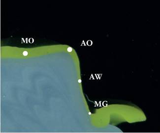
The four cross-sections of each replica were placed on a scanner (C3180 HP Photosmart, HP) for digitization. The high-resolution images (1200 dpi) were saved as jpg files and a single operator determined the thickness of the light-body PVS material using image analyzing software (ImageJ, US National Institutes of Health, Bethesda, MA, USA). Four assessment points of each cross section of the replica were evaluated (Fig. 1) 2323 Pedroche, LO; Bernardes, SR; Leao, MP; Kintopp, CC; Correr, GM; Ornaghi, BP; et al.. Marginal and internal fit of zirconia copings obtained using different digital scanning methods. Braz Oral Res 2016;30:e113.:

Marginal gap (MG): perpendicular distance at the margin from the prepared master model to the internal surface of the coping;
Axial wall (AW): distance at the mid-axial wall between the internal surface of the coping and the prepared tooth;
Axial-occlusal angle (AO): distance at the region of the axial-occlusal edge between the prepared tooth and internal surface of the coping, represented by the intersection of two straight lines, parallel to the axial wall and occlusal plane;
Mid-occlusal wall (MO): distance at the center of the occlusal region between the prepared tooth and internal surface of the coping.
For each replica, four measurements were obtained for each assessment point. The mean value for each assessment point was calculated and considered for statistical analysis.
The same master models and copings were cleaned with alcohol and then submitted to micro-CT analysis. Each coping was adapted to the master model, placed in the micro-CT chamber and scanned (SkyScan 1172, Skyscan, Kontich. Belgium). For standardization purposes, micro-CT images for the two ceramic materials were acquired at the same time. The micro-CT has an X-ray source with cone-beam geometry and small spot size (5 μm), in order to reduce dispersion. An 11-megapixel camera with CCD (charged coupled device) was used as detector. The following scanning parameters were used: Al+Cu filter, 100 uA current, accelerating voltage of 100 kV, exposure time of 4000 ms per frame, and rotation step at 0.4º with 180º rotation. The X-ray was irradiated perpendicular to the long axis of the preparation and the image pixel size was 13 μm.
Volumes were reconstructed using the NRecon software (Skyscan), employing the Feldkamp-Davis-Kress (FDK) algorithm, beam hardening of 100%, and ring artifact correction of 16. CTAnalyser software (Skyscan, Aartselaar) was used to obtain cross-section images of the central region of all copings in both buccolingual and mesiodistal directions, and the files were saved in bmp format.
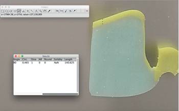
The measures for the four assessment points of the 2D micro-CT images were also performed using an image analyzing software (ImageJ, US National Institutes of Health). For each buccolingual and mesiodistal cross-section images, four measurements were obtained for each assessment point (Fig. 2). The mean value was calculated and considered for statistical analysis.
The replica cross-sections were made visually, midway between buccolingual and mesiodistal distances. The same criterion was used for the selection of micro-CT images, in an attempt to align the cross-sections and standardize the measurements for the two assessment methods. Figures 1 and 2 show one example of the silicone replica cross-section, indicating the four assessment points, and a 2D micro-CT image from which the gap widths were also measured. Figure 3 shows a representative ImageJ image illustrating how measurements were made using the straight-line selection tool.

Two-dimensional micro-CT image for measuring the marginal and internal gap of zirconia copings. The determination of assessment point AO is also shown, represented by the intersection of two straight lines, parallel to occlusal plane and axial wall.

Representative ImageJ image illustrating how measurements (MO in this example) were made with the straight-line selection tool. This tool was used to create the line selection at each assessment point. In the dialog box at the bottom left side of the image, the length (in μm) is displayed in the last column.
For each assessment point, data were statistically analyzed using factorial ANOVA (ceramic system and assessment technique) and Tukey test with a=0.05. Correlation between marginal and internal gap values determined by micro-CT and replica technique was performed using coefficient of determination (R2) and Pearson’s coefficient. All the analyses were performed with statistical software (Statistica 10; StatSoft Inc., Tulsa, OK, USA).
Results
For MG and AW, ceramic system, assessment technique and double interaction were not significant (p>0.05). However, assessment technique was statically significant for MO and AO (p=0.000825 and p=0.000044, respectively). Ceramic system and double interaction were also not significant (p>0.05).
Means and standard deviations for the assessment points evaluated by micro-CT and replica technique of the zirconia copings are shown in Table 1. For both ceramic systems, there was no statistically significant difference between the two techniques for MG and AW. On the other hand, for AO, there were significant differences between techniques for Amann copings, while for Dentsply Sirona copings similar values were observed. For MO, replica and micro-CT techniques presented significant statistically differences for both ceramic systems.
For both ceramic systems, a strong positive correlation between the marginal and internal gap values determined by silicone replica and micro-CT assessment methods was observed (Fig. 4) (R2 correlation coefficient of 0.97432 and 0.86219 for Amann and Dentsply Sirona, respectively; and Pearson correlation coefficient of 0.98708 and 0.92854 for Amann and Dentsply Sirona, respectively).

Correlation plot between marginal and internal gap values determined by micro-CT and replica techniques.
Discussion
Marginal and internal adaptation is one of the most important factors for the success and clinical longevity of ceramic crowns. This study compared two non-destructive assessment methods to evaluate marginal and internal adaptation of zirconia copings fabricated with two CAD/CAM systems. According to the results, the first test hypothesis was accepted, i.e., there was no difference in marginal and internal adaptation of zirconia copings fabricated with different ceramic systems, was accepted, while the second test hypothesis was rejected, i.e, there was a difference in marginal and internal adaptation of zirconia copings evaluated by the two methods. In the present study lower values of marginal and internal gap were observed for micro-CT in comparison to the replica technique for the two ceramic systems tested.
Recently, micro-CT has been widely applied in dentistry, and is commonly used for implant planning, evaluation of root canal preparation, forensic investigations and marginal adaptation of ceramic crowns. It is a non-destructive method of analysis that allows the evaluation of marginal and internal fit of indirect restorations through high-resolution images, permitting 2D and 3D visualization from any angle or position. By this technique, marginal and internal adaptation can be evaluated on x and y axes, enabling a more realistic perception of the space correspondent to the cement. However, in some situations, especially when there is insufficient radiographic contrast between the materials, marginal and internal gap analysis cannot be performed 1919 Borba, M; Cesar, PF; Griggs, JA; Della Bona, A. Adaptation of all-ceramic fixed partial dentures. Dent Mater 2011;27:1119-1126.,2020 Mously, HA; Finkelman, M; Zandparsa, R; Hirayama, H. Marginal and internal adaptation of ceramic crown restorations fabricated with CAD/CAM technology and the heat-press technique. J Prosthet Dent 2014;112:249-256.. Thus, to improve the contrast between metal master model and zirconia coping, in this study micro-CT scanning was performed without cementation. In previous studies, the radiopacity of the resin cement may have interfered with the results 2020 Mously, HA; Finkelman, M; Zandparsa, R; Hirayama, H. Marginal and internal adaptation of ceramic crown restorations fabricated with CAD/CAM technology and the heat-press technique. J Prosthet Dent 2014;112:249-256.. Also, cementation was not performed because all copings were adapted onto the same two master models for scanning, one for each ceramic system. If the copings were cemented for scanning, each coping would have to be removed and a new one cemented onto the same master model. This procedure could have caused damage to the master model and interfered with the results of gap measurements in subsequent samples. Other studies have also used this methodology and did not perform cementation of indirect restorations submitted to micro-CT analysis 1919 Borba, M; Cesar, PF; Griggs, JA; Della Bona, A. Adaptation of all-ceramic fixed partial dentures. Dent Mater 2011;27:1119-1126.,2020 Mously, HA; Finkelman, M; Zandparsa, R; Hirayama, H. Marginal and internal adaptation of ceramic crown restorations fabricated with CAD/CAM technology and the heat-press technique. J Prosthet Dent 2014;112:249-256.. Nevertheless, other studies have shown that when there is no cementation some factors related to the cementation process and type of cement cannot be considered 2121 Seo, D; Yi, Y; Roh, B. The effect of preparation designs on the marginal and internal gaps in Cerec3 partial ceramic crowns. J Dent 2009;37:374-382..
The CAD/CAM systems used to fabricate the zirconia copings in the present study were unable to create a homogeneous cementation gap, even though a 20 μm marginal spacer thickness and a 70 μm internal spacer thickness were previously established. Indirect restorations with homogeneous cementation gaps are important so that retention and resistance forms are not compromised, especially for ceramic restorations, due to their fragility. Some studies have already evaluated the impact of different spacer thickness settings (or luting space settings) on marginal and internal fit of ceramic restorations 2828 Hmaidouch, R; Neumann, P; Mueller, WD. Influence of preparation form, luting space setting and cement type on the marginal and internal fit of CAD/CAM crown copings. Int J Comput Dent 2011;14:219-226.,2929 Nakamura, T; Dei, N; Kojima, T; Wakabayashi, K. Marginal and internal fit of Cerec 3 CAD/CAM all-ceramic crowns. Int J Prosthodont 2003;16:244-248.. The results of these studies indicated that luting space settings might influence the marginal fit. Programming a small spacer thickness may result in premature contact points between the prepared tooth and the internal surface of the crown 2020 Mously, HA; Finkelman, M; Zandparsa, R; Hirayama, H. Marginal and internal adaptation of ceramic crown restorations fabricated with CAD/CAM technology and the heat-press technique. J Prosthet Dent 2014;112:249-256.,2929 Nakamura, T; Dei, N; Kojima, T; Wakabayashi, K. Marginal and internal fit of Cerec 3 CAD/CAM all-ceramic crowns. Int J Prosthodont 2003;16:244-248., preventing the overflow of excess cement and increasing marginal gap 2828 Hmaidouch, R; Neumann, P; Mueller, WD. Influence of preparation form, luting space setting and cement type on the marginal and internal fit of CAD/CAM crown copings. Int J Comput Dent 2011;14:219-226..
Different gap widths depending on the location (assessment point) may be related to the quality of the acquisition and processing of the CAD/CAM technology 1515 Beuer, F; Naumann, M; Gernet, W; Sorensen, JA. Precision of fit: zirconia three-unit fixed dental prostheses. Clin Oral Investig 2009;13:343-349.. Increase on the internal gap may result in rounded edges produced by the finite resolution of the scan of CAD/CAM systems. Furthermore, the overshoot phenomenon, which simulates virtual peaks near the edges, can also be responsible for higher values of internal gap 1919 Borba, M; Cesar, PF; Griggs, JA; Della Bona, A. Adaptation of all-ceramic fixed partial dentures. Dent Mater 2011;27:1119-1126..
In the present study, mean values for marginal gap varied from 68.73 to 77.90 μm, with no statistically significant difference between ceramic systems and assessment methods. All values were lower than 120 μm, considered the clinically acceptable limit for marginal gap 77 McLean, JW; von Fraunhofe,r JA. The estimation of cement film thickness by an in vivo technique. Br Dent J 1971;131:107-111.,88 Sorensen, JA. A standardized method for determination of crown margin fidelity. J Prosthet Dent 1990;64:18-24.. Literature reports that on the axial walls of prepared tooth, gap values around 122 μm could reduce fracture resistance of crowns 1010 Tuntiprawon, M, Wilson, PR. The effect of cement thickness on the fracture strength of all-ceramic crowns. Aust Dent J 1995;40:17-21.. Considering this information as well as the fact that, in this study, mean gap values for axial walls ranged from 81.00 to 85.53 μm, the evaluated ceramic systems can be considered suitable for clinical use. Axio-occlusal angle and mid-occlusal area presented the higher gap values, respectively 110.83-162.65 μm and 121.53-187.35 μm. In fact, from the clinical perspective, of all assessment points, marginal adaptation should be considered the most critical to evaluate.
Another study 2020 Mously, HA; Finkelman, M; Zandparsa, R; Hirayama, H. Marginal and internal adaptation of ceramic crown restorations fabricated with CAD/CAM technology and the heat-press technique. J Prosthet Dent 2014;112:249-256. evaluated the internal and marginal fit of lithium disilicate crowns fabricated using heat-press technique and CAD/CAM with different spacer thickness (30, 60 and 90 µm). Results showed that marginal fit values for CAD/CAM-60 µm group (49.35 µm) and for CAD/CAM-90 µm group (46.65 µm) were lower than those observed for CAD/CAM-30 µm group (55.18 µm). However, it is important to observe that all those values are low and clinically acceptable for marginal gap of ceramic crowns. On the other hand, it is interesting to note that for the axial and occlusal wall reference points, even when the spacer thickness was predetermined, the gap width obtained was higher. For example, for axial and occlusal walls respectively, the group with a 30 µm spacer thickness presented gap values of 90.04 and 160.49 µm; the 60 µm spacer group presented values of 127.68 and 152.39 µm; and 90 µm spacer group showed 147.71 and 227.38 µm. Therefore, spacer thicknesses of 30 or 60 µm were recommended for crowns fabricated using CAD/CAM technology.
The same phenomenon can be observed, in a smaller scale, in the results of the present study, when spacer thicknesses of 20 µm and 70 µm were predetermined, respectively, for the margins and for the internal axial and occlusal walls. In a previous study 2020 Mously, HA; Finkelman, M; Zandparsa, R; Hirayama, H. Marginal and internal adaptation of ceramic crown restorations fabricated with CAD/CAM technology and the heat-press technique. J Prosthet Dent 2014;112:249-256., gap widths obtained for the 60 µm-spacer group for axial and occlusal walls were approximately 113 and 152% higher than the programmed spacer thickness. The present study used a preset internal spacer thickness of 70 µm, but gap values were from 22 to 73% higher than the predetermined value for axial and occlusal walls, respectively.
The two evaluated ceramic systems showed similar values for marginal and internal fit. This result was expected, since the processing method of the copings from both systems is similar. It is important to observe that zirconia copings were machined in larger dimensions to compensate for sintering contraction, and this may have also influenced the gap values. The success of this compensation depends on the homogeneity of the pre-sintered block and on the software’s capability of estimating the real contraction of the material 1616 Colpani JT; Borba M; Della Bona, A. Evaluation of marginal and internal fit of ceramic crown copings. Dent Mater 2013;29:174-180.. Thus, both systems were able to compensate the zirconia sintering contraction providing clinically acceptable results. Different results can be expected for multiple fixed prostheses, since other difficulties and distortions on impressions and casts may occur, as well as problems with ceramic sintering contraction compensation.
Although both methods are proposed to assess the marginal and internal fit of indirect restorations by measuring the cement thickness through image analysis software, the light-body silicone might have impaired the correct adaptation of the coping onto the model or tooth, and the polymerization of the silicone could also have influenced the measurement of the cement thickness, which resulted in significant differences between cross-sections of silicone replica and of micro-CT images.
There is still no standard protocol to evaluate the fit of indirect restorations. This lack of standardization may compromise interpretation and comparison of data from different studies and to provide guidelines for clinical practice. Thus, it is important to recognize the limitations of the currently used techniques, as well as the kind of data that may obtained with each method 1616 Colpani JT; Borba M; Della Bona, A. Evaluation of marginal and internal fit of ceramic crown copings. Dent Mater 2013;29:174-180.. Few studies compared different methods used to evaluate marginal and internal fit of ceramic restorations 1414 Nawafleh, NA; Mack, F; Evans, J; Mackay ,J; Hatamleh, MM. Accuracy and reliability of methods to measure marginal adaptation of crowns and FDPs: a literature review. J Prosthodont2013;22:419-428., and only one study used silicone replica technique and micro-CT 2222 Rungruanganunt, P; Kelly, JR; Adams, DJ. Two imaging techniques for 3D quantification of pre-cementation space for CAD/CAM crowns. J Dent2010;38:995-1000.. However, it is important to note that the methodology presented in this previous study 2222 Rungruanganunt, P; Kelly, JR; Adams, DJ. Two imaging techniques for 3D quantification of pre-cementation space for CAD/CAM crowns. J Dent2010;38:995-1000. is different from the one used in the present study. Rungruanganunt et al.2222 Rungruanganunt, P; Kelly, JR; Adams, DJ. Two imaging techniques for 3D quantification of pre-cementation space for CAD/CAM crowns. J Dent2010;38:995-1000. evaluated the pre-cementation space of single zirconia copings using two methods: micro-CT and quantitative optical analysis. Impressions of the pre-cementation space were taken with low viscosity PVS and standardized thickness specimens were also fabricated from the same impression material. Micro-CT images were then obtained from the silicone specimens and standards. The quantitative optical analysis method used the same standardized thickness specimens and measured the cement space thickness by transilluminating the pre-cementation space impressions. Also, the results from the present study cannot be directly compared to a previous study 2222 Rungruanganunt, P; Kelly, JR; Adams, DJ. Two imaging techniques for 3D quantification of pre-cementation space for CAD/CAM crowns. J Dent2010;38:995-1000., because there are significant methodological differences and the values for marginal and internal adaptation are not reported and analyzed in the same way. The distribution of thicknesses in the pre-cementation space replica measured by micro-CT was reported by volume percentage and more than 90% of the measurements ranged from less than 50 µm to 170 µm.
The present study compared two increasingly used methods for the evaluation of marginal and internal fit of prosthetic restorations. Several studies and literature reviews suggested that micro-CT could be recommended as a useful tool for evaluating the adaptation of all-ceramic restorations 1313 Contrepois, M; Soenen, A; Bartala, M; Laviole, O. Marginal adaptation of ceramic crowns: a systematic review. J Prosthet Dent 2013;110:447-454 e410.,1919 Borba, M; Cesar, PF; Griggs, JA; Della Bona, A. Adaptation of all-ceramic fixed partial dentures. Dent Mater 2011;27:1119-1126.,2121 Seo, D; Yi, Y; Roh, B. The effect of preparation designs on the marginal and internal gaps in Cerec3 partial ceramic crowns. J Dent 2009;37:374-382.. Based on the results of the present study, the authors corroborate this recommendation. Also, the results suggest a strong positive correlation between the marginal and internal gap values determined by micro-CT and replica technique (Fig. 3). Therefore, both techniques can be used to determine the fit of indirect restorations.
Finally, it should be noted that long-term clinical studies should be carried out in an attempt to update what would be the clinically acceptable limit in terms of marginal and internal fit all-ceramic prosthetic restorations. The parameters still considered as references in the literature, and also for this work, are based on studies published in the 1970-1990 77 McLean, JW; von Fraunhofe,r JA. The estimation of cement film thickness by an in vivo technique. Br Dent J 1971;131:107-111.,88 Sorensen, JA. A standardized method for determination of crown margin fidelity. J Prosthet Dent 1990;64:18-24.. However, when they are established, most current ceramic systems and techniques for the fabrication of indirect restorations were not yet available. Furthermore, the selection of the ceramic system to be used should be based not only on the values of marginal and internal fit, but also on the system’s ability to produce a restoration that fulfills the existent clinical conditions as well as the aesthetic expectations of the patient 1313 Contrepois, M; Soenen, A; Bartala, M; Laviole, O. Marginal adaptation of ceramic crowns: a systematic review. J Prosthet Dent 2013;110:447-454 e410..
Based on the results of the present study, the following conclusions could be drawn: i) the two ceramic systems evaluated presented similar gap values. All marginal and internal gap values obtained were considered clinically acceptable; and ii) micro-CT showed lower marginal and internal gap values when compared to silicone replica technique. Marginal gap and axial wall assessment points showed the lowest gap values, regardless of ceramic system and assessment method used.
References
-
1Denissen, H; Dozic, A; van der Zel, J; van Waas, M. Marginal fit and short-term clinical performance of porcelain-veneered CICERO, CEREC, and Procera onlays. J Prosthet Dent 2000;84:506-513.
-
2Della Bona, A; Kelly, JR. The clinical success of all-ceramic restorations. J Am Dent Assoc 2008;139Suppl:8S-13S.
-
3Knoernschild, KL; Campbell, SD. Periodontal tissue responses after insertion of artificial crowns and fixed partial dentures. J Prosthet Dent 2000;84:492-498.
-
4Lang, NP; Kiel, RA; Anderhalden, K. Clinical and microbiological effects of subgingival restorations with overhanging or clinically perfect margins. J Clin Periodontol 1983;10:563-578.
-
5Gonzalo, E; Suarez, MJ; Serrano, B; Lozano, JF. A comparison of the marginal vertical discrepancies of zirconium and metal ceramic posterior fixed dental prostheses before and after cementation. J Prosthet Dent 2009;102:378-384.
-
6Jacobs, MS; Windeler, AS. An investigation of dental luting cement solubility as a function of the marginal gap. J Prosthet Dent 1991;65:436-442.
-
7McLean, JW; von Fraunhofe,r JA. The estimation of cement film thickness by an in vivo technique. Br Dent J 1971;131:107-111.
-
8Sorensen, JA. A standardized method for determination of crown margin fidelity. J Prosthet Dent 1990;64:18-24.
-
9Faot, F; Suzuki, D; Senna, PM; da Silva, WJ; de Mattias Sartori, IA. Discrepancies in marginal and internal fits for different metal and alumina infrastructures cemented on implant abutments. Eur J Oral Sci 2015;123:215-219.
-
10Tuntiprawon, M, Wilson, PR. The effect of cement thickness on the fracture strength of all-ceramic crowns. Aust Dent J 1995;40:17-21.
-
11An, S; Kim, S; Choi, H; Lee, JH; Moon, HS. Evaluating the marginal fit of zirconia copings with digital impressions with an intraoral digital scanner. J Prosthet Dent 2014;112:1171-1175.
-
12Alghazzawi, TF; Al-Samadani, KH; Lemons, J; Liu, PR; Essig, ME; Bartolucci, AA; et al.. Effect of imaging powder and CAD/CAM stone types on the marginal gap of zirconia crowns. J Am Dent Assoc 2015;146:111-120.
-
13Contrepois, M; Soenen, A; Bartala, M; Laviole, O. Marginal adaptation of ceramic crowns: a systematic review. J Prosthet Dent 2013;110:447-454 e410.
-
14Nawafleh, NA; Mack, F; Evans, J; Mackay ,J; Hatamleh, MM. Accuracy and reliability of methods to measure marginal adaptation of crowns and FDPs: a literature review. J Prosthodont2013;22:419-428.
-
15Beuer, F; Naumann, M; Gernet, W; Sorensen, JA. Precision of fit: zirconia three-unit fixed dental prostheses. Clin Oral Investig 2009;13:343-349.
-
16Colpani JT; Borba M; Della Bona, A. Evaluation of marginal and internal fit of ceramic crown copings. Dent Mater 2013;29:174-180.
-
17Reich, S; Uhlen, S; Gozdowski, S; Lohbauer, U. Measurement of cement thickness under lithium disilicate crowns using an impression material technique. Clin Oral Investig 2011;15:521-526.
-
18Rahme, HY; Tehini, GE; Adib, SM; Ardo, AS; Rifai, KT. In vitro evaluation of the “replica technique” in the measurement of the fit of Procera crowns. J Contemp Dent Pract 2008;9:25-32.
-
19Borba, M; Cesar, PF; Griggs, JA; Della Bona, A. Adaptation of all-ceramic fixed partial dentures. Dent Mater 2011;27:1119-1126.
-
20Mously, HA; Finkelman, M; Zandparsa, R; Hirayama, H. Marginal and internal adaptation of ceramic crown restorations fabricated with CAD/CAM technology and the heat-press technique. J Prosthet Dent 2014;112:249-256.
-
21Seo, D; Yi, Y; Roh, B. The effect of preparation designs on the marginal and internal gaps in Cerec3 partial ceramic crowns. J Dent 2009;37:374-382.
-
22Rungruanganunt, P; Kelly, JR; Adams, DJ. Two imaging techniques for 3D quantification of pre-cementation space for CAD/CAM crowns. J Dent2010;38:995-1000.
-
23Pedroche, LO; Bernardes, SR; Leao, MP; Kintopp, CC; Correr, GM; Ornaghi, BP; et al.. Marginal and internal fit of zirconia copings obtained using different digital scanning methods. Braz Oral Res 2016;30:e113.
-
24Syrek, A; Reich, G; Ranftl, D; Klein, C; Cerny, B; Brodesser, J. Clinical evaluation of all-ceramic crowns fabricated from intraoral digital impressions based on the principle of active wavefront sampling. J Dent 2010;38:553-559.
-
25Borba, M; Miranda, WG Jr; Cesar, PF; Griggs, JA; Bona, AD. Evaluation of the adaptation of zirconia-based fixed partial dentures using micro-CT technology. Braz Oral Res 2013;27:396-402.
-
26Neves, FD; Prado, CJ; Prudente, MS; Carneiro, TA; Zancope, K; Davi, LR; et al.. Micro-computed tomography evaluation of marginal fit of lithium disilicate crowns fabricated by using chairside CAD/CAM systems or the heat-pressing technique. J Prosthet Dent 2014;112:1134-1140.
-
27Pelekanos, S; Koumanou, M; Koutayas, SO; Zinelis, S; Eliades, G. Micro-CT evaluation of the marginal fit of different In-Ceram alumina copings. Eur J Esthet Dent 2009;4:278-292.
-
28Hmaidouch, R; Neumann, P; Mueller, WD. Influence of preparation form, luting space setting and cement type on the marginal and internal fit of CAD/CAM crown copings. Int J Comput Dent 2011;14:219-226.
-
29Nakamura, T; Dei, N; Kojima, T; Wakabayashi, K. Marginal and internal fit of Cerec 3 CAD/CAM all-ceramic crowns. Int J Prosthodont 2003;16:244-248.
Publication Dates
-
Publication in this collection
Jul-Aug 2017
History
-
Received
13 Feb 2017 -
Accepted
15 May 2017