Abstracts
A multiple mandible fracture was diagnosed in a mixed breed dog. After clinical examination, extra-oral radiographs were used to evaluate the trauma but, a complete visualization of the fractures extension was difficult due to the great of structures of cranium. Then, the computed tomography was accomplished to help and to find all osseous tissues fractured, their exact location, existent fragments and their positions; being superior to the radiograph exam in the accurate diagnosis realized.
computed tomography; facial; trauma; dog; mandible; fracture
Um caso de fratura múltipla de mandíbula foi diagnosticado em um cão sem raça definida. Após exame clínico, radiografias extra-orais foram utilizadas para se avaliar a extensão do traumatismo, mas uma completa visualização da extensão das fraturas estava dificultada pela grande sobreposição de estruturas do crânio. A tomografia computadorizada foi então realizada para colaborar na identificação de todos os focos de fratura, de suas exatas localizações, da presença de fragmentos e de suas posições, mostrando-se superior ao exame radiográfico na realização de um diagnóstico preciso.
tomografia computadorizada; facial; trauma; cão; mandíbula; fratura
NOTE
CLINIC AND SURGERY
Facial trauma in a dog: advantages of the use of computed tomography on diagnostic of caudal fractures of mandible
Trauma facial em um cão: vantagens do uso da tomografia computadorizada no diagnóstico de fraturas caudais da mandíbula
Vanessa Graciela Gomes CarvalhoI,1 1 Author corresponding. ; Ana Carolina Fonseca PintoII; Marco Antônio GiosoIII; Herbert Lima CorreaI; Marcelo Gusmão Paraíso CavalcantiIV
IDepartamento de Cirurgia, Faculdade de Medicina Veterinária, Universidade de São Paulo (USP), Av. Prof. Dr. Orlando Marques de Paiva, 87, 05508-900, Cidade Universitária, São Paulo, SP, Brasil. E-mail: vanggc@uol.com.br
IIDepartamento de Cirurgia, disciplina de diagnóstico por imagem FMVZ, USP, São Paulo, SP, Brasil
IIIDepartamento de Cirurgia, FMVZ, USP, São Paulo, SP, Brasil. E-mail: maggioso@usp.br
IVDepartamento de Estomatologia, Faculdade de OdontologiaUSP, São Paulo, SP, Brasil
ABSTRACT
A multiple mandible fracture was diagnosed in a mixed breed dog. After clinical examination, extra-oral radiographs were used to evaluate the trauma but, a complete visualization of the fractures extension was difficult due to the great of structures of cranium. Then, the computed tomography was accomplished to help and to find all osseous tissues fractured, their exact location, existent fragments and their positions; being superior to the radiograph exam in the accurate diagnosis realized.
Key words: computed tomography, facial, trauma, dog, mandible, fracture.
RESUMO
Um caso de fratura múltipla de mandíbula foi diagnosticado em um cão sem raça definida. Após exame clínico, radiografias extra-orais foram utilizadas para se avaliar a extensão do traumatismo, mas uma completa visualização da extensão das fraturas estava dificultada pela grande sobreposição de estruturas do crânio. A tomografia computadorizada foi então realizada para colaborar na identificação de todos os focos de fratura, de suas exatas localizações, da presença de fragmentos e de suas posições, mostrando-se superior ao exame radiográfico na realização de um diagnóstico preciso.
Palavras-chave: tomografia computadorizada, facial, trauma, cão, mandíbula, fratura.
The radiograph exam is a good method to provide the diagnosis, on the dorsal-ventral and lateral views (WIGGS & LOBPRISE, 1997). But, the complexity of the structures on this region turns the diagnosis of all the structures affected by radiographs sometimes difficult. The computed tomography (CT), currently, is the most indicated diagnostic method to discover mandible caudal lesions (MARRETA, 1998; FIKE et al., 1980; FORREST, 1999).
The CT is a high resolution imaging system (GARLAND et al., 2002) with process of production of cross-sectional images using x-ray and computers (HATHCOCK & STICKLE, 1993). They are mobile in a horizontal table through the tunnel (gantry), where the x-ray beams are radiated. Rotation movement of the radiation source around the patient obtains sequences of thin slices with 1 to 10mm (FERREIRA et al., 1998) through the area of interest. The CT images are made of the black color, white and shades of gray (called gray scale), assigned to tiny squares (pixels) arranged in columns and rows (a matrix), and can record thousands of separate opacities ranging from air to high-density metal images (CAVALCANTI et al., 1999). Each pixel represents a block of tissue (voxel). The third dimension of this block of tissue is the thickness of the tissue slice (HATHCOCK & STICKLE, 1993; HOSKINSON & TUCKER, 2001).
This clinical case reports the difficulties on facial trauma diagnosis with extra-oral radiographs in a young mixed breed dog. It was dehydrated and presented oral trauma, difficulty to eat and pain during oral examination. There were malocclusion, osseous mobility and crepitation on the left and the right mandible. The lateral oblique extra-oral radiographs showed complete and transversal bone fractures on the horizontal mandible, distal to the left canine tooth (Figure 1A) and distal to the right third molar tooth (Figure 1B). The bilateral coronoid and condilar process were difficult to evaluate because of the superimposition of osseous tissues. But, the extension of trauma and the clinical signs suggested a better investigation.
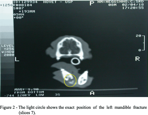
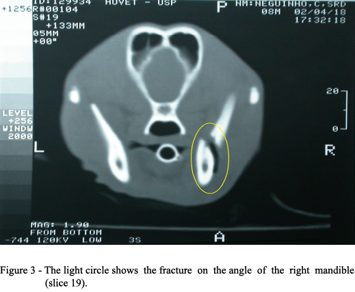
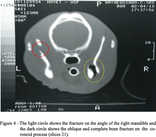
To obtain better images of the trauma, the animal was referred to the Imaging Service of the Veterinary School of the University of São Paulo to establish a computerized tomography of the cranium. General anesthesia was necessary to accomplish the exam because the patient couldnt be moved during the procedure. The ventro-dorsal was the best position for the procedure. The CT machine used was a General Electric CT max 640. At first, the scout was made to delimitate the portion of mandible to be studied. The transversal slices were made with thickness of 5mm and increments of 5mm between them. The exact diagnosis was obtained after 25 slices on the tomography. The exam showed: complete and transversal bone fracture on the left mandible, distal to the left lower canine with fragment deviation (Figure 2); complete and transversal bone fracture on the right horizontal ramous, next to the vertical angle of mandible, with low attenuance between the fracture focus (Figure 3); oblique bone fracture on the left coronoid process with caudal fragment deviation (Figure 4).
The superior soft-tissue differentiation and no superimposition of overlying structures were the major advantages of CT over conventional x-ray techniques in this case, specially at the coronoid process and on the angle of the right mandible. Each fracture could be evaluated individually, obtaining important information about the kind of fracture, the osseous fragment deviation and their exact localization.
This kind of exam is accomplished frequently in human hospitals in patients with facial trauma, being the first option on image diagnosis (COSTA E SILVA et al., 2003; SANTOS et al., 2004). Intra-oral and extra-oral radiographs are important types of imaging exam and can be performed to help the clinician on diagnosis but, all of these benefits together can not be offered together, all the times. When the CT is available, it can be used especially when the trauma is extensive (SANTOS et al., 2004), with a fast and on accurate imaging interpretation and a better planning of surgical interventions and therapy. In conclusion, we demonstrated the usefulness of CT in helping the diagnosis and evaluation outcome in a case of dogs facial trauma, showing its advantages when compared with radiograph exam.
Received 12.01.05
Approved 05.24.06
- CAVALCANTI, M.G. et al. Three-dimensional computed tomography landmark measurement in craniofacial surgical planning: experimental validation in vitro. Journal of Oral and Maxillofacial Surgery, v.57, n.6, p.690-694, 1999.
- COSTA E SILVA, A.P.A. et al. Interpretation of mandibular condyle fractures using 2D-CT and 3D-CT. Braz Dent Journal, n.14, p.203-208, 2003.
- FERREIRA, F.M. et al. A tomografia computadorizada em medicina veterinária. Clínica Veterinária, n.12, p.27-32, 1998
- FIKE, J.R. et al. Canine anatomy as assessed by computerized tomography. American Journal of Veterinary Research, v.41, n.11, p.1823-1832, 1980.
- FORREST, L.J. The head: excluding the brain and orbit. Clinical Techniques in Small Animal Practice, v.14, n.3, p.170-176, 1999.
- GARLAND, M.R. et al. Modern CT applications in veterinary medicine. Radiographics, v.22, n.1, p.399-415, 2002.
- HATHCOCK, J.T.; STICKLE, R.L. Principles and concepts of computed tomography. Veterinary Clinics of North America: Small Animal Practice, v.2, p.399-415, 1993.
- HOSKINSON, J.J., TUCKER, R.L. Diagnostic imaging of lameness in small animals. Veterinary Clinics of North America: Small Animal Practice, v.1, p.165-174, 2001.
- MARRETA, S.M. Maxillofacial surgery. Veterinary Clinics of North America: Small Animal Practice, v.29, p.1285-1288, 1998.
- SANTOS, D.T. et al. Validity of multislice computed tomography for diagnosis of maxillofacial fractures using an independent workstation. Oral Surg Oral Med Oral Pathol Oral Radiol and Endodont, v.98, p.210-216, 2004.
- WIGGS, R.B.; LOBPRISE, H.B. Oral surgery. In: ______. Veterinary dentistry, principles & practice Philadelphia: Lippincott-Raven, 1997. p.245-250.
Publication Dates
-
Publication in this collection
06 Nov 2006 -
Date of issue
Dec 2006
History
-
Accepted
24 May 2006 -
Received
01 Dec 2005