Abstracts
Laryngeal and tracheal tumors are rare in pets; some piece of information on their disease behavior, therapy and evolution are limited. Neoplasms in this area are a diagnostic challenge. In many cases, they can be biopsied and excised using endoscopic instruments, but there is no report of this in canines. The goal of this study is to report a successful case of a laryngeal neoplasm removal through endoscopy. A head and neck radiogram revealed a mass in the laryngeal lumen protruding into the trachea. The patient then underwent an endoscopy to confirm the radiographic diagnosis and to surgically remove the tumor. The histopathological diagnosis was poorly differentiated carcinoma. The most appropriate treatment for laryngeal tumors is the resection of the submucosa or a partial laryngectomy however, partial and total laryngectomies are associated with many postoperative complications. In contrast, the endoscopic approach allows for highly magnified visualization of the lesion in situ, which facilitates the surgical removal of the mass through videosurgery. With little manipulation of the affected area, the chances of postoperative complications are reduced, leading to a more rapid recovery.
tumor; larynx; canine; endoscopy
Tumores de laringe e traqueia são raros em animais de estimação e as informações sobre o comportamento, terapia e evolução destas neoplasias são limitadas. Neoplasias nesta área são desafios diagnósticos. Em muitos casos, pode ser feita a biópsia e excisão da massa utilizando instrumentos endoscópicos, mas não existe relato deste tipo de procedimento em caninos. O objetivo deste estudo foi relatar um caso de sucesso da remoção endoscópica de um tumor de laringe. A radiografia da região cervical revelou uma massa na luz da laringe invadindo a traqueia. O paciente foi então submetido a uma endoscopia para confirmar o diagnóstico radiográfico e remover cirurgicamente o tumor. O diagnóstico histopatológico foi de carcinoma pouco diferenciado. O tratamento mais adequado para os tumores da laringe é a ressecção da submucosa ou uma laringectomia parcial, no entanto, estas estão associadas a muitas complicações pós-operatórias. Em contraste, a abordagem endoscópica permite a visualização da lesão in situ, o que facilita a remoção cirúrgica da massa através de videocirurgia. Com pouca manipulação da área afetada, as chances de complicações pós-operatórias são reduzidas, levando a uma recuperação mais rápida.
tumor; laringe; canino; endoscopia
Laryngeal and tracheal tumors are rare in dogs and cats; thus, there is little information on the incidence, clinical signs, types, diagnosis, treatment and prognosis of these tumors (CLIFFORD & SORENMO, 2004CLIFFORD, C.A.; SORENMO, K.U. Tumors of the larynx and trachea. In: KING, L.G. Textbook of respiratory disease in dogs and cats. St. Louis: Saunders, 2004. p.339-345.; DUNBAR et al., 2012DUNBAR, M.D. et al. Laryngeal rhabdomyoma in a dog. Veterinary Clinical Pathology, v.41, p.590-593, 2012. Available from: <http://vet.sagepub.com/content/22/6/533.full.pdf>. Accessed: Feb. 09, 2014. doi: 10.1177/030098588502200605.
http://vet.sagepub.com/content/22/6/533....
; MACPHAIL, 2014MACPHAIL, C. Laryngeal disease in dogs and cats. Veterinary Clinics of North America: Small Animal Practice, v.44, n.2, p.19-31, 2014. Available from: <http://www.sciencedirect.com/science/article/pii/S0195561613001812>. Accessed: Feb. 08, 2014. doi: 10.1016/j.cvsm.2013.09.001.
http://www.sciencedirect.com/science/art...
). One review described 24 cases (13 dogs and 11 cats) spanning a 10-year period in which laryngeal tumors comprised 0.02% of all biopsy and necropsy specimens in dogs, comprising 11 different tumor types (SAIK et al., 1986SAIK, J.E. et al. Canine and feline laryngeal neoplasia: a 10-year survey. Journal of American Animal Hospital Association, v.22, p.359-365, 1986.; DUNBAR et al., 2012DUNBAR, M.D. et al. Laryngeal rhabdomyoma in a dog. Veterinary Clinical Pathology, v.41, p.590-593, 2012. Available from: <http://vet.sagepub.com/content/22/6/533.full.pdf>. Accessed: Feb. 09, 2014. doi: 10.1177/030098588502200605.
http://vet.sagepub.com/content/22/6/533....
). Tumors in this area are a diagnostic challenge but can be biopsied and excised with endoscopic instruments in many cases (WITHROW, 2013WITHROW, S.J. Tumors of the respiratory system. In: _____, S.J. et al. Withrow and MacEwen's small animal clinical oncology. St. Louis: Saunders Elsevier, 2013. p.451-453.). A total laryngectomy with a permanent tracheostomy is usually performed as the conventional treatment for malignant tumors in humans. Although frequently used in humans, this technique is not commonly used in veterinary medicine (SAIK et al., 1986SAIK, J.E. et al. Canine and feline laryngeal neoplasia: a 10-year survey. Journal of American Animal Hospital Association, v.22, p.359-365, 1986.; FOSSUM & ROGERS, 1998MACPHAIL, C. Laryngeal disease in dogs and cats. Veterinary Clinics of North America: Small Animal Practice, v.44, n.2, p.19-31, 2014. Available from: <http://www.sciencedirect.com/science/article/pii/S0195561613001812>. Accessed: Feb. 08, 2014. doi: 10.1016/j.cvsm.2013.09.001.
http://www.sciencedirect.com/science/art...
). Thus, the goal of this study was to report a successful case of a videosurgical approach to the excision of a laryngeal neoplasm from a dog.
A 13-year-old Boxer bitch weighing 25kg was referred to the veterinary hospital, presenting with severe dyspnea. The owner reported that the patient had exhibited respiratory changes (dyspnea) with a noticeable aggravation over the past three days. In the physical examination the dog was apathetic in an orthopneic position, severely dyspneic and cyanotic.
The patient underwent radiography of the thorax and upper airways, and a mass was observed in the larynx protruding toward the trachea. This mass was located at the laryngeal lumen and was responsible for the respiratory obstruction and consequent dyspnea (Figure 1A). In addition to the imaging exams to establish the correct stage of the tumor, an aspiration biopsy of the submandibular lymph nodes was performed, but no neoplastic cells were observed in these structures.
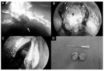
A. Tumor mass in the larynx of a Boxer bitch. On the latero-lateral radiographic image the presence of the mass (arrow) obstructing the laryngeal lumen can be seen; B. On the endoscopic image of the rostral area of the larynx, the obstruction of the lumen by the neoplasm can be seen; C. Image taken after the excision of the neoplasm; the clearing of the laryngeal lumen; D. Dissection of the neoplasm, displaying the tumor's macroscopic aspect can be observed
Subsequently, the use of endoscopy was chosen for diagnosis, and an attempt was made to excise the mass. Intravenous etomidate (1mg kg-1), midazolam (0.5mg kg-1) and lidocaine (1mg kg-1), combined in the same syringe, were used for anesthetic induction. Anesthetic maintenance was performed with isoflurane, initially with a mask because orotracheal intubation was impossible, and then, a tracheostomy was performed, thus stabilizing the cardiorespiratory parameters for the procedure.
During the endoscopic procedure, a 10mm rigid endoscope/ nephroscope 5mm with a working channel, 5-mm atraumatic Babcock forceps, bipolar cutting and bipolar coagulation forceps (LinaTripolPowerBlade(r), WEM & VIVAMED, RibeirãoPreto, SP, Brazil) were used. The instruments were inserted through the mouth, and after reaching the larynx, the mass was observed in the laryngeal lumen, confirming the radiographic findings. The areas where the mass adhered to the laryngeal mucosa were found using the forceps, and a surgical aspirator was necessary to remove the excess mucus and blood covering the field of view. After inspection, bipolar cutting and coagulation of the areas where the mass was attached to the organ's mucosa were performed, followed by the removal of the mass (Figure 1B). Then, bipolar cutting and coagulation of the vestiges of the mass were performed, followed by cleaning of the area with a 0.9% NaCl solution, aspiration of the content and final inspection of the area, which did not present significant bleeding. The tracheostomy was reversed, and the patient was kept under observation until complete anesthetic recovery. Immediately after surgery, the patient showed improvement in breathing pattern and the patient's mucous membranes had a normal color.
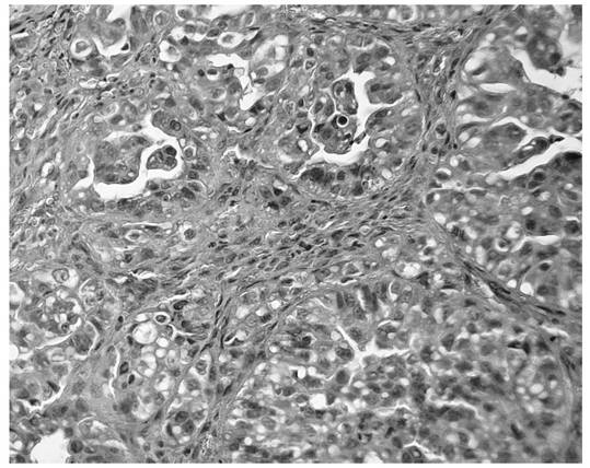
The resected tissue, which was 2.0x1.3x1.0cm, was macroscopically analyzed. It had a soft surface when cut, an irregular shape and a whitish-brown color; the tissue was stored in a formaldehyde solution (10%) for subsequent histopathological examination (Figure 1D). The histopathological analysis revealed a malignant neoplasm that was characterized by epithelial cell proliferation, high-grade pleomorphism and numerous mitotic figures and that had hemorrhagic foci, which indicated a poorly differentiated neoplasm with probable epithelial origin, thus suggesting the presence of a carcinoma (Figure 2).

Photomicrograph of poorly differentiated carcinoma in a dog, forming tubular blanks cuboidal cells with moderate pleomorphism and hyperstained cores. H.E. 400x
Due to the severe dyspnea, an abdominal ultrasound was performed after the endoscopy when the breathing pattern was normal; however, no alterations suggesting that other organs were affected were observed.
Because it was impossible to perform radiotherapy, adjuvant chemotherapy was indicated; however, due to the owner's decision, chemotherapy was not performed. Approximately 40 days after the endoscopic procedure, the patient presented with hind limb paralysis and severe dyspnea. Thoracic and lumbar spine radiography revealed miliary lung lesions and spinous process irregularity with bone rarefaction of the dorsal compartment of the sixth lumbar vertebra, which suggested pulmonary and lumbar spine metastasis. Euthanasia was performed due to the clinical condition of the patient, but the owner did not authorize the performance of a necropsy to confirm the metastasis.
Although laryngeal carcinomas are common in humans mainly due to habits such as alcohol and tobacco use, there are few reports in the veterinary literature of such neoplasms in dogs. The laryngeal neoplasms reported in dogs were rhabdomyomas, extramedullaryplasmacytomas, chondrosarcomas, carcinomas, fibrosarcomas and mastocytomas (MEUTEN et al., 1985MEUTEN, D.J. et al. Canine laryngeal rhabdomyoma. Veterinary Pathology, v.22, p.533-539, 1985. Available from: <http://vet.sagepub.com/content/22/6/533.short>. Accessed: Ago. 01, 2013. doi: 10.1177/030098588502200605.
http://vet.sagepub.com/content/22/6/533....
; HAYES et al., 2007HAYES, A.M. et al. Solitary extramedullary plasmacytoma of the canine larynx. Journal of Small Animal Practice, v.48, p.288291, 2007. Available from: <http://onlinelibrary.wiley.com/doi/10.1111/j.17485827.2006.00265.x/abstract;jsessionid=F4DD943A1374A8BD29A256060D355A97.d03t01?deniedAccessCustomisedMessage=&userIsAuthenticated=false>. Accessed: Ago. 01, 2013. doi:10.1111/j.1748-5827.2006.00265.x.
http://onlinelibrary.wiley.com/doi/10.11...
; MACPHAIL, 2014MACPHAIL, C. Laryngeal disease in dogs and cats. Veterinary Clinics of North America: Small Animal Practice, v.44, n.2, p.19-31, 2014. Available from: <http://www.sciencedirect.com/science/article/pii/S0195561613001812>. Accessed: Feb. 08, 2014. doi: 10.1016/j.cvsm.2013.09.001.
http://www.sciencedirect.com/science/art...
). Except for the rhabdomyomas, most of these neoplasms are locally invasive and have a high metastatic potential (WITHROW, 2013WITHROW, S.J. Tumors of the respiratory system. In: _____, S.J. et al. Withrow and MacEwen's small animal clinical oncology. St. Louis: Saunders Elsevier, 2013. p.451-453.).
In the present study, there were no signs of metastasis to the lung at the diagnosis, which was confirmed by radiography. Furthermore, there was no submandibular lymphadenomegaly, and the cytological analysis indicated that there was no neoplastic invasion, although no histopathological examination was performed, which is considered to be the gold standard for lymph node metastasis detection. In humans, 60% of laryngeal carcinomas are localized, 30% exhibit satellite lymph node metastasis, and 10% exhibit distant metastasis (MENDENHALL et al., 2005MENDENHALL, W.M. et al. Treatment of head and neck cancers. In: DEVITA, H. et al. Cancer: principles and practices of oncology. Philadelphia: Lippincott-Raven, 2005. p.662-732.).
The most appropriate treatment for benign laryngeal tumors is resection of the neoplasm and the submucosa or a partial laryngectomy. Aggressive surgical intervention involves complete laryngectomy with permanent tracheostomy but has been reported only in isolated cases (MACPHAIL, 2014MACPHAIL, C. Laryngeal disease in dogs and cats. Veterinary Clinics of North America: Small Animal Practice, v.44, n.2, p.19-31, 2014. Available from: <http://www.sciencedirect.com/science/article/pii/S0195561613001812>. Accessed: Feb. 08, 2014. doi: 10.1016/j.cvsm.2013.09.001.
http://www.sciencedirect.com/science/art...
). However, partial and total laryngectomies are associated with several postoperative complications that make their use more difficult. Aspiration pneumonia may occur due to the inappropriate closing of the larynx during swallowing. In addition, intermittent and excessive coughing due to the production of granulation tissue where the tumor was excised, dysphagia, nausea and fistula secondary to wound dehiscence may occur (FOSSUM & ROGERS, 1998MACPHAIL, C. Laryngeal disease in dogs and cats. Veterinary Clinics of North America: Small Animal Practice, v.44, n.2, p.19-31, 2014. Available from: <http://www.sciencedirect.com/science/article/pii/S0195561613001812>. Accessed: Feb. 08, 2014. doi: 10.1016/j.cvsm.2013.09.001.
http://www.sciencedirect.com/science/art...
; MACPHAIL, 2014MACPHAIL, C. Laryngeal disease in dogs and cats. Veterinary Clinics of North America: Small Animal Practice, v.44, n.2, p.19-31, 2014. Available from: <http://www.sciencedirect.com/science/article/pii/S0195561613001812>. Accessed: Feb. 08, 2014. doi: 10.1016/j.cvsm.2013.09.001.
http://www.sciencedirect.com/science/art...
).
In contrast, the endoscopic approach allowed the visualization of the lesion in situ with a high magnification of the image (Figure 1B), which facilitated the surgical removal of the mass with videosurgery. The removal was accomplished with minimal manipulation of the affected area and without the need to resect anatomical structures to access the tumor, thus minimizing the postoperative complications, which resulted in a safer and faster recovery for the patient (Figure 1C) (MARTINS et al., 2009MARTINS, L.G.A. et al. Biópsia do miocárdio em cães: acesso minimamente invasivo por cirurgia torácica vídeo-assistida. Arquivos Brasileiros de Medicina Veterinária e Zootecnia, v.61, p.1275-1280, 2009. Available from: <http://bases.bireme.br/cgi-bin/wxislind.exe/iah/online/?IsisScript=iah/iah.xis&src=google&base=LILACS&lang=p&nextAction=lnk&exprSearch=537251&indexSearch=ID>. Accessed: Ago. 01, 2013.
http://bases.bireme.br/cgi-bin/wxislind....
; OLIVIERI et al., 2009OLIVIERI, M. et al. Video-assisted left partial arytenoidectomy by diode laser photoablation for treatment of canine laryngeal paralysis. Veterinary Surgery, v.38, p.439-444, 2009. Available from: <http://onlinelibrary.wiley.com/doi/10.1111/j.1532-950X.2009.00546.x/abstract?deniedAccessCustomisedMessage=&userIsAuthenticated=false>. Accessed: Ago. 01, 2013. doi: 10.1111/j.1532-950X.2009.00546.x.
http://onlinelibrary.wiley.com/doi/10.11...
). Concerns regarding the use of the endoscopic procedure are related to the need of appropriate materials and difficulty to achieve free margins of the neoplasm (OLIVIERI et al., 2009OLIVIERI, M. et al. Video-assisted left partial arytenoidectomy by diode laser photoablation for treatment of canine laryngeal paralysis. Veterinary Surgery, v.38, p.439-444, 2009. Available from: <http://onlinelibrary.wiley.com/doi/10.1111/j.1532-950X.2009.00546.x/abstract?deniedAccessCustomisedMessage=&userIsAuthenticated=false>. Accessed: Ago. 01, 2013. doi: 10.1111/j.1532-950X.2009.00546.x.
http://onlinelibrary.wiley.com/doi/10.11...
; MENDENHALL et al., 2005MENDENHALL, W.M. et al. Treatment of head and neck cancers. In: DEVITA, H. et al. Cancer: principles and practices of oncology. Philadelphia: Lippincott-Raven, 2005. p.662-732.; CLIFFORD & SORENMO, 2004CLIFFORD, C.A.; SORENMO, K.U. Tumors of the larynx and trachea. In: KING, L.G. Textbook of respiratory disease in dogs and cats. St. Louis: Saunders, 2004. p.339-345.).
In veterinary medicine, there are no reports of endoscopic treatments in cases of laryngeal tumors, but it is a resource, together with electrosurgery which has been shown effective for the treatment of this condition. In humans the use of transoral laser surgery performed by laryngoscopy has been developing in recent years. This approach does not follow one of the basic principles of oncologic surgery because the tumor is segmented and then removed in pieces in some cases. However, the cutting part reveals the depth of penetration of the tumor and allows a clear view of the resection margins during surgery (RODRIGO et al., 2011RODRIGO, J.P. et al. The current role of partial surgery as a strategy for functional preservation in laryngeal carcinoma. Acta Otorrinolaringologica Espanola, v.62, p.231-238, 2011. Available from: <http://www.sciencedirect.com/science/article/pii/S2173573511000135>. Accessed: Feb. 08, 2014. doi: 10.1016/j.otoeng.2010.06.002.
http://www.sciencedirect.com/science/art...
).
The primary clinical symptoms in patients with laryngeal tumors are due to the physical presence of the mass, which obstructs the airway as it grows, or due to the accumulation of secretions in the airway (SAIK et al., 1986SAIK, J.E. et al. Canine and feline laryngeal neoplasia: a 10-year survey. Journal of American Animal Hospital Association, v.22, p.359-365, 1986.). Thus, the immediate improvement of the patient's respiratory distress after the removal of the mass through videosurgery (during the postsurgical period) is explained. In some cases, in addition to the removal of the neoplasm, there is the possibility of performing radiotherapy as a way to locally control the disease when dealing with a radiosensitive tumor (WITHROW, 2013WITHROW, S.J. Tumors of the respiratory system. In: _____, S.J. et al. Withrow and MacEwen's small animal clinical oncology. St. Louis: Saunders Elsevier, 2013. p.451-453.), but this treatment is still difficult to access in Brazil.
In this case, treatment using videosurgery was shown to be efficient and minimally invasive when treating a laryngeal neoplasm, providing an important improvement to the patient in the immediate postsurgical period.
REFERENCES
- CLIFFORD, C.A.; SORENMO, K.U. Tumors of the larynx and trachea. In: KING, L.G. Textbook of respiratory disease in dogs and cats. St. Louis: Saunders, 2004. p.339-345.
- DUNBAR, M.D. et al. Laryngeal rhabdomyoma in a dog. Veterinary Clinical Pathology, v.41, p.590-593, 2012. Available from: <http://vet.sagepub.com/content/22/6/533.full.pdf>. Accessed: Feb. 09, 2014. doi: 10.1177/030098588502200605.
» https://doi.org/10.1177/030098588502200605» http://vet.sagepub.com/content/22/6/533.full.pdf - FOSSUM, T.W.; ROGERS, K.S. Sistema respiratório: oncologia [Respiratory system: oncology]. In: SLATTER, D. Manual de cirurgia de pequenos animais. São Paulo: Manole, 1998. V.2, p.2635-2641.
- HAYES, A.M. et al. Solitary extramedullary plasmacytoma of the canine larynx. Journal of Small Animal Practice, v.48, p.288291, 2007. Available from: <http://onlinelibrary.wiley.com/doi/10.1111/j.17485827.2006.00265.x/abstract;jsessionid=F4DD943A1374A8BD29A256060D355A97.d03t01?deniedAccessCustomisedMessage=&userIsAuthenticated=false>. Accessed: Ago. 01, 2013. doi:10.1111/j.1748-5827.2006.00265.x.
» https://doi.org/10.1111/j.1748-5827.2006.00265.x» http://onlinelibrary.wiley.com/doi/10.1111/j.17485827.2006.00265.x/abstract;jsessionid=F4DD943A1374A8BD29A256060D355A97.d03t01?deniedAccessCustomisedMessage=&userIsAuthenticated=false - MACPHAIL, C. Laryngeal disease in dogs and cats. Veterinary Clinics of North America: Small Animal Practice, v.44, n.2, p.19-31, 2014. Available from: <http://www.sciencedirect.com/science/article/pii/S0195561613001812>. Accessed: Feb. 08, 2014. doi: 10.1016/j.cvsm.2013.09.001.
» https://doi.org/10.1016/j.cvsm.2013.09.001» http://www.sciencedirect.com/science/article/pii/S0195561613001812 - MARTINS, L.G.A. et al. Biópsia do miocárdio em cães: acesso minimamente invasivo por cirurgia torácica vídeo-assistida. Arquivos Brasileiros de Medicina Veterinária e Zootecnia, v.61, p.1275-1280, 2009. Available from: <http://bases.bireme.br/cgi-bin/wxislind.exe/iah/online/?IsisScript=iah/iah.xis&src=google&base=LILACS&lang=p&nextAction=lnk&exprSearch=537251&indexSearch=ID>. Accessed: Ago. 01, 2013.
» http://bases.bireme.br/cgi-bin/wxislind.exe/iah/online/?IsisScript=iah/iah.xis&src=google&base=LILACS&lang=p&nextAction=lnk&exprSearch=537251&indexSearch=ID - MENDENHALL, W.M. et al. Treatment of head and neck cancers. In: DEVITA, H. et al. Cancer: principles and practices of oncology. Philadelphia: Lippincott-Raven, 2005. p.662-732.
- MEUTEN, D.J. et al. Canine laryngeal rhabdomyoma. Veterinary Pathology, v.22, p.533-539, 1985. Available from: <http://vet.sagepub.com/content/22/6/533.short>. Accessed: Ago. 01, 2013. doi: 10.1177/030098588502200605.
» https://doi.org/10.1177/030098588502200605» http://vet.sagepub.com/content/22/6/533.short - OLIVIERI, M. et al. Video-assisted left partial arytenoidectomy by diode laser photoablation for treatment of canine laryngeal paralysis. Veterinary Surgery, v.38, p.439-444, 2009. Available from: <http://onlinelibrary.wiley.com/doi/10.1111/j.1532-950X.2009.00546.x/abstract?deniedAccessCustomisedMessage=&userIsAuthenticated=false>. Accessed: Ago. 01, 2013. doi: 10.1111/j.1532-950X.2009.00546.x.
» https://doi.org/10.1111/j.1532-950X.2009.00546.x» http://onlinelibrary.wiley.com/doi/10.1111/j.1532-950X.2009.00546.x/abstract?deniedAccessCustomisedMessage=&userIsAuthenticated=false - RODRIGO, J.P. et al. The current role of partial surgery as a strategy for functional preservation in laryngeal carcinoma. Acta Otorrinolaringologica Espanola, v.62, p.231-238, 2011. Available from: <http://www.sciencedirect.com/science/article/pii/S2173573511000135>. Accessed: Feb. 08, 2014. doi: 10.1016/j.otoeng.2010.06.002.
» https://doi.org/10.1016/j.otoeng.2010.06.002» http://www.sciencedirect.com/science/article/pii/S2173573511000135 - SAIK, J.E. et al. Canine and feline laryngeal neoplasia: a 10-year survey. Journal of American Animal Hospital Association, v.22, p.359-365, 1986.
- WITHROW, S.J. Tumors of the respiratory system. In: _____, S.J. et al. Withrow and MacEwen's small animal clinical oncology. St. Louis: Saunders Elsevier, 2013. p.451-453.
-
PROTOCOL COMMITTEE OF ETHICS 017022/13.
Publication Dates
-
Publication in this collection
09 Sept 2014 -
Date of issue
Jan 2015
History
-
Received
10 Aug 2013 -
Accepted
14 Mar 2014