ABSTRACT:
We described a case of fatal septicemic yersiniosis in a young adult brown titi monkey (Plecturocebus brunneus) which presented lethargy and severe anemia. Postmortem external assessment revealed marked dehydration and pale pink mucous membranes. The main gross findings included enlarged liver with yellow pinpoints, enlarged spleen with yellow nodules, mucosal ulcerations in the large intestine, enlarged mesenteric lymph nodes, and pulmonary hemorrhage. Histology revealed necrosuppurative hepatosplenitis with intralesional colonies of rod-shaped gram-negative bacteria, ulcerative colitis, reactive lymphoid hyperplasia, and fibrinous and hemorrhagic pneumonia. Bacterial culture and identification using matrix-assisted laser desorption/ionization time-of-flight mass spectrometry confirmed the diagnosis of yersiniosis by Yersinia enterocolitica. This study indicated that yersiniosis should be considered as a differential diagnosis of death in brown titi monkeys.
Key words:
colitis; hepatitis; splenitis; nonhuman primate
RESUMO:
Descrevemos um caso de yersiniose septicêmica fatal em um zogue-zogue (Plecturocebus brunneus) jovem adulto que apresentava um quadro de letargia e anemia severa. Macroscopicamente, havia acentuada desidratação e as mucosas estavam pálidas. Notou-se hepatomegalia com múltiplos pontos amarelos e esplenomegalia com múltiplos nódulos amarelos pelo parênquima. Ainda, ulcerações da mucosa do intestino grosso, linfonodos mesentéricos aumentados e hemorragia pulmonar foram observados. A avaliação histológica revelou hepatite e esplenite necrossupurativas associadas a agregados bacterianos bacilares gram-negativos intralesionais, colite ulcerativa, hiperplasia linfoide reativa e pneumonia fibrino-hemorrágica. A cultura bacteriana e identificação através do método de espectrometria de massa por ionização e dessorção a laser assistida por matriz associada ao tempo de voo confirmou o diagnóstico de yersiniose por Yersinia enterocolitica. Este estudo demonstra que a yersiniose deve ser considerada como um diagnóstico diferencial de causa de morte em zogue-zogues.
Palavras-chave:
colite; hepatite; esplenite; primata não-humano
Yersiniosis is a zoonotic disease caused by a gram-negative enteric bacterium of the genus Yersinia (MÄTZ-RENSING & LOWENSTINE, 2018MÄTZ-RENSING K.; LOWENSTINE, L.J. New World and Old World Monkeys. In: TERIO, K.A. et al. Pathology of Wildlife and Zoo Animals. London: Elsevier Academic Press, 2018. Chap.14, p.343-373.). Enteric species of this genus have been isolated from several animals, including mammals, birds, and reptiles; although, not necessarily associated with clinical disease (SHAYEGANI et al., 1986SHAYEGANI, M. et al. Yersinia enterocolitica and related species isolated from wildlife in New York State. Applied and Environmental Microbiology, v.52, n.3, p.420-424, 1986. Available from: <Available from: https://journals.asm.org/doi/abs/10.1128/aem.52.3.420-424.1986 >. Accessed: Nov. 13, 2021. doi: 10.1128/aem.52.3.420-424.1986.
https://journals.asm.org/doi/abs/10.1128...
; KWAGA & IVERSEN, 1993KWAGA, J.; IVERSEN, J. O. Isolation of Yersinia enterocolitica (0: 5, 27 biotype 2) from a common garter snake. Journal of Wildlife Diseases, v.29, n.1, p.127-129, 1993. Available from: <Available from: https://meridian.allenpress.com/jwd/article/29/1/127/120967/Isolation-of-Yersinia-enterocolitica-0-5-27 >. Accessed: Nov. 15, 2021. doi: 10.7589/0090-3558-29.1.127.
https://meridian.allenpress.com/jwd/arti...
). Yersinia enterocolitica and Yersinia pseudotuberculosis infections have been reported in several nonhuman primates (NHP) species, e.g., gibbons, lemurs, animals of the genus Chlorocebus, marmosets, and squirrel monkeys (POELMA et al., 1977POELMA, F. G. et al. Yersinia enterocolitica infections in non-human primates. Acta Zoologica et Pathologica Antverpiensia, n.69, p.3-9, 1977. Available from: <Available from: https://pubmed.ncbi.nlm.nih.gov/418633/ >. Accessed: Nov. 13, 2021.
https://pubmed.ncbi.nlm.nih.gov/418633/...
; TAFFS & DUNN, 1983TAFFS, L. F.; DUNN, G. An outbreak of Yersinia pseudotuberculosis infection in a small indoor breeding colony of red-bellied (Saguinus labiatus) tamarins. Laboratory Animals, v.17, n.4, p.311-320, 1983. Available from: <Available from: https://journals.sagepub.com/doi/abs/10.1258/002367783781062280 >. Accessed: Nov. 13, 2021. doi: 10.1258/002367783781062280.
https://journals.sagepub.com/doi/abs/10....
; BRESNAHAN et al., 1984BRESNAHAN, J. F. et al. Yersinia enterocolitica infection in breeding colonies of ruffed lemurs. Journal of the American Veterinary Medical Association, v.185, n.11, p.1354-1356, 1984. Available from: <Available from: https://pubmed.ncbi.nlm.nih.gov/6511581/ >. Accessed: Nov. 15, 2021.
https://pubmed.ncbi.nlm.nih.gov/6511581/...
; BAKKER et al., 2007BAKKER, J. et al. A report on Yersinia-related mortality in a colony of New World Monkeys. Laboratory Primate Newsletter, v.46, p.11-16, 2007. Available from: <Available from: https://www.brown.edu/Research/Primate/lpn46-3.pdf#page=13 >. Accessed: Nov. 15, 2021.
https://www.brown.edu/Research/Primate/l...
; HABLOLVARID et al., 2008HABLOLVARID, M. H. et al. Yersinia enterocolitica infection in a vervet monkey (Cercopithecus aethiops) in Iran. Archives of Razi Institute, v.63, n.1, p.53-57, 2008. Available from: <Available from: https://archrazi.areeo.ac.ir/article_103752_0.html >. Accessed: Nov. 13, 2021. doi: 10.22092/ARI.2008.103752.
https://archrazi.areeo.ac.ir/article_103...
; NAKAMURA et al., 2010NAKAMURA, S. et al. Pathological changes in captive monkeys with spontaneous yersiniosis due to infection by Yersinia enterocolitica serovar O8. Journal of Comparative Pathology, v.143, n.2-3, p.150-156, 2010. Available from: <Available from: https://www.sciencedirect.com/science/article/pii/S0021997510000253?via%3Dihub >. Accessed: Nov. 15, 2021. doi: 10.1016/j.jcpa.2010.01.017.
https://www.sciencedirect.com/science/ar...
; SOTO et al., 2013SOTO, E. et al. An outbreak of Yersinia enterocolitica in a captive colony of African green monkeys (Chlorocebus aethiops sabaeus) in the Caribbean. Comparative Medicine, v.63, n.5, p.439-444, 2013. Available from: <Available from: https://www.ncbi.nlm.nih.gov/pmc/articles/PMC3796755/ >. Accessed: Nov. 13, 2021.
https://www.ncbi.nlm.nih.gov/pmc/article...
; LEMOS et al., 2021LEMOS, G. A. A. et al. Spontaneous outbreak of Yersinia enterocolitica infection and co-infection with Escherichia coli in black-tufted marmosets (Callithrix penicillata). Brazilian Journal of Veterinary Pathology, v.14, n.3, p.173-179, 2021. Available from: <Available from: https://bjvp.org.br/wp-content/uploads/2021/11/v14-n3-5.pdf >. Accessed: Nov. 21, 2021. doi: 10.24070/bjvp.1983-0246.v14i3p173-179.
https://bjvp.org.br/wp-content/uploads/2...
). Wild birds and rodents act as reservoirs of the etiological agent and the main route of infection is fecal-oral (MAIR, 1973MAIR, N. S. Yersiniosis in wildlife and its public health implications. Journal of Wildlife Diseases, v.9, n.1, p.64-71, 1973. Available from: <Available from: https://meridian.allenpress.com/jwd/article/9/1/64/73625/YERSINIOSIS-IN-WILDLIFE-AND-ITS-PUBLIC-HEALTH/ >. Accessed: Nov. 15, 2021. doi: 10.7589/0090-3558-9.1.64.
https://meridian.allenpress.com/jwd/arti...
; BOTTONE, 1999BOTTONE, E. J. Yersinia enterocolitica: overview and epidemiologic correlates. Microbes and Infection, v.1, n.4, p.323-333, 1999. Available from: <Available from: https://pubmed.ncbi.nlm.nih.gov/10602666/ >. Accessed: Nov. 13, 2021. doi: 10.1016/s1286-4579(99)80028-8.
https://pubmed.ncbi.nlm.nih.gov/10602666...
; MÄTZ-RENSING & LOWENSTINE, 2018). In NHP, the infection usually starts as ulcerative enterocolitis that often progresses to systemic disease (NAKAMURA et al., 2010; MÄTZ-RENSING & LOWENSTINE, 2018). However, depending on the affected species, a wide range of clinical presentations may be observed (SIMMONS & GIBSON, 2012SIMMONS, J.; GIBSON, S. Bacterial and Mycotic Diseases of Nonhuman Primates. In: ABEE, C.R. et al. Nonhuman Primates in Biomedical Research: Biology and Management. Academic Press. Chap.2, p.105-172, 2012.). Also, proximity between captive NHP and humans can increase the risk for yersiniosis zoonotic potential (BURGOS-RODRIGUEZ, 2011BURGOS-RODRIGUEZ, A. G. Zoonotic diseases of primates. Veterinary Clinics: Exotic Animal Practice, v.14, n.3, p.557-575, 2011. Available from: <Available from: https://www.vetexotic.theclinics.com/article/S1094-9194(11)00046-6/fulltext >. Accessed: Feb. 04, 2022. doi: 10.1016/j.cvex.2011.05.006.
https://www.vetexotic.theclinics.com/art...
). These factors highlighted the importance of species-specific knowledge about the disease. We now describe the clinical, hematological, pathological, and bacteriological findings of fatal yersiniosis by Y. enterocolitica in a captive brown titi monkey (Plecturocebus brunneus).
A 5-year-old (estimated age), male brown titi monkey, weighing 0.72 kg, was clinically assessed for a history of lethargy with 8 days of duration. The brown titi monkey was kept in a wildlife conservation institution located in Morro Reuter, metropolitan mesoregion of Porto Alegre, Southern Brazil (29º32’17”S 51º04’51”W). Four years prior to this, the animal had been seized from illegal trading in the state of Rondônia, Northern Brazil. In the wildlife conservation institution, the brown titi monkey was housed in an indoor-outdoor, two crimped wire mesh enclosure with a currently healthy individual of the genus Callicebus. Also, the institution maintains other animals (mammals, including other primates, birds, and reptiles) in nearby enclosures with similar diets.
Complete blood count (CBC) was performed using an automatic blood analyzer Procyte Dx (IDEXX®), and severe anemia was observed (red blood cells, 1.57 x 10¹²/L; reference range [RR], 3.7 ± 8.4 x 10¹²/L; hematocrit, 10%; [RR], 28 ± 61%; hemoglobin, 2.5 g/dL; [RR], 9.7 ± 19.2 g/dL) (ISIS, 2002INTERNATIONAL SPECIES INFORMATION SYSTEM (ISIS). Callicebus moloch physiological reference ranges. Apple Valley, 2002.). Schistocytes and acanthocytes were observed. The leukogram had no quantitative abnormalities; however, neutrophils had toxic characteristics (i.e., chromatin degeneration, and cytoplasmic vacuolation). Biochemical results were within RR for the evaluated markers (alanine aminotransferase, albumin, alkaline phosphatase, and creatinine). The brown titi monkey underwent a blood transfusion procedure using whole blood; however, the animal had a cardiac arrest during the blood transfusion and died. The primate was referred for a postmortem examination at the Department of Veterinary Pathology of Universidade Federal do Rio Grande do Sul.
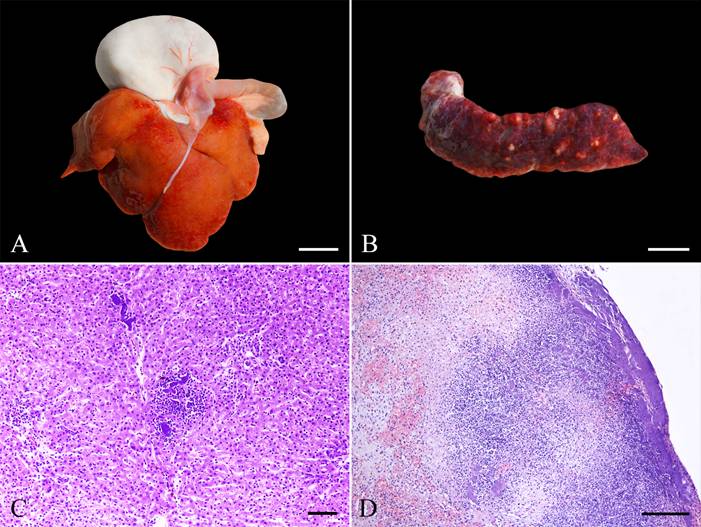
At gross examination, marked dehydration and pale pink mucous membranes were observed. There was mild hepatomegaly with multifocal hemorrhage and multiple yellowish pinpoints (<1 mm in diameter) in the hepatic parenchyma (Figure 1A) and marked splenomegaly with multiple yellowish nodules (1 to 3 mm in diameter) in the splenic parenchyma (Figure 1B). The mucosal surface of the colon had scattered blackish areas with a yellowish center (1 to 3 mm in diameter), and mesenteric lymph nodes were mildly enlarged. Additionally, the lung had multifocal dark-red areas. No gross lesions were observed in other organs. Tissue samples of the main organs of the thoracic and abdominal cavities and the brain were collected, fixed in 10% neutral buffered formalin, routinely processed for histopathological evaluation, and stained with hematoxylin and eosin (HE). Selected sections of liver and spleen were stained with Gram stain.

Pathological findings of yersiniosis in a brown titi monkey. (A) Liver with hepatomegaly, multifocal hemorrhage, and multiple yellowish pinpoints throughout the hepatic surface. Bar, 2 cm. (B) Spleen presenting splenomegaly and multiple yellowish nodules throughout the splenic surface. Bar, 1 cm. (C) Photomicrograph of the liver with hepatic necrosis associated with neutrophil infiltration and numerous intralesional colonies of bacteria. HE. Bar, 200 µm. (D) Photomicrograph of the spleen showing necrosis admixed with neutrophil infiltration, hemorrhage, and numerous intralesional colonies of bacteria. HE. Bar, 400 µm.
Histology revealed marked, random necrotizing and suppurative hepatitis (Figure 1C) and splenitis (Figure 1D) associated with intralesional colonies of rod-shaped gram-negative bacteria, fibrin deposition, and multifocal hemorrhage. Multifocal thrombosis was observed in the lumen of spleen vessels. In the colon mucosa, there were areas of moderate multifocal ulceration accompanied by necrosis and infiltration of inflammatory cells (neutrophils, lymphocytes, plasma cells, and macrophages). Numerous blood vessels in the colon mucosa and submucosa had mural inflammatory infiltration of degenerate neutrophils, with necrotic debris and fibrin deposition (vasculitis). The medullary sinuses of mesenteric lymph nodes were expanded by moderate edema and numerous macrophages. In the lung, there were multifocal areas of moderate deposition of fibrin, macrophages, and hemorrhage within alveolar spaces, and vasculitis. Similar colonies of rod-shaped bacteria were observed in the lumen of numerous blood vessels in the lung parenchyma.
Fresh samples of liver and spleen collected during the postmortem examination were submitted for bacterial culture. The samples were inoculated in Mueller Hinton agar (Kasvi®, Brazil) supplemented with 5% sheep blood and MacConkey agar (Kasvi®, Brazil) plates and incubated for 48 hours at 37 °C. The bacteria, isolated in pure cultures from the liver and spleen; were subsequently, identified through matrix-assisted laser desorption/ionization time-of-flight mass spectrometry (MALDI-TOF) Microflex Biotyper 4.0 (Bruker Daltonics, Bremen, Germany) as Yersinia enterocolitica, with a score of 2.33. Therefore, based on the clinical, pathological, and microbiological findings, a diagnosis of yersiniosis by Y. enterocolitica was made.
In NHP, acute septicemic yersiniosis is more commonly observed than chronic cases. A triad of lesions in the gastrointestinal tract, liver, and spleen characterizes the disease (MÄTZ-RENSING & LOWENSTINE, 2018MÄTZ-RENSING K.; LOWENSTINE, L.J. New World and Old World Monkeys. In: TERIO, K.A. et al. Pathology of Wildlife and Zoo Animals. London: Elsevier Academic Press, 2018. Chap.14, p.343-373.). The pathogenesis of these triad lesions is directly related to the main route of infection, the fecal-oral route (BOTTONE, 1999BOTTONE, E. J. Yersinia enterocolitica: overview and epidemiologic correlates. Microbes and Infection, v.1, n.4, p.323-333, 1999. Available from: <Available from: https://pubmed.ncbi.nlm.nih.gov/10602666/ >. Accessed: Nov. 13, 2021. doi: 10.1016/s1286-4579(99)80028-8.
https://pubmed.ncbi.nlm.nih.gov/10602666...
; MÄTZ-RENSING & LOWENSTINE, 2018). Once Y. enterocolitica reaches the intestinal lumen and penetrates the epithelial barrier, its replication occurs in reticuloendothelial cells, followed by systemic dissemination through blood and/or lymph (FINLAY & FALKOW, 1988FINLAY, B. B.; FALKOW, S. Comparison of the invasion strategies used by Salmonella cholerae-suis, Shigella flexneri and Yersinia enterocolitica to enter cultured animal cells: endosome acidification is not required for bacterial invasion or intracellular replication. Biochimie, v.70, n.8, p.1089-1099, 1988. Available from: <Available from: https://www.sciencedirect.com/science/article/pii/0300908488902714 >. Accessed: Nov. 15, 2021. doi: 10.1016/0300-9084(88)90271-4.
https://www.sciencedirect.com/science/ar...
; HANDLEY et al., 2005HANDLEY, S. A. et al. Yersinia enterocolitica invasin-dependent and invasin-independent mechanisms of systemic dissemination. Infection and Immunity, v.73, n.12, p.8453-8455, 2005. Available from: <Available from: https://www.ncbi.nlm.nih.gov/pmc/articles/PMC1307041/ >. Accessed: Nov. 15, 2021. doi: 10.1128/IAI.73.12.8453-8455.2005.
https://www.ncbi.nlm.nih.gov/pmc/article...
; NAKAMURA et al., 2010NAKAMURA, S. et al. Pathological changes in captive monkeys with spontaneous yersiniosis due to infection by Yersinia enterocolitica serovar O8. Journal of Comparative Pathology, v.143, n.2-3, p.150-156, 2010. Available from: <Available from: https://www.sciencedirect.com/science/article/pii/S0021997510000253?via%3Dihub >. Accessed: Nov. 15, 2021. doi: 10.1016/j.jcpa.2010.01.017.
https://www.sciencedirect.com/science/ar...
; MÄTZ-RENSING & LOWENSTINE, 2018).
Clinical presentation of acute cases of yersiniosis is largely variable across different NHP species. Affected animals can die without clinical signs or may have diarrhea, lethargy, and/or dehydration (POELMA et al., 1977POELMA, F. G. et al. Yersinia enterocolitica infections in non-human primates. Acta Zoologica et Pathologica Antverpiensia, n.69, p.3-9, 1977. Available from: <Available from: https://pubmed.ncbi.nlm.nih.gov/418633/ >. Accessed: Nov. 13, 2021.
https://pubmed.ncbi.nlm.nih.gov/418633/...
; BAKKER et al., 2007BAKKER, J. et al. A report on Yersinia-related mortality in a colony of New World Monkeys. Laboratory Primate Newsletter, v.46, p.11-16, 2007. Available from: <Available from: https://www.brown.edu/Research/Primate/lpn46-3.pdf#page=13 >. Accessed: Nov. 15, 2021.
https://www.brown.edu/Research/Primate/l...
; HABLOLVARID et al., 2008HABLOLVARID, M. H. et al. Yersinia enterocolitica infection in a vervet monkey (Cercopithecus aethiops) in Iran. Archives of Razi Institute, v.63, n.1, p.53-57, 2008. Available from: <Available from: https://archrazi.areeo.ac.ir/article_103752_0.html >. Accessed: Nov. 13, 2021. doi: 10.22092/ARI.2008.103752.
https://archrazi.areeo.ac.ir/article_103...
; NAKAMURA et al., 2010NAKAMURA, S. et al. Pathological changes in captive monkeys with spontaneous yersiniosis due to infection by Yersinia enterocolitica serovar O8. Journal of Comparative Pathology, v.143, n.2-3, p.150-156, 2010. Available from: <Available from: https://www.sciencedirect.com/science/article/pii/S0021997510000253?via%3Dihub >. Accessed: Nov. 15, 2021. doi: 10.1016/j.jcpa.2010.01.017.
https://www.sciencedirect.com/science/ar...
; SOTO et al., 2013SOTO, E. et al. An outbreak of Yersinia enterocolitica in a captive colony of African green monkeys (Chlorocebus aethiops sabaeus) in the Caribbean. Comparative Medicine, v.63, n.5, p.439-444, 2013. Available from: <Available from: https://www.ncbi.nlm.nih.gov/pmc/articles/PMC3796755/ >. Accessed: Nov. 13, 2021.
https://www.ncbi.nlm.nih.gov/pmc/article...
; LEMOS et al., 2021LEMOS, G. A. A. et al. Spontaneous outbreak of Yersinia enterocolitica infection and co-infection with Escherichia coli in black-tufted marmosets (Callithrix penicillata). Brazilian Journal of Veterinary Pathology, v.14, n.3, p.173-179, 2021. Available from: <Available from: https://bjvp.org.br/wp-content/uploads/2021/11/v14-n3-5.pdf >. Accessed: Nov. 21, 2021. doi: 10.24070/bjvp.1983-0246.v14i3p173-179.
https://bjvp.org.br/wp-content/uploads/2...
). Lethargy was the only clinical sign seen in our case. The hematological alterations can range from leukocytosis with neutrophilia to leukopenia (SIMMONS & GIBSON, 2012SIMMONS, J.; GIBSON, S. Bacterial and Mycotic Diseases of Nonhuman Primates. In: ABEE, C.R. et al. Nonhuman Primates in Biomedical Research: Biology and Management. Academic Press. Chap.2, p.105-172, 2012.). In the present case, we identified toxic changes in neutrophils, and anemia with morphological abnormalities in erythrocytes. Bacterial infections, particularly those involving the production of bacterial leukotoxins, are associated with the most pronounced toxic changes in neutrophils and reflect accelerated or stress granulopoiesis (VALLI et al., 2016VALLI, V. E. O. et al. Hematopoietic System. In: MAXIE, M.G. Jubb, Kennedy and Palmer’s Pathology of Domestic Animals. 6.ed. St. Louis: Elsevier Publisher, 2016, v.3. Chap. 2, p. 102-268.). Schistocytes and acanthocytes observed among normal erythrocytes demonstrate the occurrence of physical damage (e.g., through disseminated intravascular coagulation, vasculitis) to red blood cells in vessels with turbulent blood flow, which may lead to anemia from mechanical trauma (VALLI et al., 2016). Many conditions and agents, including gram-negative bacteria, may initiate a disseminated intravascular coagulation (VALLI et al., 2016), which we suspected occurred in our case.
The disease caused by Y. enterocolitica in NHP occurs mainly in captive monkeys in outbreaks originated from a common infection source, such as contaminated food and water (IWATA et al., 2005IWATA, T. et al. Yersinia enterocolitica serovar O: 8 infection in breeding monkeys in Japan. Microbiology and Immunology, v.49, n.1, p.1-7, 2005. Available from: <Available from: https://onlinelibrary.wiley.com/doi/full/10.1111/j.1348-0421.2005.tb03630.x >. Accessed: Nov. 13, 2021. doi: 10.1111/j.1348-0421.2005.tb03630.x.
https://onlinelibrary.wiley.com/doi/full...
; BAKKER et al., 2007BAKKER, J. et al. A report on Yersinia-related mortality in a colony of New World Monkeys. Laboratory Primate Newsletter, v.46, p.11-16, 2007. Available from: <Available from: https://www.brown.edu/Research/Primate/lpn46-3.pdf#page=13 >. Accessed: Nov. 15, 2021.
https://www.brown.edu/Research/Primate/l...
; FREDRIKSSON-AHOMAA et al., 2007FREDRIKSSON-AHOMAA, M. et al. Yersiniosis in zoo marmosets (Callitrix jacchuss) caused by Yersinia enterocolitica 4/O: 3. Veterinary microbiology, v.121, n.3-4, p.363-367, 2007. Available from: <Available from: https://www.sciencedirect.com/science/article/pii/S037811350600530X?via%3Dihub >. Accessed: Nov. 13, 2021. doi: 10.1016/j.vetmic.2006.12.010.
https://www.sciencedirect.com/science/ar...
; NAKAMURA et al., 2010NAKAMURA, S. et al. Pathological changes in captive monkeys with spontaneous yersiniosis due to infection by Yersinia enterocolitica serovar O8. Journal of Comparative Pathology, v.143, n.2-3, p.150-156, 2010. Available from: <Available from: https://www.sciencedirect.com/science/article/pii/S0021997510000253?via%3Dihub >. Accessed: Nov. 15, 2021. doi: 10.1016/j.jcpa.2010.01.017.
https://www.sciencedirect.com/science/ar...
; SOTO et al., 2013SOTO, E. et al. An outbreak of Yersinia enterocolitica in a captive colony of African green monkeys (Chlorocebus aethiops sabaeus) in the Caribbean. Comparative Medicine, v.63, n.5, p.439-444, 2013. Available from: <Available from: https://www.ncbi.nlm.nih.gov/pmc/articles/PMC3796755/ >. Accessed: Nov. 13, 2021.
https://www.ncbi.nlm.nih.gov/pmc/article...
). Unfortunately, the infection source could not be determined in our case. Even though the wildlife conservation institution maintains other animals, only the brown titi monkey developed the disease. Isolated cases of yersiniosis are commonly related to factors that can lead the individual to a poor immune response, such as antibiotics and antiparasitic therapy, environmental stressors, or dietary imbalances (BAKKER et al., 2007). Thus, we suggested that this isolated case may be related to individual challenges that could be aggravated due to captivity.
Gross and histological findings of this case are similar to the current literature of yersiniosis in NHP (POELMA et al., 1977POELMA, F. G. et al. Yersinia enterocolitica infections in non-human primates. Acta Zoologica et Pathologica Antverpiensia, n.69, p.3-9, 1977. Available from: <Available from: https://pubmed.ncbi.nlm.nih.gov/418633/ >. Accessed: Nov. 13, 2021.
https://pubmed.ncbi.nlm.nih.gov/418633/...
; BAKKER et al., 2007BAKKER, J. et al. A report on Yersinia-related mortality in a colony of New World Monkeys. Laboratory Primate Newsletter, v.46, p.11-16, 2007. Available from: <Available from: https://www.brown.edu/Research/Primate/lpn46-3.pdf#page=13 >. Accessed: Nov. 15, 2021.
https://www.brown.edu/Research/Primate/l...
; HABLOLVARID et al., 2008HABLOLVARID, M. H. et al. Yersinia enterocolitica infection in a vervet monkey (Cercopithecus aethiops) in Iran. Archives of Razi Institute, v.63, n.1, p.53-57, 2008. Available from: <Available from: https://archrazi.areeo.ac.ir/article_103752_0.html >. Accessed: Nov. 13, 2021. doi: 10.22092/ARI.2008.103752.
https://archrazi.areeo.ac.ir/article_103...
; NAKAMURA et al., 2010NAKAMURA, S. et al. Pathological changes in captive monkeys with spontaneous yersiniosis due to infection by Yersinia enterocolitica serovar O8. Journal of Comparative Pathology, v.143, n.2-3, p.150-156, 2010. Available from: <Available from: https://www.sciencedirect.com/science/article/pii/S0021997510000253?via%3Dihub >. Accessed: Nov. 15, 2021. doi: 10.1016/j.jcpa.2010.01.017.
https://www.sciencedirect.com/science/ar...
; SOTO et al., 2013SOTO, E. et al. An outbreak of Yersinia enterocolitica in a captive colony of African green monkeys (Chlorocebus aethiops sabaeus) in the Caribbean. Comparative Medicine, v.63, n.5, p.439-444, 2013. Available from: <Available from: https://www.ncbi.nlm.nih.gov/pmc/articles/PMC3796755/ >. Accessed: Nov. 13, 2021.
https://www.ncbi.nlm.nih.gov/pmc/article...
; MÄTZ-RENSING & LOWENSTINE, 2018MÄTZ-RENSING K.; LOWENSTINE, L.J. New World and Old World Monkeys. In: TERIO, K.A. et al. Pathology of Wildlife and Zoo Animals. London: Elsevier Academic Press, 2018. Chap.14, p.343-373.; LEMOS et al., 2021LEMOS, G. A. A. et al. Spontaneous outbreak of Yersinia enterocolitica infection and co-infection with Escherichia coli in black-tufted marmosets (Callithrix penicillata). Brazilian Journal of Veterinary Pathology, v.14, n.3, p.173-179, 2021. Available from: <Available from: https://bjvp.org.br/wp-content/uploads/2021/11/v14-n3-5.pdf >. Accessed: Nov. 21, 2021. doi: 10.24070/bjvp.1983-0246.v14i3p173-179.
https://bjvp.org.br/wp-content/uploads/2...
). Necrosuppurative hepatosplenitis, ulcerative colitis, and reactive lymphoid hyperplasia are frequently described (BAKKER et al., 2007; NAKAMURA et al., 2010; SOTO et al., 2013). Pneumonia without the presence of necrosis or bacteria has also been described in an NHP with Y. enterocolitica infection (POELMA et al., 1977). Fibrinous and hemorrhagic pneumonia observed in this study represent a common finding observed in cases of death associated with septic shock (ACKERMANN, 2017ACKERMANN, M. R. Inflammation and Healing. In: ZACHARY J.F. Pathologic Basis of Veterinary Disease, 6.ed. St. Louis : Elsevier, 2017. Chap.3, p.73-131.).
The differential diagnosis includes other bacterial agents with similar pathological findings in NHP, such as Francisella tularensis and Y. pseudotuberculosis (MÄTZ-RENSING & LOWENSTINE, 2018MÄTZ-RENSING K.; LOWENSTINE, L.J. New World and Old World Monkeys. In: TERIO, K.A. et al. Pathology of Wildlife and Zoo Animals. London: Elsevier Academic Press, 2018. Chap.14, p.343-373.). The disease caused by F. tularensis, tularemia, cannot be differentiated from yersiniosis macroscopically; however, tularemia is characterized microscopically by pyogranulomas predominantly in the liver, spleen, respiratory tract, and lymph nodes (MÄTZ-RENSING & LOWENSTINE, 2018). For differentiation between Yersinia species, complementary tests are recommended for correct species designation (STEPHAN et al., 2011STEPHAN, R. et al. Rapid species specific identification and subtyping of Yersinia enterocolitica by MALDI-TOF mass spectrometry. Journal of Microbiological Methods, v.87, n.2, p.150-153, 2011. Available from: <Available from: https://www.sciencedirect.com/science/article/pii/S0167701211003162?via%3Dihub >. Accessed: Nov. 16, 2021. doi: 10.1016/j.mimet.2011.08.016.
https://www.sciencedirect.com/science/ar...
). In this case, these differential diagnoses were excluded based on histological and microbiological findings.
Our study described the clinical, hematological, pathological, and bacteriological features of yersiniosis in a different primate species using a modern and rapid method of identification of the etiological agent involved. This study indicated that yersiniosis should be considered as a differential diagnosis of death in brown titi monkeys.
ACKNOWLEDGEMENTS
This study was supported by the Conselho Nacional de Desenvolvimento Científico e Tecnológico (CNPq, PQ-2021 - 307277/2021-6), Pró-Reitoria de Pesquisa da Universidade Federal do Rio Grande do Sul (PROPESQ/UFRGS), and Ministério da Educação (MEC), and was financed in part by the Coordenação de Aperfeiçoamento de Pessoal de Nível Superior (CAPES), Brasil - Finance code 001.
REFERENCES
- ACKERMANN, M. R. Inflammation and Healing. In: ZACHARY J.F. Pathologic Basis of Veterinary Disease, 6.ed. St. Louis : Elsevier, 2017. Chap.3, p.73-131.
- BAKKER, J. et al. A report on Yersinia-related mortality in a colony of New World Monkeys. Laboratory Primate Newsletter, v.46, p.11-16, 2007. Available from: <Available from: https://www.brown.edu/Research/Primate/lpn46-3.pdf#page=13 >. Accessed: Nov. 15, 2021.
» https://www.brown.edu/Research/Primate/lpn46-3.pdf#page=13 - BOTTONE, E. J. Yersinia enterocolitica: overview and epidemiologic correlates. Microbes and Infection, v.1, n.4, p.323-333, 1999. Available from: <Available from: https://pubmed.ncbi.nlm.nih.gov/10602666/ >. Accessed: Nov. 13, 2021. doi: 10.1016/s1286-4579(99)80028-8.
» https://doi.org/10.1016/s1286-4579(99)80028-8.» https://pubmed.ncbi.nlm.nih.gov/10602666/ - BRESNAHAN, J. F. et al. Yersinia enterocolitica infection in breeding colonies of ruffed lemurs. Journal of the American Veterinary Medical Association, v.185, n.11, p.1354-1356, 1984. Available from: <Available from: https://pubmed.ncbi.nlm.nih.gov/6511581/ >. Accessed: Nov. 15, 2021.
» https://pubmed.ncbi.nlm.nih.gov/6511581/ - BURGOS-RODRIGUEZ, A. G. Zoonotic diseases of primates. Veterinary Clinics: Exotic Animal Practice, v.14, n.3, p.557-575, 2011. Available from: <Available from: https://www.vetexotic.theclinics.com/article/S1094-9194(11)00046-6/fulltext >. Accessed: Feb. 04, 2022. doi: 10.1016/j.cvex.2011.05.006.
» https://doi.org/10.1016/j.cvex.2011.05.006.» https://www.vetexotic.theclinics.com/article/S1094-9194(11)00046-6/fulltext - FINLAY, B. B.; FALKOW, S. Comparison of the invasion strategies used by Salmonella cholerae-suis, Shigella flexneri and Yersinia enterocolitica to enter cultured animal cells: endosome acidification is not required for bacterial invasion or intracellular replication. Biochimie, v.70, n.8, p.1089-1099, 1988. Available from: <Available from: https://www.sciencedirect.com/science/article/pii/0300908488902714 >. Accessed: Nov. 15, 2021. doi: 10.1016/0300-9084(88)90271-4.
» https://doi.org/10.1016/0300-9084(88)90271-4.» https://www.sciencedirect.com/science/article/pii/0300908488902714 - FREDRIKSSON-AHOMAA, M. et al. Yersiniosis in zoo marmosets (Callitrix jacchuss) caused by Yersinia enterocolitica 4/O: 3. Veterinary microbiology, v.121, n.3-4, p.363-367, 2007. Available from: <Available from: https://www.sciencedirect.com/science/article/pii/S037811350600530X?via%3Dihub >. Accessed: Nov. 13, 2021. doi: 10.1016/j.vetmic.2006.12.010.
» https://doi.org/10.1016/j.vetmic.2006.12.010» https://www.sciencedirect.com/science/article/pii/S037811350600530X?via%3Dihub - HABLOLVARID, M. H. et al. Yersinia enterocolitica infection in a vervet monkey (Cercopithecus aethiops) in Iran. Archives of Razi Institute, v.63, n.1, p.53-57, 2008. Available from: <Available from: https://archrazi.areeo.ac.ir/article_103752_0.html >. Accessed: Nov. 13, 2021. doi: 10.22092/ARI.2008.103752.
» https://doi.org/10.22092/ARI.2008.103752» https://archrazi.areeo.ac.ir/article_103752_0.html - HANDLEY, S. A. et al. Yersinia enterocolitica invasin-dependent and invasin-independent mechanisms of systemic dissemination. Infection and Immunity, v.73, n.12, p.8453-8455, 2005. Available from: <Available from: https://www.ncbi.nlm.nih.gov/pmc/articles/PMC1307041/ >. Accessed: Nov. 15, 2021. doi: 10.1128/IAI.73.12.8453-8455.2005.
» https://doi.org/10.1128/IAI.73.12.8453-8455.2005.» https://www.ncbi.nlm.nih.gov/pmc/articles/PMC1307041/ - INTERNATIONAL SPECIES INFORMATION SYSTEM (ISIS). Callicebus moloch physiological reference ranges. Apple Valley, 2002.
- IWATA, T. et al. Yersinia enterocolitica serovar O: 8 infection in breeding monkeys in Japan. Microbiology and Immunology, v.49, n.1, p.1-7, 2005. Available from: <Available from: https://onlinelibrary.wiley.com/doi/full/10.1111/j.1348-0421.2005.tb03630.x >. Accessed: Nov. 13, 2021. doi: 10.1111/j.1348-0421.2005.tb03630.x.
» https://doi.org/10.1111/j.1348-0421.2005.tb03630.x.» https://onlinelibrary.wiley.com/doi/full/10.1111/j.1348-0421.2005.tb03630.x - KWAGA, J.; IVERSEN, J. O. Isolation of Yersinia enterocolitica (0: 5, 27 biotype 2) from a common garter snake. Journal of Wildlife Diseases, v.29, n.1, p.127-129, 1993. Available from: <Available from: https://meridian.allenpress.com/jwd/article/29/1/127/120967/Isolation-of-Yersinia-enterocolitica-0-5-27 >. Accessed: Nov. 15, 2021. doi: 10.7589/0090-3558-29.1.127.
» https://doi.org/10.7589/0090-3558-29.1.127.» https://meridian.allenpress.com/jwd/article/29/1/127/120967/Isolation-of-Yersinia-enterocolitica-0-5-27 - LEMOS, G. A. A. et al. Spontaneous outbreak of Yersinia enterocolitica infection and co-infection with Escherichia coli in black-tufted marmosets (Callithrix penicillata). Brazilian Journal of Veterinary Pathology, v.14, n.3, p.173-179, 2021. Available from: <Available from: https://bjvp.org.br/wp-content/uploads/2021/11/v14-n3-5.pdf >. Accessed: Nov. 21, 2021. doi: 10.24070/bjvp.1983-0246.v14i3p173-179.
» https://doi.org/10.24070/bjvp.1983-0246.v14i3p173-179.» https://bjvp.org.br/wp-content/uploads/2021/11/v14-n3-5.pdf - MAIR, N. S. Yersiniosis in wildlife and its public health implications. Journal of Wildlife Diseases, v.9, n.1, p.64-71, 1973. Available from: <Available from: https://meridian.allenpress.com/jwd/article/9/1/64/73625/YERSINIOSIS-IN-WILDLIFE-AND-ITS-PUBLIC-HEALTH/ >. Accessed: Nov. 15, 2021. doi: 10.7589/0090-3558-9.1.64.
» https://doi.org/10.7589/0090-3558-9.1.64.» https://meridian.allenpress.com/jwd/article/9/1/64/73625/YERSINIOSIS-IN-WILDLIFE-AND-ITS-PUBLIC-HEALTH/ - MÄTZ-RENSING K.; LOWENSTINE, L.J. New World and Old World Monkeys. In: TERIO, K.A. et al. Pathology of Wildlife and Zoo Animals. London: Elsevier Academic Press, 2018. Chap.14, p.343-373.
- NAKAMURA, S. et al. Pathological changes in captive monkeys with spontaneous yersiniosis due to infection by Yersinia enterocolitica serovar O8. Journal of Comparative Pathology, v.143, n.2-3, p.150-156, 2010. Available from: <Available from: https://www.sciencedirect.com/science/article/pii/S0021997510000253?via%3Dihub >. Accessed: Nov. 15, 2021. doi: 10.1016/j.jcpa.2010.01.017.
» https://doi.org/10.1016/j.jcpa.2010.01.017.» https://www.sciencedirect.com/science/article/pii/S0021997510000253?via%3Dihub - POELMA, F. G. et al. Yersinia enterocolitica infections in non-human primates. Acta Zoologica et Pathologica Antverpiensia, n.69, p.3-9, 1977. Available from: <Available from: https://pubmed.ncbi.nlm.nih.gov/418633/ >. Accessed: Nov. 13, 2021.
» https://pubmed.ncbi.nlm.nih.gov/418633/ - SHAYEGANI, M. et al. Yersinia enterocolitica and related species isolated from wildlife in New York State. Applied and Environmental Microbiology, v.52, n.3, p.420-424, 1986. Available from: <Available from: https://journals.asm.org/doi/abs/10.1128/aem.52.3.420-424.1986 >. Accessed: Nov. 13, 2021. doi: 10.1128/aem.52.3.420-424.1986.
» https://doi.org/10.1128/aem.52.3.420-424.1986» https://journals.asm.org/doi/abs/10.1128/aem.52.3.420-424.1986 - SIMMONS, J.; GIBSON, S. Bacterial and Mycotic Diseases of Nonhuman Primates. In: ABEE, C.R. et al. Nonhuman Primates in Biomedical Research: Biology and Management. Academic Press. Chap.2, p.105-172, 2012.
- SOTO, E. et al. An outbreak of Yersinia enterocolitica in a captive colony of African green monkeys (Chlorocebus aethiops sabaeus) in the Caribbean. Comparative Medicine, v.63, n.5, p.439-444, 2013. Available from: <Available from: https://www.ncbi.nlm.nih.gov/pmc/articles/PMC3796755/ >. Accessed: Nov. 13, 2021.
» https://www.ncbi.nlm.nih.gov/pmc/articles/PMC3796755/ - STEPHAN, R. et al. Rapid species specific identification and subtyping of Yersinia enterocolitica by MALDI-TOF mass spectrometry. Journal of Microbiological Methods, v.87, n.2, p.150-153, 2011. Available from: <Available from: https://www.sciencedirect.com/science/article/pii/S0167701211003162?via%3Dihub >. Accessed: Nov. 16, 2021. doi: 10.1016/j.mimet.2011.08.016.
» https://doi.org/10.1016/j.mimet.2011.08.016.» https://www.sciencedirect.com/science/article/pii/S0167701211003162?via%3Dihub - TAFFS, L. F.; DUNN, G. An outbreak of Yersinia pseudotuberculosis infection in a small indoor breeding colony of red-bellied (Saguinus labiatus) tamarins. Laboratory Animals, v.17, n.4, p.311-320, 1983. Available from: <Available from: https://journals.sagepub.com/doi/abs/10.1258/002367783781062280 >. Accessed: Nov. 13, 2021. doi: 10.1258/002367783781062280.
» https://doi.org/10.1258/002367783781062280.» https://journals.sagepub.com/doi/abs/10.1258/002367783781062280 - VALLI, V. E. O. et al. Hematopoietic System. In: MAXIE, M.G. Jubb, Kennedy and Palmer’s Pathology of Domestic Animals. 6.ed. St. Louis: Elsevier Publisher, 2016, v.3. Chap. 2, p. 102-268.
-
CR-2021-0866.R1
BIOETHICS AND BIOSSECURITY COMMITTEE APPROVAL
-
We authors of the article entitled “Fatal yersiniosis by Yersinia enterocolitica in a brown titi monkey (Plecturocebus brunneus)” declared, for all due purposes, the project that gave rise to the present data of the same has not been submitted for evaluation to the Ethics Committee of the Universidade Federal do Rio Grande do Sul (UFRGS), but we are aware of the content of the Brazilian resolutions of the National Council for Control of Animal Experimentation - CONCEA “http://www.mct.gov.br/index.php/content/view/310553.html” if it involves animals. Thus, the authors assume full responsibility for the presented data and are available for possible questions, should they be required by the competent authorities.
Edited by
Publication Dates
-
Publication in this collection
26 July 2022 -
Date of issue
2023
History
-
Received
07 Dec 2021 -
Accepted
18 Apr 2022 -
Reviewed
07 June 2022