Abstracts
Citrus variegated chlorosis (CVC), caused by Xylella fastidiosa, is an important disease of citrus in Brazil. X. fastidiosa is restricted to xylem vessels of plants and knowledge regarding xylem colonization is still limited. Our goal was to verify how this bacterium colonizes and spreads within xylem vessels of sweet orange Citrus sinensis cv. Pêra. Petioles and pieces of leaf blades from naturally infected plant exhibiting characteristic symptoms were prepared for light microscopy (LM), scanning electron microscopy (SEM), transmission electron microscopy (TEM) and immunogold labeling (IGL). Petioles from healthy plants were used as control. IGL results, using an antibody against wall hemicelluloses, revealed that the pit membrane of vessels was altered. Bacterial cells were observed in the pit between adjacent vessels. Results support the contention that X. fastidiosa produces cellulases to reach adjacent vessels. SEM revealed that colonization of sweet orange started with X. fastidiosa cells attaching to the xylem wall, followed by an increase in the number of bacterial cells, the production of fibrous material, and finally vessel occlusion by biofilm composed of copious amounts of amorphous material, strands and cells. Phenolic materials, hyperplasia and hypertrophy were noticed in leaves with gummy material. Xylem vessels frequently contained an unknown needle-like, crystallized matter blocking the vessel.
ultrastructure; scanning electron microscopy; transmission electron microscopy; immunolabel
A clorose variegada dos citrus (CVC), causada por uma bactéria restrita ao xilema (Xylella fastidiosa), é uma importante doença de citros no Brasil, entretanto, pouco se sabe sobre a colonização dos vasos do xilema pela bactéria. O objetivo deste trabalho foi estudar como X. fastidiosa invade os vasos adjacentes do xilema e algumas das alterações expressas por plantas de laranja Pêra. Foram coletadas 15 amostras de pecíolos e áreas das folhas de plantas com sintomas característicos da doença, as quais foram preparadas para microscopia de luz, microscopia eletrônica de varredura (MEV) e transmissão (MET) e imunomarcação de polissacarídeos da parede primária. Pecíolos de folhas sadias foram utilizados como controle. Os resultados da imunomarcação para hemicelulose mostraram alterações na integridade da membrana da pontuação. A bactéria foi localizada nas imediações e no interior das pontuações entre dois vasos. Estas observações suportam a hipótese de que X. fastidiosa produz celulases para alcançar os vasos adjacentes do xilema. Imagens de MEV revelaram que a colonização dos vasos do xilema de laranja doce inicia-se com as células da bactéria aderidas às paredes do xilema, seguida pelo aumento no número de células bacterianas, produção de material extracelular e finalmente a obstrução do vaso. Também, nos tecidos das folhas, verificou-se a deposição de goma, compostos fenólicos, hiperplasia e alterações no citoplasma das células da bainha e dos parênquimas paliçádico e esponjoso. Foi também observada presença de material cristalino formado por estruturas em forma de agulhas nos vasos do xilema, freqüentemente causando sua obstrução.
ultra-estrutura; microscopia eletrônica de varredura; microscopia eletrônica de transmissão; imunomarcação
PLANT PATHOLOGY
Citrus sinensis leaf petiole and blade colonization by Xylella fastidiosa: details of xylem vessel occlusion
Colonização de pecíolo e folha de Citrus sinensis por Xylella fastidiosa: detalhes da obstrução de vasos do xilema
Eduardo AlvesI, * * Corresponding author < ealves@ufla.br> ; Breno LeiteII; Sérgio Florentino PascholatiIII; Maria Lúcia IshidaIV; Peter Craig AndersenV
IUFLA - Depto. de Fitopatologia, C.P. 3037 - 37200-000 - Lavras, MG - Brasil
IIThermo Scientific Instruments, 1400 - Northpointe Parkway - Suite 10 -West Palm Beach - FL - 33407 - USA
IIIUSP/ESALQ - Depto. de Entomologia, Fitopatologia e Zoologia Agrícola, C.P. 9 - 13418-900 - Piracicaba, SP - Brasil
IVFlorida Department of Agriculture, 3125 - Conner Blvd - Building 9 - Tallahassee - FL 32399-1650 - USA
VNFREC-IFAS, University of Florida, 155 - Research Road - Quincy - FL - 32351 - USA
ABSTRACT
Citrus variegated chlorosis (CVC), caused by Xylella fastidiosa, is an important disease of citrus in Brazil. X. fastidiosa is restricted to xylem vessels of plants and knowledge regarding xylem colonization is still limited. Our goal was to verify how this bacterium colonizes and spreads within xylem vessels of sweet orange Citrus sinensis cv. Pêra. Petioles and pieces of leaf blades from naturally infected plant exhibiting characteristic symptoms were prepared for light microscopy (LM), scanning electron microscopy (SEM), transmission electron microscopy (TEM) and immunogold labeling (IGL). Petioles from healthy plants were used as control. IGL results, using an antibody against wall hemicelluloses, revealed that the pit membrane of vessels was altered. Bacterial cells were observed in the pit between adjacent vessels. Results support the contention that X. fastidiosa produces cellulases to reach adjacent vessels. SEM revealed that colonization of sweet orange started with X. fastidiosa cells attaching to the xylem wall, followed by an increase in the number of bacterial cells, the production of fibrous material, and finally vessel occlusion by biofilm composed of copious amounts of amorphous material, strands and cells. Phenolic materials, hyperplasia and hypertrophy were noticed in leaves with gummy material. Xylem vessels frequently contained an unknown needle-like, crystallized matter blocking the vessel.
Key words: ultrastructure, scanning electron microscopy, transmission electron microscopy, immunolabel
RESUMO
A clorose variegada dos citrus (CVC), causada por uma bactéria restrita ao xilema (Xylella fastidiosa), é uma importante doença de citros no Brasil, entretanto, pouco se sabe sobre a colonização dos vasos do xilema pela bactéria. O objetivo deste trabalho foi estudar como X. fastidiosa invade os vasos adjacentes do xilema e algumas das alterações expressas por plantas de laranja Pêra. Foram coletadas 15 amostras de pecíolos e áreas das folhas de plantas com sintomas característicos da doença, as quais foram preparadas para microscopia de luz, microscopia eletrônica de varredura (MEV) e transmissão (MET) e imunomarcação de polissacarídeos da parede primária. Pecíolos de folhas sadias foram utilizados como controle. Os resultados da imunomarcação para hemicelulose mostraram alterações na integridade da membrana da pontuação. A bactéria foi localizada nas imediações e no interior das pontuações entre dois vasos. Estas observações suportam a hipótese de que X. fastidiosa produz celulases para alcançar os vasos adjacentes do xilema. Imagens de MEV revelaram que a colonização dos vasos do xilema de laranja doce inicia-se com as células da bactéria aderidas às paredes do xilema, seguida pelo aumento no número de células bacterianas, produção de material extracelular e finalmente a obstrução do vaso. Também, nos tecidos das folhas, verificou-se a deposição de goma, compostos fenólicos, hiperplasia e alterações no citoplasma das células da bainha e dos parênquimas paliçádico e esponjoso. Foi também observada presença de material cristalino formado por estruturas em forma de agulhas nos vasos do xilema, freqüentemente causando sua obstrução.
Palavras-chave: ultra-estrutura, microscopia eletrônica de varredura, microscopia eletrônica de transmissão, imunomarcação
INTRODUCTION
Xylella fastidosa (Wells et al., 1987) is a xylem-limited bacterium that has affected many economically-important crops worldwide (Purcell, 1997). X. fastidiosa is the causal agent of citrus variegated chlorosis (CVC) (Chang et al., 1993), which has had a serious negative impact on Brazilian citrus production. Characteristic symptoms of CVC include interveinal chlorosis and necrosis, variegation on older leaves with chlorotic areas on the upper side and corresponding light brown lesions with gum-like material on the lower side (Rosseti et al., 1990). Stem dieback and reduction of fruit size are also observed (Feichtenberger et al., 2005). These symptoms are a result of vessel obstruction due to systemic colonization of bacteria (Sherald & Lei, 1991) and the production of fastidian gum within the xylem vessel lumen (Silva et al., 2001). The blockage of vessels by bacterial cells has been associated with subsequent physiological alterations. Accumulation of toxins (Hopkins, 1989), hormonal deficiency (Simpson et al., 2000) and sequestration of key nutrients (Leite et al., 2002) have been invoked to explain the disease epidemiology. Xylem fluid chemistry is under investigation for its possible role to induce cell aggregation and biofilm formation (Leite et al., 2004b; Andersen, et al., 2005). Aggregation and biofilm formation were shown to be controlled by media composition (Leite et al., 2004a). Hopkins (1989) suggested that X. fastidiosa cells could degrade the pit membrane between two xylem vessels to promote intervessel migration. The pit membrane consists of a primary cell wall of cellulose, hemicellulose, pectin and protein. Precursor genes of the polygalacturonase and celullase were identified by the bacterial genome analysis (Simpson et al., 2000) suggesting that X. fastidiosa is capable of degrading pit membranes. Wuff et al. (2003) cloned and expressed X. fastidiosa cellulase genes.
In the present study, light microscopy (LM), scanning electron microscopy (SEM), transmission electron microscopy (TEM) and immunogold labeling (IGL) were used to verify how this bacterium colonizes and spreads within sweet orange petioles. A description of some xylem structural alterations in plant tissues infected by X. fastidiosa is also included.
MATERIAL AND METHODS
Plant materials and sample collection
Fifteen leaf samples of Citrus sinensis cultivar Pêra exhibiting CVC symptoms were collected during two periods (April and May, 2001) in three distinct regions of the São Paulo State, Brazil - Neves Paulista (20°50' S; 49°37' W), Gavião Peixoto (21°50' S; 48°29' W) and Santa Rita do Passa Quatro (21°42' S; 47°28' W). Leaves from healthy plants were collected as controls.
Sample preparation for scanning electron microscopy (SEM)
Petioles of five leaves in each sample were cut and submitted to a fixative procedure in a modified Karnovsky solution (glutaraldehyde 2.5%, formaldehyde 2.5% in sodium cacodylate buffer 0.05 M, pH 7.2, CaCl2 0.001 M), for 24 hours and infiltrated with a cryo-protectant solution (glycerol 30% in water) for 30 min and cross-sectioned with a scalpel blade after being immersed in liquid nitrogen. Sections were transferred to a 1% aqueous solution of osmium tetroxide for 1 h at room temperature and subsequently dehydrated for 10 min each in a series of acetone solutions (30, 50, 70, 90, and 100%) and dried in a critical-point drier (Baltec CPD 050). Processed materials were mounted on aluminum stubs, fractured side up, sputter coated with gold (Baltec MED 010) and observed in a LEO 435 VP SEM. Petioles of healthy plants of citrus were used as controls. Images of the vascular region of the petioles were generated at random for each sample on several magnifications and digitally recorded at a working distance of 9 mm. Images were processed using the software COREL Photopaint 9.0. Some samples were also observed under a DSM 940 - Zeiss SEM attached to a X ray Microanalysis System (EDS - Oxford Instrument LINK ISIS) to analyze xylem lumen crystals.
Sample preparation for light microscopy (LM) and transmission electron microscopy (TEM)
Petioles and leaves from symptomatic areas of five leaves were immersed in a modified Karnovsky solution for 24 h. The fixed tissue was rinsed three times (10 min each) with cacodylate buffer 0.05 M and transferred to a 1% aqueous solution of osmium tetroxide for 1 h at room temperature. The fixed tissue was rinsed twice for 15 min each in distilled water. The plant tissue was en bloc stained with an aqueous solution of uranyl acetate 0.5% over night at 4°C and subsequently dehydrated for 10 min each in a series of acetone solutions (30, 50, 70, 90, and 100%). The dehydrated tissue was gradually infiltrated with low viscosity epoxy Spurr resin (30% resin for 8 h, 70% resin for 12 h and twice at pure resin for 24 h). The infiltrate tissue was transferred to silicone molds (EMS) containing fresh pure Spurr resin for embedding. Polymerization of the resin was accomplished at 70°C for 24 h.
The blocks were trimmed with a Leica ultratrim EM-Specimen Trimmer and then thick sections (0.85 µm) were cut in a Reichert-Jung (Ultracut E) ultramicrotome equipped with a diamond knife. Samples were collected on gold slot ring, put on microscopy slides, stained with Toluidine Blue filtrated in a Millipore (0.2 µm) and permanently mounted in a Permount Mounting Media. Thin sections (<100 nm) were cut with the same ultramicrotome equipped with a diamond knife, collected on gold slot grids, and allowed to dry onto Formvar-coated aluminum racks (Rowley & Moran, 1975). Sections were post-stained with uranyl acetate followed by lead citrate (3 min each) and examined in a Zeiss EM 902A transmission electron microscope operating at 80 kV.
Antibodies
Two antibodies were used for immunolabeling. The primary antibody CCRC-M1 has been described by Puhlmann et al. (1994). The main characteristics of this antibody are: i) the ability to recognize two polysaccharides, xyloglucan and Rhamnogalacturonan I (primary cell-wall polysaccharides), and, ii) the epitope is the terminal α-fucosyl residue (1→ 2)-linked to a galactosyl residue. The secondary antibody is a goat anti-mouse IgG-gold conjugate (10 nm) (Sigma. G-7652).
Immunolabel procedures
Thin sections mounted on gold slot grids were first hydrated by floating the grids for 10 min, section side down, on 10 µL droplets of potassium phosphate-buffered saline (KPBS) containing 0.02% (w/v) PEG. Nonspecific antibody-binding sites on the sections were blocked by incubating the sections for 45 min on droplets of 3% (w/v) nonfat dried milk in KPBS. The sections were then incubated for 60 min on droplets of CCRC-M1 antibody diluted in KPBS, followed by a 30 s rinse with KPBS. The sections were labeled by incubating them for 60 min on droplets of goat anti-mouse IgG conjugated to 10 nm colloidal gold, diluted as recommended by Sigma. The sections were washed with one 30 s rinse each of KPBS and distilled water. All incubations were carried out at room temperature. The primary antibody was omitted from some samples for control.
RESULTS
LM and SEM of X. fastidiosa in citrus xylem vessels
Thick cross sections of the petioles observed by LM revealed that vessel occlusion by X. fastidiosa occurred mainly in the external xylem vessels. Adjacent vessels appeared to be colonized as a group. Once a vessel was colonized, the surrounding xylem vessels also became invaded by X. fastidiosa cells (Figures 1A). Studies with SEM showed that four types of xylem vessels were present in citrus petioles: spiral, scalariform, reticulate and pitted (Figures 1B and C). Scalariform vessels were located more internally, near the medullar parenchyma. Pitted vessels were found more externally and were higher in number. Reticulated vessels were found in the middle portion of the vascular bundle (Figure 1B). X. fastidiosa cells were mainly located within the pitted vessels (Figures 1A, C and D), and were often found inside pits (Figures 1C-D, 2B-C). The observations suggest that the colonization involve the initial invasion of pitted vessels (Figures 1C and 1D). These vessels are separated from neighbor vessels by the pit membrane within the pits, which consists of a middle lamella and a primary cell wall.

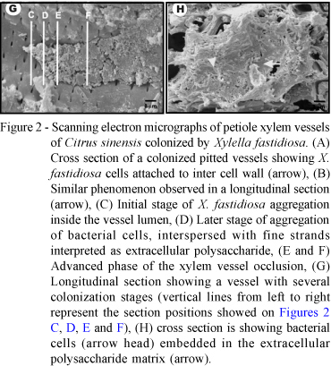
A great number of bacteria were found to exhibit polar attachment (Figure 2A). Bacterial cells first interacted with the vessel wall and then were joined by more cells and the colonization process is initiated. A sequential process was observed considered to be steps prior to the xylem vessel occlusion (Figures 2C-F). Initially, X. fastidiosa cells aggregate without extracellular material (Figure 2C). As the process advanced and a microcolony was established, cells became covered by a fibrillar material, presumably extracellular polysaccharides (Figure 2D). This material increased in size filling the entire vessel lumen (Figure 2E) causing occlusion (Figure 2F). Longitudinal sections of the xylem vessel reveal that this colonization pattern occurs along large portions of the vessels (Figure 2G). We interpret this complex of bacterial cells and fibrillar material as the biofilm, which in some cases was so extensive that anatomical features of vessel wall become barely discernable (Figure 2H).
TEM and Immunogold studies in citrus xylem vessels colonized by X. fastidiosa
The indication that X. fastidiosa once infecting pitted vessels colonizes adjacent vessels accessed by rupturing pit membranes was supported by TEM. In longitudinal sections of the occluded pitted vessels, many X. fastidiosa cells were observed in the proximity, and inside the pits (Figure 3A). Pit membrane frequently showed signs of disruption, probably due to enzymatic digestion (Figures 3B-D), an indication that bacterial cells are using the pit as a pathway to gain access to adjacent vessels. Immunogold labeling with an antibody against primary cell wall hemicelluloses, identified the degraded material within the pits as the pit membrane (Figures 3E-F).

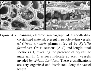
SEM and LM of the general plant reaction to vessel colonization by X. fastidiosa large, parallels arrays of needle-like, crystalline material were observed within the xylem vessels by SEM of leaf blade and petiole preparations from X. fastidiosa-infected plants (Figure 4). They occurred commonly in vessels colonized by the bacteria (Figure 4C). In several instances these crystalline structures totally obstructed the vessel lumen (Figures 4A-B), and were deposited along the vessel length (Figure 4D). These crystals were not observed in healthy plants. X-ray microanalysis of the crystallizations revealed that the main signal detected is carbon; however, we could not determine the nature of this material.

Yellowish areas from symptomatic leaves that showed gum oozing spots were observed by light microscopy. These yellowish areas were associated with an expansion of upper and lower epidermis. This expansion was due to the production of mucilaginous substances. A deposition of a dark material was observed within the intercellular space accompanied by hypertrophy of some palisade parenchyma cells (Figures 5A-B). The cells also exhibited a great number of vesicles and the presence of an osmiophilic material (Figure 5C). In the bundle sheath, cells were exhibiting hypertrophy with material deposited within the intercellular space (Figure 5D).

DISCUSSION
The colonization process of X. fastidiosa in petioles of C. sinensis cultivar Pêra tissues was determined. Bacterial colonization started on the most external petiole vessels consisting mainly of pitted vessels (Figure 1A), which are apparently more accessible to leafhoppers during feeding and bacterial inoculation. Pitted vessels have a thin cell wall, low lignin, and high hydraulic conductance when compared to other vessels (Leperen, 2000).
Once inside pitted vessels, SEM images showed strong evidence that X. fastidiosa is capable of degrading the primary cell wall and migrating to adjacent vessels. This passageway could explain how radial migration occurs. This contention is supported by the following results: i) presence of bacterial cells in the interior of pits (Figures 1C-D, Figure 2C, and Figure 3B); ii) degradation of the primary cell wall at the pit membranes by X. fastidiosa cells (Figures 3B-C), and iii) absence of colloidal gold immunolabeling against primary cell wall components. These observations of radial bacterial migration are supported by Fry et al. (1994), who verified the production of proteases by X. fastidiosa from grapevines; by Simpson et al. (2000) that identified precursors for poly-galacturonases and cellulases in the X. fastidiosa genome and by Wulff et al. (2003) who expressed cellulase genes from X. fastidiosa in E. coli. These studies all support the hypothesis that X. fastidiosa utilizes pit to facilitate intercellular migration. Pseudomonas syringae pv. syringae cells were also capable of systemic movement in plum leaves and shoots by degrading pit membranes (Roos & Hattingh, 1987).
After intervessel migration, bacterial cells initiate biofilm formation in the new pitted vessel. The first cells did not accompany any strands or EPS accumulation. However as the colony grew, strands and EPS accumulation appeared (Figures 2C, D and E). This pattern of biofilm formation was previous proposed for X. fastidiosa after results of in vitro experiments (Leite et al., 2002). The model proposes that EPS is only important to establish the biofilm architecture/complexity and does not influence the attachment phase. The presence of strands and amorphous material associated with X. fastidiosa in grapevine xylem vessels was previous observed by Tyson et al. (1985).
The colonization of other portions of the same vessel seems to require rearrangement of the biofilm organization. Some cells might get detached from the biofilm and start other colonies away from the initial location. Some SEM images indicated that some area of the xylem vessels had unstable biofilm, which did not respond to regular fixation techniques (Figure 2H). This would cause rearrangement of the biofilm, allowing systemic migration of the released cells. This process is in agreement with the model proposed by Leite et al. (2002) for X. fastidiosa, based on a general scheme published by Watnick & Kolter (2000).
The detection of crystallized forms inside xylem vessels of plants infected by X. fastidiosa was somewhat surprising (Figure 4). Our first hypothesis was that the crystallized material was calcium oxalate crystals (McConn & Nakata, 2004). This hypothesis was elaborated after observations that has been reported the presence of calcium oxalate crystals inside grapevine xylem vessels of plants with Pierce's disease (Tyson et al., 1985) and coffee leaf scorch (Queiroz-Voltan et al., 1998), both diseases caused by X. fastidiosa. Accumulation of needle-like crystal formations were observed in adjacent vessels to those colonized by X. fastidiosa, but not in the same vessel lumen (Figure 4C). In addition, Leite et al. (2002) found that calcium and magnesium accumulated in occluded vessels along with X. fastidiosa cells. Presence of calcium oxalate crystals is usually a plant physiological response to reduce calcium availability (Webb, 1999; McConn & Nakata, 2004). However, the presence of these elements was not confirmed by X-ray microanalysis (data not shown).
An alternative hypothesis is that the needle-like crystal is hesperidin. This substance is a common flavone produced by citrus plants and also forms needle-like crystals leaf inside blade petiole (Erickson, 1968). Hesperidin was previously associated with lesions caused by X. fastidiosa (Queiroz-Voltan & Paradela Filho, 1999). This flavone was present in areas where tissues were disrupted by X. fastidiosa. Hesperidin was recently invoked as an important defense tool of Citrus sinensis cv. Valencia Late against Phytophthora citrophthora, the causal agent of brown rot lesion (Del Rio et al, 2004). We assume that tissue disruption is also a rational explanation for the fact that hesperidin was observed inside leaf xylem vessels. Hesperidin is produced by leaf gland cells (Queiroz-Voltan & Paradela Filho, 1999), and was in the proximity to gain access to the vessel within the leaf blade, but away from petioles. Similar crystallization was not observed in petiole vessels from health plants of citrus, coffee and plum (Alves et al., 2004).
Hypertrophy of parenchyma cells, the presence of unknown material in intercellular spaces and cytoplasm alterations were also observed (Figure 5), which is in agreement with observations performed by Queiroz-Voltan & Paradela Filho (1999). Hypertrophy may result from a hormone imbalance already reported for grapevines infected with X. fastidiosa (Goodwin et al., 1988; Purcell & Hopkins, 1996).
In conclusion, we have shown that initially X. fastidiosa attach to the cell wall followed by an increase in the number of bacteria, the production of strand-like material and the formation of biofilm. Radial spread of X. fastidiosa occurred via digestion of pit membranes. Several instances were observed where the bacteria were inside pits between two vessels. Needle-like crystallized material was often present in xylem vessels of C. sinensis infected by X. fastidiosa.
ACKNOWLEDGMENTS
To Conselho Nacional de Desenvolvimento Científico (CNPq) and Fundação de Amparo a Pesquisa do Estado de São Paulo (FAPESP) for support. To Dr. Elliot Watanabe Kitajima (NAP-MEPA, ESALQ, University of São Paulo, Piracicaba, SP, Brazil) and Dr. Charles W. Mims (University of Georgia, Athens, GA, USA) for the use of microscopy facilities, support and suggestions.
ROWLEY, C.R.; MORAN, D.T. A simple procedure for mounting wrinkle-free sections on formvar-coated slot grids. Ultramicrotomy, v.1, p.151-155, 1975.
Received October 25, 2007
Accepted July 21, 2008
- ALVES, E.; MARUCCI, R.C.; LOPES, J.R.S.; LEITE, B. Relationship between the proportion of colonized vessels by Xylella fastidiosa in plum, coffee and citrus and the leaf symptomatology exhibited. Journal of Phytopathology, v.152, p.291-297, 2004.
- ANDERSEN, P.C.; LEITE, B.; ISHIDA, M.L. Xylem chemistry mediation of resistance to Pierce's disease. In: PIERCE'S DISEASE RESEARCH SYMPOSIUM, San Diego, 2004. Proceedings. San Diego: California Department of Food and Agriculture, 2005. p.141-144.
- CHANG, C.J.; GARNIER, M.; ZREIK, L.; ROSSETTI, V.; BOVÉ, J.M. Citrus variegated chlorosis: cultivation of the causal bacterium and experimental reproduction of the disease. In: CONFERENCE OF THE INTERNATIONAL ORGANIZATION OF CITRUS VIROLOGISTS, 12., New Delhi, 1993. Proceedings Riverside: International Organization of Citrus Virologists, 1993. p.294-300.
- DEL RIO, J.A.; GOMEZM, P.; BAIDEZ, A.G.; ARCAS, M.C.; BOTIA, J.M.; ORTUNO, A. Changes in the levels of polymethoxyflavones and flavanones as part of the defense mechanism of Citrus sinensis cv. Valencia Late fruits against Phytophthora citrophthora Journal of Agriculture and Food Chemistry, v.7, p.1913-1917, 2004.
- ERICKSON, L.C. The general physiology of citrus. In: REUTHER, W; BATCHELOR, L.D.; WEBBER, H.J. (Ed.) The citrus industry: anatomy, physiology, genetics, and reproduction. Riverside: University of California Press, 1968. p.86-122.
- FEICHTENBERGER, E.; BASSANEZI, R.B.; SPÓSITO, M.B.; BELASQUE JR., J. Doenças dos citros (Citrus spp). In: KIMATI, H.; AMORIM, L.; REZENDE, J.A.M; BERGAMIN FILHO, A.; CAMARGO, L.E.A. (Ed..) Manual de fitopatologia: doenças das plantas cultivadas. São Paulo: Ceres. 2005. p.239-269.
- FRY, S.M.; HUANG, J.S.; MILHOLLAND, R.D. Isolation and preliminary characterization of extracellular proteases produced by strains of Xylella fastidiosa from grapevines. Phytopathology, v.84, p.357-363, 1994.
- GOODWIN, P.H.; DEVAY, J.E.; MEDEDITH, C.P. Roles of water stress and phytotoxins in the development of Pierce's disease of the grapevine. Physiological and Molecular Plant Pathology, v.32, p.1-15, 1988.
- HOPKINS, D.L. Xylella fastidiosa xylem-limited bacterial pathogen of plants. Annual Review of Phytopathology, v.27, p.271-290, 1989.
- LEITE, B.; ANDERSEN, P.C.; ISHIDA, M.L. Colony aggregation and biofilm formation in xylem chemistry-based media for Xylella fastidiosa FEMS Microbiology Letters, v.230, p.283-290, 2004a.
- LEITE, B.; ISHIDA, M.L.; BRODBECK, B.; MARQUES, L.; OLSON, L.; BRAGA, M.R.; ANDERSEN, P.C. Calcium bridging is critical for Xylella fastidiosa aggregation. Phytopathology, v.94, p.S44, 2004b.
- LEITE, B.; ISHIDA, M.L.; ALVES, E.; CARRER, H.; PASCHOLATI, S.F.; KITAJIMA, E.W. Genomic and X-ray microanalysis indicate that Ca 2+ and thiols mediate the aggregation and adhesion of Xylella fastidiosa Brazilian Journal of Medical and Biological Research, v.35, p.645-650, 2002.
- LEPEREN, W. van. Fluid composition influences hydraulic conductance of xylem conduits. Journal of Experimental Botany, v.51, p.769-776, 2000.
- McCONN, M.M.; NAKATA, P.A. Oxalate reduces calcium availability in the pads of the prickly pear cactus through formation of calcium oxalate crystals. Journal of Agriculture and Food Chemistry, v.52, p.1371-1374, 2004.
- PUHLMANN, J.; BUCHELI, E.; SWAIN, M.J.; DUNNING, N.; ALBERSHEIM, P.; DARVILL, A.G.; HAHN, M.G. Generation of monoclonal antibodies against plant cell wall polysaccharides. I. Characterization of a monoclonal antibody to a terminal α-(1→2)-linked fucosyl-containing epitope. Plant Physiology, v.104, p.699-710, 1994.
- PURCELL, A.H. Xylella fastidiosa, a regional problem or global threat? Journal of Plant Pathology, v.79, p.99-105, 1997.
- PURCELL, A.H.; HOPKINS, D.L. Fastidious xylem-limited bacterial plant pathogens. Annual Review of Phytopathology, v.34, p.131-151, 1996.
- QUEIROZ-VOLTAN, R.B.; PARADELA FILHO, O. Caracterização de estruturas anatômicas de citros infectados com Xylella fastidiosa Laranja, v.20, p.55-76, 1999.
- QUEIROZ-VOLTAN, R.B.; FILHO, O.P.; CARELLI, M.L.C.; FAHL, J.I. Aspectos estruturais de cafeeiro infectado com Xylella fastidiosa Bragantia, v.57, p.23-33, 1998.
- ROOS, I.M.M.; HATTINGH, M.J. Systemic invasion of plum leaves and shoots by Pseudomonas syringae pv. syringae introduced into petioles. Phytopathology, v.77, p.1253-1257, 1987.
- ROSSETI, V.; GARNIER, M.; BOVÉ, J.M.; BERETTA, M.J.G.; TEIXEIRA, A. R.; QUAGGIO, J.A.; DE NEGRI, J.D. Présence de bactéries dans le xyléme dórangers atteints de chlorose variégée, une nouvelle maladie des agrumes au Brésil. Comptes Rendus de l´Academie des Sciences de Paris, v.310, p.345-349, 1990.
- SHERALD, J.L.; LEI, J.D. Evaluation of a rapid ELISA test kit for detection of Xylella fastidiosa in landscaping trees. Plant Disease, v.75, p.200-203, 1991.
- SILVA, F.R.; VETTORE, A.L.; KEMPER, E.L.; LEITE, A.; ARRUDA, P. Fastidian gum: the Xylella fastidiosa exopolysaccharide possibly involved in bacterial pathogenicity. FEMS Microbiology Letters, v.203, p.165-171. 2001.
- SIMPSON, A.J.G.; REINACH, F.C.; ARRUDA, P.; ABREU, F.A.; ACENCIO, M.; ALVARENGA, R.; ALVES, L.M.C.; ARAYA, J.E.; BAIA, G.S.; BAPTISTA, C.S.; BARROS, M.H.; BONACCORSI, E.D.; BORDIN, S.; BOVÉ, J.M.; BRIONES, M.R.S.; BUENO, M.R.P.; CAMARGO, A.A.; CAMARGO, L.E.A.; CARRARO, D.M.; CARRER, H.; COLAUTO, N.B.; COLOMBO, C.; COSTA, COSTA, M.C.R.; COSTA-NETO, C.M.; COUTINHO, L.L.; CRISTOFANI, M.; DIAS-NETO, E.; DOCENA, C.; EL-DORRY, H.; FACINCANI, A.P.; FERREIRA, A.J.S.; FERREIRA, V.C.A.; FERRO, J.A.; FRAGA, J.S.; FRANÇA, S.C.; FRANCO, M.C.; FROHME, M.; FURLAN, L.R.; GARNIER, M.; GOLDMAN, G.H.; GOLDMAN, M.H.S.; GOMES, S.L.; GRUBER, A.; HO, P.L.; HOHEISEL, J.D.; JUNQUEIRA, M.L.; KEMPER, E.L.; KITAJIMA, J.P.; KRIEGER, J.E.; KURAMAE, E.E.; LAIGRET, F.; LAMBAIS, M.R.; LEITE, L.C.C.; LEMOS, E.G.M.; LEMOS, M.V.F.; LOPES, S.A.; LOPES, C.R.; MACHADO, J.A.; MACHADO, M.A.; MADEIRA, A.M.B.N.; MADEIRA, H.M.F.; MARINO, C.L.; MARQUES, M.V.; MARTINS, E.A.L.; MARTINS, E.M.F.; MATSUKUMA, A.Y.; MENCK, C.F.M.; MIRACCA, E.C.; MIYAKI, C.Y.; MONTEIRO-VITORELLO, C.B.; MOON, D.H.; NAGAI, M.A.; NASCIMENTO, A.L.T.O.; NETTO, L.E.S.; NHANI, A.; NOBREGA, F.G.; NUNES, L.R.; OLIVEIRA, M.A.; DE OLIVEIRA, M.C.; DE OLIVEIRA, R.C.; PALMIERI, D.A.; PARIS, A.; PEIXOTO, B.R.; PEREIRA, G.A.G.; PEREIRA, H.A.; PESQUERO, J.B.; QUAGGIO, R.B.; ROBERTO, P.G.; RODRIGUES, V.; DE M. ROSA, A.J.; DE ROSA, V.E.; DE SÁ, R.G.; SANTELLI, R.V.; SAWASAKI, H.E.; DA SILVA, A.C.R.; DA SILVA, A.M.; DA SILVA, F.R.; SILVA, W.A.; DA SILVEIRA, J.F.; SILVESTRI, M.L.Z.; SIQUEIRA, W.J.; DE SOUZA, A.A.; DE SOUZA, A.P.; TERENZI, M.F.; TRUFFI, D.; TSAI, S.M.; TSUHAKO, M.H.; VALLADA, H.; VAN SLUYS, M.A.; VERJOVSKI-ALMEIDA, S.; VETTORE, A.L.; ZAGO, M.A.; ZATZ, M.; MEIDANIS, J.; SETUBAL, J.C. The genome sequence of the plant pathogen Xylella fastidiosa Nature, v.406, p.151-157, 2000.
- TYSON, G.E.; STOJANOVIC, B.J.; KUKLINSKI, R.F.; DIVITTORIA, T.J.; SULLIVAN, M.L. Scanning electron microscopy of Pierce's disease bacterium in petiolar xylem of grape leaves. Phytopathology, v.75, p.264-269, 1985.
- WATNICK, P.; KOLTER, R. Biofilm, city of microbes. Journal of Bacteriology, v.182, p.2675-2679, 2000.
- WEBB, M.A. Cell-mediated crystallization of calcium oxalate in plants. Plant Cell, v.11, p.751-61, 1999.
- WELLS, J.M.; RAJU, B.C.; HUNG, H.Y.; WEISBERG, W.G.; MANDELCO-PAUL, L.; BRENNER, D.J. Xylella fastidiosa gen. nov., sp. nov: gram-negative, xylem-limited, fastidious plant bacteria related to Xanthomonas spp.International Journal of Systematic Bacteriology, v.37, p.136-143, 1987
- WULFF, N.A.; CARRER, H.; PASCHOLATI, S.F. Cloning and expression of cellulase XF-818 of Xylella fastidiosa in Eschericha coli Scientia Agrícola, v.60, p.715-721, 2003.

Publication Dates
-
Publication in this collection
31 Mar 2009 -
Date of issue
Apr 2009
History
-
Accepted
21 July 2008 -
Received
25 Oct 2007