Abstract
In Costa Rica, approximately 700 snakebite cases occur each year, 5 to 10 of which result in death. At the Hospital Nacional de Niños (HNN), 6 to 10 cases are reported annually, more than half of these cases and nearly all deaths are result from Bothrops asper snakebite. This venomous snake, popularly known as the "terciopelo", most often attacks the lower or upper limbs and characteristically produces local tissue damage, which can be severe. The following is a report of the first case of a non-fatal and unusual facial bite caused by Bothrops asper in our country.
snakebite; abscess; Bothrops asper
Abscess secondary to facial snakebite
M. QUIROGA , M. L. AVILA-AGÜERO
 CORRESPONDENCE TO:
M. L. AVILA-AGÜERO - SJO 1978, P. O. Box 025216, Miami, Fla. 33102-5216, USA.
Telephone: (506) 258-2173 - Fax: (506) 221-6821
E-mail:
mavila@hnn.sa.cr
maluvi@sol.racsa.co.cr
, I. FAINGEZICHT
CORRESPONDENCE TO:
M. L. AVILA-AGÜERO - SJO 1978, P. O. Box 025216, Miami, Fla. 33102-5216, USA.
Telephone: (506) 258-2173 - Fax: (506) 221-6821
E-mail:
mavila@hnn.sa.cr
maluvi@sol.racsa.co.cr
, I. FAINGEZICHT
1 General Practitioner; 2 Pediatric Infectious Diseases Specialist, Professor of Pediatrics Universidad Autónoma de Centro América; 3 Head of Department, Pediatric Infectious Diseases, Professor of Pediatrics Universidad de Costa Rica
ABSTRACT. In Costa Rica, approximately 700 snakebite cases occur each year, 5 to 10 of which result in death. At the Hospital Nacional de Niños (HNN), 6 to 10 cases are reported annually, more than half of these cases and nearly all deaths are result from Bothrops asper snakebite. This venomous snake, popularly known as the "terciopelo", most often attacks the lower or upper limbs and characteristically produces local tissue damage, which can be severe. The following is a report of the first case of a non-fatal and unusual facial bite caused by Bothrops asper in our country.
KEY WORDS: snakebite, abscess, Bothrops asper.
INTRODUCTION
Inflammation is a main characteristic of snakebite envenoming by the Viperidae family. In Latin America, most snakebites are caused by species of the genus Bothrops. The venom from these snakes induces a prominent local edema in humans and experimental animals, producing significant fluid loss that indirectly contributes to other detrimental effects, such as tissue compression and ischemia.(2,3,9).
In Costa Rica, 700 snake envenomings are reported every year, 90% of them are due to the Viperidae family. Nearly all children who have been bitten by snakebites are treated at the National Children Hospital. The most important complications reported in these patients are infections produced by Gram-negative and Gram-positive bacteria organisms, which are found in snake fauces.
The following illustrates a facial snakebite with an unusual complication subgaleal abscess complication.
CASE REPORT
A 3 year-old male patient, who had been bitten on the face by a Bothrops asper, was referred from a rural hospital to the HNN. This patient had no significant medical history and his immunizations were up-to-date. At the rural hospital, he received 10 vials of polyvalent antivenom by the intravenous route and was intubated prior to sedation, in order to prevent ventilatory failure due to the serious facial edema.
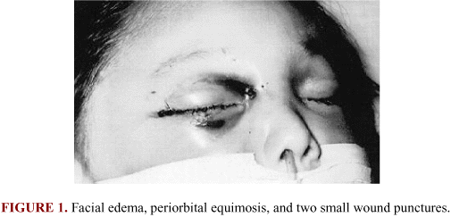
The patient arrived at the HNN approximately 3 hours after the snakebite. He was hemodynamically stable, intubated, under the effects of muscle-relaxants, afebrile. He had an important facial edema, with periorbital ecchymoses, subconjunctival hemorrhage in his right eye, and two small puncture wounds, one on the right side of the nasal bridge and the second on the medial part of the inferior palpebrae (Figure 1). The patient also showed mild and diffuse wheezing. The rest of his physical examination was normal.
In the Emergency Room, the patient received fresh plasma and the first doses of intravenous antibiotics (gentamicin and clindamycin). Chest X-ray was normal. A CT scan of the orbits showed evidence of subcutaneous tissue compromise, without orbital lesion. Laboratory findings revealed: 28,600/mm leukocytes, 80% neutrophils, 15% lymphocytes, 434,000 platelets/mm and hemoglobin 11.1 g/dl, prothrombin time less than 10%, and partial thromboplastin time more than 180 seconds. Later, he received cryoprecipitate and was transferred to the Pediatric Intensive Care Unit (PICU). There, the edema was described as involving the entire head, including important conjunctivae edema of the right eye. He also presented hematomas and bleeding from the bite site wounds. He received 5 additional vials of polyvalent antivenom. During the following 5 days, he remained hemodynamically stable. However, two days after the envenoming the patient began showing spikes of fever.
Three days later, he was transferred to the Infectious Diseases ward because of a gangrenous ecthyma of the wound was suspected. Ceftazidime was initiated and gentamicin discontinued. During the following 5 days, the facial edema decreased. On the seventh day, the wound showed necrotic tissue and abundant seropurulent secretion from which methicillin resistant Staphylococcus coagulase negative was isolated, so vancomycin was initiated and clindamycin suspended.
The following day, he was taken to the Operating Room for debridement of the wound. Two wound areas showed necrotic tissue in their bases. One area was located in the superior palpebrae, which compromised the orbicularis oculi muscle, and the second was in the inferior palpebrae, which perforated the orbital septum. There were several abscesses: one in the superior maxillary, another in the malar region, and the third, a subgaleal abscess that extended from the right frontal region to the vertex. Several incisions were made: one in the preauricular area, the second in the retro auricular region, the third in the occipital area, and the last in the vertex. Abundant purulent material was drained from these incisions. The CT scan showed a large subgaleal abscess, which did not affect either the bone structures nor the central nervous system (Figure 2). The patient was transferred to the Plastic Surgery ward, where surgical cleavage and debridement of the wounds were performed daily. Two weeks later, there was no purulent material or necrotic tissue to be seen in the area (Figure 3). The patient was discharged in very good health, without sequelae.
DISCUSSION
One hundred and thirty-five species of snakes have been described in Costa Rica, of which only 18 are venomous. These 18 are divided into 3 families: Hydrophiidae or "sea snakes", Elapidae or "coral" snakes, and Viperidae or "pit-viper snakes". The species of the Viperidae family cause 85% of the snakebites and 99% of the deaths. Of the Viperidae family reported in Costa Rica, Bothrops is the most common genus in which B. asper is the most frequently involved in human envenomings (12).
Pit viper venom is predominantly cytolytic, damaging the endothelial lining of the blood vessels causing hemorrhage and tissue destruction. Local symptoms, such as swelling, pain, and blood extravasations occur immediately. Systemic symptoms may include hemorrhage, coagulopathy, renal failure, and cardiovascular shock (6,10,13).
The severity of the envenoming varies greatly and it is determined by 3 factors: 1. Quantity of venom inoculated - B. asper inoculates the highest quantity of venom per bite, which makes it extremely dangerous (6); 2. Anatomic site of inoculation - It is well known that snakebites in the thorax or head are more severe than those in the extremities (5). In our Hospital, from 1985 to 1997, 67% of the patients were bitten in the lower extremities, 26% in the upper extremities, and 7% on the face. Facial snakebites, especially in the periorbital region, as in this case, not only increase morbidity and mortality due to the venom, but also increase the risk of CNS infections and lesions to structures of the vision system. To date, all patients admitted to our hospital with facial bites died due to complications. One of these cases involved a 24-month-old patient who was admitted with disseminated intravascular coagulation of unknown cause. The diagnosis of snakebite was only made when the mother mentioned that the child had complained about ants biting his head. His hair was shaved and fang marks were found on the scalp; 3.Weight, height, and general physiological state - Since children have lower weight and height measurements, snakebites and their complications are more severe than in adults (7,11,14,15).
Several complications can arise after a pit viper bite. The most frequent is the compartment syndrome, which is probably related to the fact that more than 90% of snakebites occur in the extremities (14). The second most frequent complication is infection due to the inoculation of venom, as well as a wide variety of bacteria, which colonize the oral cavity of snakes (1,7,8). Also, skin lesion allows human skin bacteria to enter the wound. In snakebite wounds, anaerobic,
Gram-positive, and Gram-negative bacteria can be found. In a retrospective review of snakebite cases treated at the Infectious Disease ward, approximately 50% of these patients had positive cultures from their wounds. The most frequent microorganisms isolated were Pseudomonas aeruginosa, Aeromona hydrophiliae, Klebsiella, and Serratia sp. Bolaños & Brunker (4) reported a concentration of 3 x 10 CFU/ml of anaerobic and aerobic bacteria in the venom of both B. asper and C. durissus durissus from Costa Rica. Since the most frequent pathogens are known, we recommend the initiation of antimicrobial therapy with clindamycin or penicillin plus an aminoglycoside, such as gentamicin. Further treatment includes drainage of abscesses and debridement of necrotic tissue, while repeated cultures from the wound should be taken in order to make a more specific selection of antibiotics.
In conclusion, Bothrops envenoming injuries are relatively common. Successful treatment of these potentially devastating lesions requires a thorough understanding of the complex nature of the local and systemic effects caused by envenoming (16). Early aggressive systemic support with judicious use of antivenom and antibiotics will provide satisfactory treatment in the majority of the cases. However, when the face is involved, the outcome is poor, and a good clinical management plays a critical role in the prevention of sequelae and death in these patients.
REFERENCES
Received 09 August 1999
Accepted 13 December 1999
- 01 ANDRADE JG., PINTO RN., ANDRADE AL.,MARTELLI CMT., ZICKER,F. Estudo bacteriológico de abcessos causados por picada de serpentes do gênero Bothrops. Rev. Inst. Med. Trop. São Paulo, 1989, 31, 363-7.
- 02 BARRAVIERA B. Acute-phase reactions in snakebites. Toxicon, 1994, 32, 861-2.
- 03 BARRAVIERA B., LOMONTE B., TARKOWSKI A., HANSON LA., MEIRA DA. Acute-phase reactions, including cytokines, in patients bitten by Bothrops and Crotalus snakes in Brazil. J. Venom. Anim. Toxins, 1995, 1,11-22.
- 04 BOLAÑOS R., BRUNKER T. Bacteriología del veneno y de las glándulas veneníferas de Bothrops asper y Crotalus durissus durissus de Costa Rica. Rev. Cost. Cienc. Med., 1983, 4, S27-S30.
- 05 GOLD BS., BARISH RA. Venomous snakebites: current concepts in diagnosis, treatment and management. Emerg. Med. Clin. North Am., 1992, 2, 249-67.
- 06 GUTIÉRREZ JM. Clinical toxicology of snake bites in Central America. In: MEIER J., WITE J. Eds. Handbook of clinical toxicology of animal venomous and poisons Boca Raton: CRS, 1995: 646-63.
- 07 JORGE MT., MENDONÇA JS., RIBEIRO LA., SILVA MLR., HUSANO EJU., CORDEIRO CLS. Flora bacteriana da cavidade oral, presas e veneno de Bothrops jacaraca: possível fonte de infecção no local da picada. Rev. Inst. Med. Trop. São Paulo, 1990, 32, 6-10.
- 08 JORGE MT., ADRIANO L., SILVA ML., URO EJ., SILVA J. Microbiological studies of abscesses complicating Bothrops snake bite in humans: a prospective study. Toxicon, 1994, 32, 743-8
- 09 LOMONTE B., TARKOWSKI A., HANSON LA. Host response to Bothrops asper snake venom. Inflammation, 1993,17, 93-105.
-
10NELSON BK. Snake envenomation: incidence, clinical presentation and management. Med. Toxicol., 1989, 4, 17-31.
-
11PARRISH H., GOLDNER J., SILBERG S. Comparation between snakebite in children and adults. Pediatrics, 1965, 36, 251-5.
-
12PICADO-TWIGHT C. Serpientes venenosas de Costa Rica Costa Rica: Tecnológica, 1987 v.3.
-
13SEILER JG., SAGERMAN SD., GELLER RJ., ELDRIDGE JC., FLEMING LL. Venomous snake bite. Curr. Concepts Treatm. Orthop., 1994, 8, 707-14.
-
14SUTHERLAND SK., LEONARD RL. Snake bite deaths in Australia 1992-1994 and a management update. Med. J. Aust, 1995, 163, 616-8.
-
15VINCENT JL., CRÉTEUR J. Morsures de serpent. Rev. Med. Brux., 1995, 5, 349-52.
-
16WAGNER CW., GOLLADAY ES. Crotalid envenomation in children: selective conservative management. J. Pediatr. Surg., 1989, 24, 128-31.
CORRESPONDENCE TO:Publication Dates
-
Publication in this collection
22 Sept 2000 -
Date of issue
2000
History
-
Received
09 Aug 1999 -
Accepted
13 Dec 1999