Abstract
Secções ultrafinas do ácaro Brevipalpus phoenicis Geijskes, vetor da leprose dos citros, foram examinadas ao microscópio eletrônico. Partículas semelhantes a vírus (rhabdovírus ou badnavírus) foram observadas no corpo do vetor. As partículas são similares às já noticiadas em tecidos foliares de citros que apresentavam sintomas de leprose.
Badnavirus; microscopia eletrônica; Rhabdoviridae
Badnavirus; microscopia eletrônica; Rhabdoviridae
SCIENTIFIC NOTE
Virus-like particles associated with Brevipalpus phoenicis Geijskes (Acari: Tenuipalpidae), vector of citrus leprosis virus
Partículas semelhantes a vírus associadas a Brevipalpus phoenicis Geijskes (Acari: Tenuipalpidae), vetor do vírus da leprose dos citros
José C. V. RodriguesI,II; Neusa L. NogueiraI; Deisi S. FreitasIII; Heloisa S. PratesIV
IFitopatologia e Microscopia Eletrônica - CENA/USP, Caixa postal 96, 13400-970, Piracicaba, SP
IICentro de Citricultura Sylvio Moreira/Instituto Agronômico de Campinas, Caixa postal 04, 13490-970, Cordeirópolis, SP
IIIUniversidade Federal de Santa Maria, 97119-900, Santa Maria, RS
IVCoordenadoria de Assistência Técnica Integral/SAA/SP, 13073-001, Campinas, SP
RESUMO
Secções ultrafinas do ácaro Brevipalpus phoenicis Geijskes, vetor da leprose dos citros, foram examinadas ao microscópio eletrônico. Partículas semelhantes a vírus (rhabdovírus ou badnavírus) foram observadas no corpo do vetor. As partículas são similares às já noticiadas em tecidos foliares de citros que apresentavam sintomas de leprose.
Palavras-chave:Badnavirus, microscopia eletrônica, Rhabdoviridae.
Citrus leprosis is the main viral disease in the Brazilian citrus; however, its etiology has not been defined. The disease compromises seriously the plant production and its lifetime; causing chlorotic spots and premature leaf and fruit drop, dryness of branches and death of the plant when the mite attack is severe (Rodrigues et al. 1994).
The leprosis symptoms have been associated with mites of the genus Brevipalpus. According to Haramoto (1969) and Gonzáles (1975), Brevipalpus is cosmopolite. In Argentina, this disease was associated with Brevipalpus obovatus (=Tenuipalpus pseudocuneatus) (Frezzi 1940, Vergani 1945); in Florida, with B. californicus (Knorr 1950); and in Brazil, with B. phoenicis (Musumeci & Rossetti 1963). Two hypothesis could be tested to explain the transmissibility of the disease: the lesions of leprosis were caused by the mite injection of a toxin into the plant tissue; and the mite was a vector of a pathogen, possibly a virus, responsable for the lesions.
In Florida, Knorr (1968) reported that leprosis could be transmitted by the insertion of a piece of bark tissue from plants that had leprosis symptoms into the bark of healthy potted plants. In Brazil, Chagas & Rossetti (1983) also transmitted leprosis by grafting. These results support the hypothesis that the disease is caused by a pathogen. Knorr (1968) also demonstrated the transmition of leprosis symptoms by the mites (B. obovatus and B. californicus) obtained from larvae eclosioned on Petri dishes without any contact with sick tissues. Then, it was possible to hypothesize that the virus multiplies inside the mite tissue and may be transmitted through the egg. However, Rossetti et al. (1969) verified that only B. phoenicis mites from plants with leprosis symptoms were able to produce lesions in infested plants; and mites collected from healthy plants were able to transmit the disease only after feeding on plant tissues with the disease.
In Brazil, Kitajima et al. (1971) and Colariccio et al. (1995) supported the viral ethiology theory. They related the occurrence of a virus-like particles, type rhabdovirus (Franki et al. 1981) in the foliar tissues with leprosis lesions. These particles were not found in the healthy adjacent areas in the plants, indicating a non-systemic characteristic of the virus. This characteristic increases the importance of the vector in the disease epidemiology, because of the mite presence is a sine qua non condition to the disease dissemination in the plant and throughout the citrus grove. Elucidations are urgent regarding to the possible relation between the virus and the vector. This report relates the results obtained by analysis ultrathin sections of B. phoenicis tissues observed under electron microscope and the occurrence of particles like virus closely related to those described by Kitajima et al. (1971, 1972) in foliar tissues.
Colonies of B. phoenicis were originated from one female and maintained, under laboratory conditions, on plants of Citrus sinensis showing typical symptoms of leprosis disease. The female adult mites were fixed with 5% glutaraldehyde and post fixed with 2% OsO4; both solutions were prepared in phosphate buffer at 0.2 M and pH 7.2. Fixed material was dehydrated in acetone, embedded in epoxy resin (Luft 1961) and the ulthathin sections (60-90 nm) stained with uranyl acetate and lead citrate before examination in a Zeiss EM 900 electron microscope.
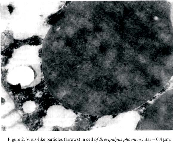
An oval shape structures were observed when ultrathin cross section was performed in the median portion between anterior medioventral metapodosomal and posterior medioventral setae (1-3 sections in this region) of the ten adult female mites examinated. The digestive and reproductive system was probably located in the sectioned portion. Electron dense portions were also found associated with all these cells (Fig. 1) which contained many cylindrical particles with different arrangements at higher magnification (Fig. 2). The particles were tubular with 40 nm wide and 110-130 nm long. These measurements were very similar to those reported by Kitajima et al. (1971) in citrus foliar tissues with leprosis lesions. These characteristics are similar to those described to the plant virus Badnavirus and Rhabdovirus groups (Brunt et al. 1990, Matthews 1991). Similar fat body cells of spider Pissaura mirabilis are designed as hepatopancreatic cells or only fat body cells in spider mite Panonychus ulmi (Morel 1991).
The amount and sites of the particles found in B. phoenicis suggest that the virus multiply inside the vector, suggesting the ocurrence of trans-stadial trasmission of the virus in this mite (Rodrigues 1995). Trans-stadial transmission have been widely used to describe not only the passage but also the development of a pathogen from one instar to the next (Burgdorfer & Varma 1967). On the other hand, B. phoenicis does not transfer the citrus leprosis virus to its progeny (Boaretto et al. 1993). More biochemical and molecular studies should be conducted to verify the association of these particles with citrus leprosis and to grouping them in Badnavirus or Rhabdovirus group.
Acknowledgments
To Dr. Carlos H.W. Flechtmann, Paulo A.V. Barroso and Marli F. Fiori without the generous assistance of these specialists, this paper could not have been written. NAP-MEPA/ESALQ/USP for the use transmission-electron-microscope.
Received 10/IV/97. Accepted 14/VI/97.
- Boaretto, M.A.C., L.G. Chiavegato, C.A. D.da Silva. 1993. Transmissão da leprose através de fêmeas de Brevipalpus phoenicis (Geijskes, 1939)(Acari: Tenuipalpidae) e de seus descendentes, em condicões de laboratório.Científica 21:245-253.
- Brunt, A., K. Crabtree & A. Gibbs. 1990. Viruses of tropical plants. Wallingford, UK, CAB International, 707 p.
- Burgdorfer, W. & M.G.R. Varma. 1967. Trans-stadial and transovarial development of disease agents in arthropods. Annu. Rev. Entomol. 11:347-376.
- Chagas, C.M. & V. Rossetti. 1983 Transmission of leprosis by grafting. In : Proc. IOCV, Riverside, p. 215-217.
- Colariccio, A., O. Lovisolo, C.M. Chagas, S.R. Galleti; V. Rossetti & E.W. Kitajima. 1995. Mechanical transmission and ultrastructural aspects of citrus leprosis disease. Fitopatol. Bras. 20:208-213.
- Francki, R.I.B., E.W. Kitajima & D. Peters. 1981 Rhabdoviruses. In E. Kurstak. Handbook of plant virus infections and comparative diagnosis. Elsevier, North-Holland Biomedical Press.
- Frezzi, M.J. 1940 La lepra explosiva del naranjo. Min. Agric. Nacion. Bol. Frutas y Hortalizas, Buenos Ayres, 15p.
- Gonzáles, R.H. 1975. Revision of the Brevipalpus phoenicis 'complex' with descriptions of a new species from Chile and Thailand (Acarina, Tenuipalpidae). Acarologia 27: 81-91.
- Hamaroto, F.H. 1969 Biology and control of Brevipalpus phoenicis (Geijskes) (Acarina: Tenuipalpidae). Hawaii Agr. Exp. Sta., Univ. Hawaii, Tech. Bull. nº 68, 63p.
- Kitajima, E.W., G.M. Müller, A.S. Costa. 1971 Partículas baciliformes associadas à leprose dos citros. In An. Congr. Brasil. Frut. 1: 419-438.
- Kitajima, E.W., G.M. Müller, A.S. Costa & W. Yuki. 1972 Short, Rod-like particles associated with citrus leprosis. Virology 50:254-258.
- Knorr, L.C. 1950 Etiological association of a Brevipalpus mite with Florida scaly bark of citrus. Phytopathology 40:15.
- Knorr, L.C. 1968 Studies on the etiology of leprosis in citrus. Proc. IOCV, p. 332-341.
- Luft, J.H. 1961. Improvements in epoxy resin embedding methods. Jour. Bioch. Cyt. 9:409.
- Matthews, R.E.F. 1991. Plant virology. 3.ed. San Diego, Academic Press, 835p.
- Morel, G. 1991. Unclassified viruses of arachnida. p.623-635. In J.R. Adams, & J.R. Bonani. Atlas of invertebrate viruses. Boca Raton, CCR.
- Musumeci, M.R. & V. Rossetti. 1963 Transmissão dos sintomas da leprose dos citros pelo ácaro Brevipalpus phoenicis Ciên. Cult. 15:228.
- Rodrigues, J.C.V., N.L. Nogueira, H.S. Prates & D.S. Freitas. 1994. Leprose dos citros: importância, histórico, distribuição e relações com o ácaro vetor. Laranja 15:123-138.
- Rodrigues, J.C.V. 1995. Leprose dos citros: relação vetor x patógeno. In: C.A.L. de Oliveira & L.C. Donadio. Leprose dos citros. Jaboticabal, FUNEP, 1995. 219p.
- Rossetti, V., C.C. Lasca & S. Negretti, 1969 New developments regarding leprosis and zonate clorosis of citrus. Proc. 1st. Int. Citr. Symp. 3:1453-1456.
- Vergani, A.R. 1945 Transmission y naturaleza de la "lepra explosiva" del naranjo. Buenos Aires. Min. Agric. Nacion, 45p.
Publication Dates
-
Publication in this collection
26 Oct 2006 -
Date of issue
Aug 1997
History
-
Received
10 Apr 1997 -
Accepted
14 June 1997