Abstracts
Despite continued progress obtained recently in treating the massively burned, mortality and morbidity rates remain quite high. Implementation of preventive strategies has still not managed to significantly alter the dramatic epidemiological picture of burns patients. Survivors of severe burns still carry a heavy load of physical and psychosocial sequelae, which engenders a considerable share of suffering to overcome. There is little doubt that the final prognosis of a burn essentially depends on prompt and adequate inicial management. This is why it is important for all physicians, including dermatologists, to be able to guide first aid for and manage initially a burns victim. The aim of this article is to review the pathophysiology of burns and the principles according to which a burns victim is assessed. In addition, it is discussed a single and objective way of carrying out the emergency medical care until the victim be moved to a burns unit, if needed.
burns; burns; burns; burns
Apesar dos crescentes progressos obtidos ultimamente no tratamento dos grandes queimados, ainda são consideráveis as taxas de mortalidade e morbidade. As estratégias preventivas implementadas ainda não foram capazes de alterar significativamente o dramático quadro epidemiológico das queimaduras. Os sobreviventes de queimaduras graves ainda carregam um pesado fardo de seqüelas físicas e psicossociais que geram grande sofrimento a ser superado. Não há dúvida de que o prognóstico final de uma queimadura depende essencialmente de um pronto e adequado primeiro atendimento. Daí a importância de todo médico, inclusive o dermatologista, estar habilitado a orientar os primeiros socorros e a prestar o primeiro atendimento médico à vítima de queimadura. Este artigo procura fazer uma revisão das bases fisiopatológicas das queimaduras e dos princípios de avaliação do queimado, além de discutir de forma simples e objetiva a abordagem médica de urgência até a remoção da vítima para uma unidade de queimados, se houver indicação.
Queimaduras; Queimaduras; Queimaduras; Queimaduras
CONTINUING MEDICAL EDUCATION
Inicial management of burns: approach by dermatologists* * Work done at Dermatology Service, Hospital das Clínicas, Federal University of Minas Gerais state.
Everton Carlos Siviero do Vale
Assistant Professor of Dermatology, Department of Clinical Medicine, Federal University of Minas Gerais (UFMG), Master's Degree in Dermatology (UFMG), Graduate studies in Immunodermatology, University of Munich, Germany
Correspondence Correspondence to Everton Carlos Siviero do Vale Rua Orenoco, 51/502 Belo Horizonte MG 30310-060 Phone: (31) 3241-2535 Fax: (31) 3241-6827 E-mail: everton.vale@terra.com.br
ABSTRACT
Despite continued progress obtained recently in treating the massively burned, mortality and morbidity rates remain quite high. Implementation of preventive strategies has still not managed to significantly alter the dramatic epidemiological picture of burns patients. Survivors of severe burns still carry a heavy load of physical and psychosocial sequelae, which engenders a considerable share of suffering to overcome. There is little doubt that the final prognosis of a burn essentially depends on prompt and adequate inicial management. This is why it is important for all physicians, including dermatologists, to be able to guide first aid for and manage initially a burns victim. The aim of this article is to review the pathophysiology of burns and the principles according to which a burns victim is assessed. In addition, it is discussed a single and objective way of carrying out the emergency medical care until the victim be moved to a burns unit, if needed.
Key words: burns/diagnosis; burns/epidemiology; burns/pathophysiology; burns/therapy.
INTRODUCTION
A burn can be defined as the condition resulting from direct or indirect action of heat on the human organism. Prognosis of burns has dramatically improved, thanks especially to recognizing the importance of an early debridement1 and to progress made in using biological skin substitutes.2 By contrast, burns still remain an important cause of death.3 This is due mainly to infections that are able to evolve into septicemia or provoke systemic repercussions with possible kidney, cardiovascular, lung, musculoskeletal, hematologic and gastrointestinal complications. In addition, burns lead to considerable morbidity by developing sequelae, the most severe of which are functional incapacities, especially those affecting the hands,4 and cosmetic deformities, of the face foremost, as well as psychosocial sequelae. Depending on their localization, burns might even cause neurological, ophthalmologic and genitourinary complications. The conclusion to draw from this is that correctly handling a burn from the outset is essential for short and long-term prognosis.5
Although not a common situation in daily practice for dermatologists working in large cities, they may at times be solicited to apply first aid to a burns patient owing to the fact that a burn affects the skin. However, in outlying areas where specialized resources for the care of burns victims are not readily available, dermatologists might be frequently called upon. Therefore they have to be properly trained in order to evaluate and provide emergency care to patients until they can be moved to a hospital burns unit, if and when necessary.
The objective of this article is to review the physiological bases of burns and the principles according to which burns victims are assessed so as to adequately provide first aid and treatment to a patient. The article does not discuss later treatment with curative dressings applied to the burn area, procedures by which destroyed tissues are repaired nor treatment of sequelae which may result from burns.
ETIOLOGY, EPIDEMIOLOGY AND PREVENTIVE MEASURES
The most frequent causes of burns are flames, contact with boiling water or other hot liquids, and contact with hot objects. Less common are burns provoked by electric currents that are transformed into heat when coming into contact with the body. As for chemical burns, this is an inappropriate name given to caustic lesions provoked by chemical agents in which tissue damage is not always the result of heat production.6
In most cases, burns affecting children occur in the household environment and are provoked by hot liquids spilt on the body, such as boiling water in the kitchen, hot bath water, drinks and other hot liquids, such as cooking oil. These cases tend to be more superficial, yet extensive. Another common cause of burns in Brazil are flames stemming from the manipulation of liquid ethyl alcohol, which is responsible for the majority of cases in teenagers and is the second major cause for treating children at the emergency ward of the Minas Gerais State7 reference hospital. It is also the cause for 40% of burns in children between the ages of seven and 11 years at a hospital-school in the State of Sao Paulo.8
By contrast adults burn themselves most frequently with flames and mainly in the professional environment. However, the resulting burns tend to be deeper. They usually incur additional damage due to smoke inhalation.
Burns from contact with hot objects, such as ovens, toasters, grills and heaters, are most common in patients experiencing convulsions, or in patients under the effects of alcohol or illicit drugs, as well as during episodic losses of consciousness. They tend to be deep owing to prolonged contact with the heat source.9 Electrical injuries might be caused by the passage of an electric current through the body or by exposure to heat generated by a high-voltage current. In the first case, in addition to heat damage, there is a risk of altering cardiac electric conduction, which must be duly monitored.9
In the United Kingdom annually, 250 thousand persons suffers from some kind of burn, 175 thousand are treated at emergency units, 13 thousand hospitalized, with 1,000 requiring measures of hydroelectrolytic resuscitation and 300 who die. Meanwhile, the numbers are much more alarming in developing countries like Nepal, where mortality may reach 17 times the British rate.
10
According to the Brazilian Burn Society, millions of burn cases occur every year in Brazil. Two hundred thousand are treated at emergency wards, and 40 thousand require hospitalization. Burns are among the main external causes of death recorded in Brazil, losing out only to other violent causes of death, including road accidents and homicides.11 A study conducted by the Federal District of Brazil demonstrated a mortality rate of 6.2% among interned burns victims in hospital emergency wards.12
In response to recent attacks and terrorist threats, particularly in the United States, strategic and logistics systems have been implemented and focus on training specialized teams that can provide care to large groups of people in the event of catastrophes, including to burns victims.13
Based on the assumption that 90% of burns can be avoided, preventive measures have been implemented in order to reduce their incidence, though they rely on education and legislation. Educative means of prevention consist in instructing children from an early age on how to avoid risk situations that might incur burns in the household. Teaching general preventive measures that are geared to the whole population should be included in school curricula.14 Private educational campaigns, to be more effective, have to be based on reliable epidemiological data identifying the specific causes of burns and the respective risk populations - toward whom these campaigns must be periodically aimed.10
Legislative norms basically contain the compulsory measures for implementing fire prevention equipment in public and private buildings, as well as specific work safety equipment. In Brazil, a legal measure of extreme importance was the prohibition of liquid ethyl alcohol, following a resolution from the National Sanitary Vigilance Agency. However, attempts to apply the legislation have clashed with the resistance of producers themselves. By means of court injunctions they have insisted upon maintaining the distribution of this product in commercial outlets.
Regrettably, the implementation of all of these prevention strategies has not succeeded in producing the expected impact on the dramatic epidemiological picture regarding burns.
PATHOPHYSIOLOGY OF BURNS
Burns prevent the skin from functioning completely. They may therefore affect hydroelectrolytic homeostasis, internal temperature control, flexibility and body surface lubrication, for which the skin is responsible. However, the magnitude of involvement of these functions depends on the extension and depth of the burn.15
Thermal injuries provoke a local response in the organism, which is translated into necrosis of tissue coagulation and a progressive thrombosis of the adjacent vessels over a 12-to-4 hour period. A burn wound is sterile in principle, though the necrotic tissue quickly becomes colonized by endogenous and exogenous bacteria and proteases producers, which lead to liquefaction and separation of the scar. This gives rise to granulation in the tissue, which is responsible for healing the wound. In third degree burns, the latter process is characterized by a high capacity of retraction and fibrosis.6,15
In the massively burned, in addition to a local response, thermal damage triggers a systemic reaction in the organism as a result of the injured tissue releasing mediators. Damage occurs to the whole capillary with accelerated loss of fluids, either by evaporation through the injury or by sequestration in the interstices, which is aggravated by the subproducts of bacterial colonization. Furthermore, with extensive burns, that is, those covering over 40% of the body surface, the immune system is unable to delimit infection. As infections spread systemically, this condition can dramatically reduce chances of survival. Systemic response is manifested by fever, hyperdynamic blood circulation, and an accelerated metabolic rhythm, with increased muscular catabolism resulting from the alteration of the hypothalamic function (increased secretion of glucagons, cortisol and catecholamines), of gastrointestinal barrier deficiency (passage of bacteria and its subproducts to the circulatory system), bacterial contamination of the burn area (systemic release of bacteria and subproducts), heat loss (evaporation through the wound leading to hypothermia) and fluid loss (hydroelectrolytic imbalance).9,15
ASSESSMENT OF BURNS
There are many factors involved in burns that must be observed in an assessment. Depth, extension and burn localization, victim's age, existence of previous diseases, concomitance of aggravating conditions, and smoke inhalation have to be considered when assessing burns. The assessment environment must be heated so as to minimize liquid loss in the skin through evaporation, as the skin has to be left uncovered and be examined in sections.
Depth
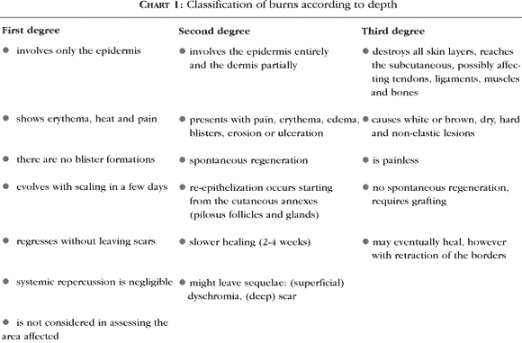
Depends on the intensity of the thermal agent, whether it is a heat generator or transmitter, and time of contact with the tissue. This is the determining factor of the cosmetic and functional outcome of the burn, and may be evaluated in degrees (Chart 1).
Extension
The general risks to a burns patient within the first hours depend fundamentally on the extension of the burn area. Systemic repercussions tend to increase with losses of skin function, which in turn increases with the size of the area affected. The extension is calculated as a percentage of total body surface (TBS). Only areas with a depth of second or third degree burns are considered.
A practical method for calculating the burn area takes the palm of the victim's hand as a benchmark by considering the palmar surface with the fingers put together and stretched out. This corresponds to roughly 1% of body surface. Excluding the fingers, the palmar surface represents 0.5% of the TBS, irrespective of age.15,16 Though this is a rough measurement, the method is quite useful for immediately determining whether the area with mainly irregular burns exceeds 15% of the adult TBS and 10% of a child's--a situation in which rehydration must begin with utmost urgency. On the other hand, the method to be most used is Wallace's Rule of Nines (Chart 2), which is easy to memorize. This method has to be adjusted for children under the age of 10 years (Chart 3). The most accurate method to evaluate the burn area utilizes the Lund and Browder diagram,17,18 which considers variations in body shape with respect to age. It is best suited for children, but additional, hard to memorize information must be available, such as the victim's medical file.
Localization of burns
Owing to cosmetic and functional risks, burns involving the face, throat, and hands are highly unpleasant. Furthermore, burns localized on the face and throat tend to be more frequently associated with smoke inhalation. They may also cause considerable edema, and may further damage the permeability of the respiratory tract and lead to respiratory insufficiency. On the other hand, burns close to natural orifices show a higher risk of septic contamination.
Age of the burns patient
This must be considered when evaluating the severity of burns. The elderly and children tend to experience more critical systemic repercussions. The elderly do so since their organism has a harder time to readapt, while children tend to because of the disproportion of body surface in relation to weight. This is why in these age ranges complications are more common and more severe.
Associated diseases and conditions
These conditions worsen the prognosis for concomitant traumas, mainly neurological, orthopedic and abdominal, or even polytraumatism, like the presence of preexisting diseases, such as cardiac insufficiency, renal insufficiency, arterial hypertension, diabetes and ethylism. Patients under the effect of alcohol or illicit drugs tend to have a worse prognosis. These situations have to be considered and confronted appropriately. In such cases, the recuperation of defects resulting from burns can turn out to be substantially compromised.
Inhalation of fuel products
In addition to the damage provoked by toxic gas inhalation, like carbon monoxide, fuel products are irritants and cause inflammation with edema of the tracheobronchial mucosa. This is manifested by hoarseness, stridor, dyspnea, bronchospasm and grey saliva. These lesions tend to be severe, and worsen the prognosis a lot. They are responsible for increasing the mortality rate among burns patients.5
FIRST AID
Appropriate first aid provided to a burns victim is vital for determining the final outcome of treatment. It contributes decisively to reducing morbidity and mortality. To achieve the latter reduction, it is important to educate the population in general and train risk groups in particular on proper modes of action to pursue when faced with burns. In this sense, health education programs must include teaching first-aid procedures for burns.
Like any physician, a dermatologist must take action in these cases if and when solicited to provide guidance to family members on how to proceed with first aid treatment for burns victims. Dermatologists may be called upon to provide emergency assistance or even hospitalization at the ambulatory level when being far away from large centers of specialized emergency care for burns victims.
Removal of the heat source
The first measure to be taken is removal of the heat source, thereby freeing the victim from the flame or withdrawing the burning object. If a person's clothing has caught fire, the victim must be rolled on the ground, but never run or be covered in cloth as this may only end up reactivating the flames. Clothing has to be removed as soon as it stops adhering to the skin. Otherwise it can only be removed under anesthesia at the moment of the debridement of the wounded person. In cases of electrical burns, a switch or circuit breaker has to be triggered so as to cut the power source prior to coming into contact with the victim. When this is not possible, try separating the victim from the source by using an insulated object, such as dry wood.5,19
Cooling of the burned area
Next, the burnt area has to be cooled with cold running tap or shower water. Never use ice water or other coolants, such as toothpaste or hydrating lotions. In addition to cleansing the victim and removing harmful agents, cold water can interrupt the progression of heat. It thus limits any deepening of the lesion, provided it is carried out within the first few seconds or minutes. It can alleviate pain, even if applied a few minutes later, as well as possibly reduce edema. Therefore cooling the burn site with running water has to be done as soon as possible, that is, roughly within the first 10 minutes, and up to 20 minutes if need be. However, it ought to be done sooner if the burn is more extensive owing to the risk of hypothermia. This approach is not recommended for burns affecting over 15% of TBS. After cooling, the burn area, if less than 5% of TBS, can be protected with humid gauzes, compresses, or cotton towels, and covered afterward with plastic or another impermeable material. Finally, the patient must be wrapped with a cloth or blanket. In light of these measures, let us bear in mind the basic guideline: "Cool the burns, but keep the patient warm."20
MEDICAL APPROACH
First degree burns
In these cases, medical treatment is ambulatory and basically consists of controlling pain and providing local care to the burn area. Analgesics might be administered orally with 50 mg daily tramadol chlorhydrate for adults, and 2 mg/kg/dose for children every four to six hours. Another alternative for adults is paracetamol/codeine phosphate in a 500 mg/30 mg dose every four to six hours. Cold water compresses might also help to relieve pain. Topical corticosteroid might also be used in lotion or cream form to reduce inflammation. It is important to recommend protection from sunlight so as to avoid residual dyschromias.
Second and third degree burns
While the patient is being examined, gather detailed background on the burns, and try to identify any possible concomitant injuries, smoke inhalation and previously instituted treatment. If possible, a short account of past medical history should be taken, including diseases, medication, allergies and vaccinations.21 The patient must be kept calm at all times while the burns are treated. It is important not to be overwhelmed at the chocking aspect of the burn, as it might disturb your concentration as a professional. At times, this may lead to very severe lesions being left unnoticed, such as neurological, orthopedic or visceral traumas. If the background of the accident suggests trauma with involvement of the spinal column, you must give special attention to your assessment and provide the patient with due stabilization. Whenever the face and throat are burned, the burns are considered severe since they might affect the permeability of the respiratory tract. In cases of electrical burns, the risk of cardio-respiratory arrest must always be borne in mind due to cardiac arrhythmias. Once the assessment of the burn and of the respiratory and circulatory conditions is finished, it becomes imperative to distinguish a benign burn from a severe burn.5
Benign burns
- absence of installed respiratory insufficiency
- no risk of future respiratory insufficiency (face and throat burns)
- second or third degree burns below 10% of TBS (children) and 15% TBS (adults)
Although benign burns are seldom life threatening, there are situations in which their removal is indicated which call for hospital level treatment. This is especially the case when specialized care is required, as with third degree lesions covering over 1% of TBS and hand lesions, or in cases that pose greater risk due to the victim's condition, like critical age range (the elderly and children) and associated diseases (diabetes, arterial hypertension, cardiac insufficiency, renal insufficiency, among others).
On the other hand, ambulatory assistance consists of immediately administering analgesia intravenously with meperidine in a 100 mg dose diluted in 20 ml of distilled water, administered for 30 minutes, or tramadol chlorhydrate in a 100 mg dose. Later, pain control may be maintained orally with tramadol chlorhydrate in a 50 mg dose every four to six hours.
Once the pain is controlled, excision of the large blisters can begin. Leave the smaller ones and any devitalized tissues intact, and cleanse the wound thoroughly with diluted chlorohydrins or polyvinylpyrrolidone iodine (PVPI), rinsing it with water or a physiological solution.22,23 The PVPI must be left for five minutes or so for the release of iodine to occur since the latter has an antimicrobial property. In this case, rinsing with a physiological solution must be avoided as it might partially inactivate the PVPI. The curative is next. Preferably, a sterilized vaseline gauze should be applied, and covered with gauze bandages, bound thereafter with a crepe bandage without pressing very hard. Silver sulfadiazine must be avoided in principle, because it may hinder the assessment of the burn area, which could progress within the first 48 hours.19
One must not forget to check the state of the patient's antitetanus protection. If safely immunized (at minimum three doses), there is a need for a booster shot only if the last dose was give over five years in the past. In case of unknown or incomplete immunization, a booster shot has to be taken with tetanic toxoid by applying 250 mg of hyperimmune gamma-globulin for tetanus.24
Severe burns
- installed or potential respiratory insufficiency (face or throat)
- second or third degree burns on over 10% of TBS (children) and 15% TBS (adults)
In these cases, all and any medication must be administered exclusively intravenously, except the tetanic toxoid booster, if and when necessary, which has to be administered intramuscularly. Therefore, a superficial venous access has to immediately be prepared with a polyethylene catheter needle. The assistance given to a severe burns victim must be given in a hospital environment and include four chronologically ordered stages:5 1. Control of respiratory function (permeability of the air tract); 2. Parenteral rehydration and monitoring of the hemodynamic state; 3. Analgesic treatment; 4. Preparation of the burns patient to be moved to a Burns Unit.
1. Control of respiratory function
To maintain control of respiratory functions, oxygenotherapy has to be instituted with a nasal catheter by administering three to five liters per minute of humidified oxygen. An intubation is required when faced with acute respiratory insufficiency. This is highly recommended in cases of smoke inhalation, extensive facial burns, and in circular throat burns. Late edema might survive in either of these situations and obstruct the respiratory tract, making intubation difficult and at times even impossible at a later point.5,21
2. Hemodynamic control
With severe burns, plasmatic losses are considerable. The delay in replacing them exposes the victim to a great risk of developing hypovolemic shock. Therefore, it is of utmost urgency to perform parenteral rehydration in second and third degree burns exceeding 10% of TBS in children and 15% in adults, that is, greater than 10 or 15 palms of the victim's hand, respectively.18 To make calculations of the hydroelectrolytic replacement easier, Parkland's formula is used:
Daily volume (ml) = 2 to 4 x weight (kg) x burns area (% TBS).
For example, a 50-kg patient with 20% of TBS burned must received 2000-4000 ml in the first 24 hours, counting from the moment of the burn. This rule is valid for a burns area of up to 50% of TBS, which is the maximum volume allowed. Isotonic lactated ringer is used preferably, thus reserving the hypertonic ringer for cases of hypovolemic shock and in burns exceeding 40% of TBS. Half of the volume calculated must be administered in the first eight hours, and the rest within the next 16 hours. From thereon, the volume must be oriented by hemodynamic variables. A vessel probe must be installed so as to measure urinary flow, which must be maintained at a minimum of 30-50 ml/h for adults, and 0.5-1 ml/kg/h for children; the ideal is 2 mg/kg/h for both.5
3. Pain control
Morphinic agonists, like meperidine in a dose of 100 mg per adult or 2 mg/kg per child, must be administered intravenously and diluted in 20 ml of distilled water or physiological solution for 30 minutes. Later, if and when necessary, a 50 mg dose can be maintained, orally if possible, every four to six hours. In refractory cases, benzodiazepines may be used in combination.25
4. Preparation of patient to be moved
In the event that transporting the patient to a Burns Unit is necessary, a gastric probe has to be installed and the burns patient's wound must be protected in a sterile field and thermal protection provided with blankets, so as to prevent hypothermia.
At the Burns Unit, the primary objective of intensive care is to limit the progression of any systemic repercussions that may arise from severe burns, thereby preventing the onset of organ failure, especially respiratory, cardiac, kidney and brain failure. Moreover, nutritional support and infection control must be maintained. Infections are the main cause of death once the period of resuscitation has been overcome.26
With survival rates improving among massive burns victims, the fields of repairing destroyed areas, of functional and psychological rehabilitation, and of socially reintegrating burns victims have all shown signs of increased progress.27-31 q
REFERENCES
Received on January 03, 2005.
Approved by the Consultive Council and accepted for publication on January 11, 2005.
- 1. Barret JP, Herndon, DN. Effects of burn wound excision on bacterial colonization and invasion. Plast Reconstr Surg. 2003;111:744-50; discussion 751-2.
- 2. Ramos-e-Silva M, Ribeiro de Castro MC . New dressings, including tissue-engineered living skin. Clin Dermatol. 2002;20:715-23.
- 3. Sheridan RL, Hinson MI, Liang MH, Nackel AF, Schoenfeld DA, Ryan CM, et al. Long-term outcome of children surviving massive burns. JAMA. 2000;283:69-73.
- 4. Barillo DJ, Paulsen SM. Management of burns to the hand. Wounds. 2003;15:4-9.
- 5. Wassermann D. Évaluation et premiers soins d'une brűlure thermique. Rev Prat. 2002;52:2228-33.
- 6. Leão CEG. Queimaduras. In: Fonseca FP, Rocha PRS, editors. Cirurgia ambulatorial. 3Ş ed. Rio de Janeiro: Guanabara Koogan; 1999. p.122-8.
- 7. Costa DM, Abrantes MM, Lamounier JA, Lemos ATO. Estudo descritivo de queimaduras em crianças e adolescentes. J Pediatr. 1999;75:181-6.
- 8. Rossi LA, Barruffini RCP, Garcia TR, Chianca TCM. Queimaduras: características dos casos tratados em um hospital escola em Ribeirão Preto (SP), Brasil. Rev Panam Salud Publica. 1998; 4:401-4.
- 9. Hettiaratchy S, Dziewulski P. ABC of burns: pathophysiology and types of burns. BMJ. 2004;328:1427-9.
- 10. Hettiaratchy S, Dziewulski P. ABC of burns. Introduction. BMJ. 2004;328:1366-8.
-
11Brasil e a mortalidade por causas externas no ano 2000. Disponivel em: http://www.cip.saude.sp.gov.br/Brasil2000.htm Acessado em: 29 de Dezembro de 2004.
- 12. Macedo JLS, Rosa SC. Estudo epidemiológico dos pacientes internados na Unidade de Queimados: Hospital Regional da Asa Norte, Brasília, 1992-1997. Brasilia Med. 2000; 37:87-92.
- 13. Sheridan RL, Tompkins RG. What's new in burns and metabolism. J Am Coll Surg. 2004;198:243-63.
- 14. Grant EJ. Burn prevention. Crit Care Nurs Clin North Am. 2004;16:127-38.
- 15. Sheridan R. Evaluation and management of the thermally injured patient. In: Freedberg IM, Eisen AZ, Wolff K, Austen KF, Goldsmith LA, Katz SI, editors. Fitzpatrick's dermatology in general medicine. 6th ed. New York: McGraw-Hill;2003. p.1220-9.
- 16. Jose RM, Roy DK, Vidyadharan R, Erdmann M. Burns area estimation-an error perpetuated. Burns. 2004;30:481-2.
- 17. Novaes FN. Primeiro atendimento ao paciente queimado. Bras Med. 2003;84:56-62.
- 18. Hettiaratchy S, Papini R. Initial management of a major burn: II - assessment and resuscitation. BMJ. 2004;329:101-3.
- 19. Hudspith J, Rayatt S. First aid and treatment of minor burns. BMJ. 2004;328:1487-9.
- 20. Allison K, Porter K. Consensus on the pre-hospital approach to burns patient management. Accid Emerg Nurs. 2004;12:53-7.
- 21. Hettiaratchy S, Papini R. Initial management of a major burn: I - overview. BMJ. 2004;328:1555-7.
- 22. Dhennin C. Traitment local des brűlures. Pathol Biol (Paris). 2002;50:109-17.
- 23. Ferreira E, Lucas R, Rossi LA, Andrade D. Curativo do paciente queimado: uma revisão de literatura. Rev Esc Enferm USP. 2003; 37:44-51.
- 24. Starling CEF, Silva EU. Antimicrobianos e síndromes infecciosas. Guia prático. Rio de Janeiro: Medsi; 2002. p. 394.
- 25. Latarjet J. La douleur du brűlé. Pathol Biol (Paris). 2002;50:127-33.
- 26. Ansermino M, Hemsley C. Intensive care management and control of infection. BMJ. 2004;329:220-3.
- 27. Bello YM, Falabella AF, Eaglstein WH. Tissue-engineered skin. Current status in wound healing. Am J Clin Dermatol. 2001;2:305-13.
- 28. Papini R. Management of burn injuries of various depths. BMJ. 2004;329:158-60.
- 29. Barret JP. Burns reconstruction. BMJ. 2004;329:274-6.
- 30. Edgar D, Brereton M. Rehabilitation after burn injury. BMJ. 2004;329:343-5.
- 31. Wiechman SA, Patterson DR. ABC of burns. Psychosocial aspects of burn injuries. BMJ. 2004;329:391-3.
Publication Dates
-
Publication in this collection
25 Nov 2005 -
Date of issue
Feb 2005
History
-
Received
03 Jan 2005 -
Accepted
11 Jan 2005