Abstracts
The authors report a case of pityriasis versicolor circinata whose isolated etiologic agent was Malassezia sympodialis in a 34-year-old woman. The isolation and identification of Malassezia sympodialis were accomplished with modified Dixon's agar, and the molecular method used to confirm the species was polymerase chain reaction and restriction fragment length polymorphism analysis (PCR-RFLP).
Clinical Evolution; Malassezia; Pityriasis
Os autores descrevem caso de pitiríase versicolor circinada, cujo agente etiológico isolado foi Malassezia sympodialis em uma mulher de 34 anos. O isolamento e identificação da Malassezia sympodialis foi em ágar Dixon modificado e o método molecular para confirmação da espécie foi PCR-RFLP (polymerase chain reaction and restriction fragment length polymorphism analysis).
Evolução Clínica; Malassezia; Pitiríase
CASE REPORT
Pityriasis versicolor circinata: isolation of Malassezia sympodialis - Case report
Valéria Maria de Souza FramilI; Márcia S.C. MelhemII; Maria Walderez SzeszsIII; Elaine Cristina CornetaIV; Clarisse ZaitzV
IAssistant Professor at the Faculty of Medical Sciences of Santa Casa de Sao Paulo, and Second-year Assistant Physician at the Dermatology Clinic of Santa Casa de Sao Paulo - São Paulo (SP), Brazil
IILevel IV Scientific Researcher at Adolfo Lutz Institute - Sao Paulo (SP), Brazil
IIIScientific Researcher at Adolfo Lutz Institute- Sao Paulo (SP), Brazil
IVMaster's degree student in Microbiology/ICB-USP with a scholarship from CAPES - Coordination for the Improvement of Higher Education Personnel - Sao Paulo (SP), Brazil
VProfessor, Ph.D., at the Faculty of Medical Sciences of Irmandade Misericordia da Santa Casa de Sao Paulo - São Paulo (SP), Brazil
Mailing Address
ABSTRACT
The authors report a case of pityriasis versicolor circinata whose isolated etiologic agent was Malassezia sympodialis in a 34-year-old woman. The isolation and identification of Malassezia sympodialis were accomplished with modified Dixon's agar, and the molecular method used to confirm the species was polymerase chain reaction and restriction fragment length polymorphism analysis (PCR-RFLP).
Keywords: Clinical evolution; Malassezia; Pityriasis
INTRODUCTION
Pityriasis versicolor is a disease of universal distribution. Until recently, Malassezia furfur was considered the only etiologic agent of pityriasis versicolor. Several species of Malassezia have been described since the 1990s. The genus Malassezia is part of the normal skin microbiota, especially of the hair follicle. It needs predisposing factors for multiplication and subsequent conversion into its pseudo-filamentous parasitic form.
An epidemiologic study involving the frequency and distribution of each species is not sufficient for clarification of the pathogenicity of the yeast in pityriasis versicolor.1
CASE REPORT
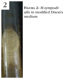
Female patient, 34 years old, white, has been showing pruriginous macules on her skin for 5 years. Dermatologic examination revealed hypochromic follicular lesions that evolved into erythematous, scaly lesions of circinate aspect, pruriginous, affecting her trunk and upper extremities. Lesions were positive for Zirelis sign (Figure 1). Direct microscopic examination (potassium hydroxide_20%) revealed the presence of yeast-like cells resembling a bunch of grapes and pseudohyphae. The clinical material was cultivated in modified Dixons agar. The isolation of Malassezia was achieved after 15 days (Figure 2) and the identification of the species was performed through the molecular biology method PCR-RFLP (polymerase chain reaction and restriction fragment length polymorphism analysis)2 (Figure 3). The patient was treated with ketoconazole (200 mg/day for 30 days) with total regression of lesions.
DISCUSSION
Clinical manifestations of pityriasis versicolor are characterized by multiple macular lesions, perifollicular at first, with light scaling. Stretching of the affected skin may help see scaling of lesions. This procedure is known as Zirelis sign. The clinical variants of pityriasis versicolor show variable coloration, from white to chestnut- brown. It can rarely become erythematous, which justifies the denomination of pityriasis versicolor.3
Among the clinical variants of pityriasis versicolor, the majority of cases described in the literature replicate the classical clinical symptoms of the disease, such as hypochromic macules. There are reports of less frequent clinical forms of the disease; for instance, hyperchromic macules and association between hypochromic and hyperchromic macules. 4-6
A few authors describe follicular lesions of pityriasis versicolor in the literature.5-7 A clinical variant with intense skin depigmentation may occur in melanodermic individuals, called parasitic achromia. Pityriasis versicolor atrophicans is also described, a rare form of the disease in which lesions are depressed by the prolonged used of topical corticosteroids.8,9
There are reports in the literature of pityriasis versicolor in the inguinocrural region, which simulate erythrasma10, and one case of pityriasis versicolor mimicking pityriasis rubra pilaris.11
The present case report shows a clinical variant of pityriasis versicolor with hypochromic follicular lesions chronically evolving into pruriginous, erythematous, scaly lesions of circinate aspect. Malassezia sympodialis, the isolated etiologic agent, needs proper culture medium and temperature for growth and identification. After the clinical and laboratory diagnosis of pityriasis versicolor, appropriate treatment was implemented with an excellent therapeutic response. Differential diagnosis for pityriasis versicolor circinata includes tinea corporis, pityriasis rosea, erythema annulare centrifugum, and even secondary syphilis.
REFERENCES
- 1. Crespo Erchiga V, Delgado Florêncio V. Malassezia species in skin diseases. Curr Opin Infect Dis. 2006;15:133-42.
- 2. Corneta , Melhem MSC, Chioccola VLP, Pires MC, Keiko LO, Framil VMS, et al. Molecular identification of Malassezia species isolated from pityriasis versicolor and seborrheic dermatitis Brazilian patients. Paris: ISHAM; 2006. (International Society for Human & Mycology.
- 3. Zaitz C. Micoses superficiais propriamente ditas. In: Zaitz C, Campbell I, Marques AS, Ruiz LR, Souza VM. Compêndio de micologia médica. São Paulo: Medsi; 1998. p.65-79.
- 4. Chetty GN, Kamalam A, Thambiah AS. Pityriasis versicolor: a study of 200 cases in a tropical skin clinic. Mykosen. 1979;22:234-6.
- 5. Forjaz MHH, Freire EL, Gama MP, Fischman O, De Lamonica Freire E. Pitiríase versicolor: estudo epidemiológico em voluntários da Universidade Federal de MatoGrosso (Brasil). An Bras Dermatol. 1983;58:249-52.
- 6. Framil VMS. Pitiríase versicolor: influência de fatores etiológicos, imunológicos, familiares, constitucionais, clínicos e de hábitos pessoais no seu desencadeamento e na sua recidiva: estudo de uma amostra ambulatorial [tese]. São Paulo: Faculdade de Ciências Médicas da Santa Casa de São Paulo; 2006.
- 7. Maeda M, Makimura KC, Yamaguchi H. Pityriasis versicolor rubra. Eur J Dermatol. 2002;12:160-4.
- 8. Aspiroz Sancho MC, Saenz de Santamaria MC, Moreno Borraz LA. Afecciones cutâneas relacionadas con Malassezia furfur. Rev Clin Esp. 1997;197:420-8.
- 9. Romano C, Maritati E, Ghilardi A, Miracco C, Mancianti F. A case of pityriasis versicolor atrophicans. Mycoses. 2005;48:439-41.
- 10. Aste N, Pau M, Aste N. Pityriasis versicolor on the groin mimicking erythrasma. Mycoses. 2004;47:249-51.
- 11. Darling MJ, Lambiase MC, Young RJ. Tinea versicolor mimicking pityriasis rubra pilaris. Cutis. 2005;75:265-7.
Publication Dates
-
Publication in this collection
23 June 2010 -
Date of issue
Apr 2010
History
-
Received
28 Oct 2008 -
Accepted
27 Nov 2009