Abstracts
This case report is about a 48-year-old female patient with systemic amyloidosis and multiple myeloma simultaneously. Amyloid cutaneous infiltrative lesions like papules, nodules, or plaques with a serous-hemorrhagic aspect were found in the eyelids, neck and retroauricular region, among others. She had presented intermittent papular lesions on the upper eyelids one year before, which worsened following local trauma. A local skin biopsy showed amorphous and eosinophilic substance in the dermis. Congo red staining confirmed the amyloid deposits. Abnormal exams: proteinuria (570mg/24h), Bence-Jones proteinuria and clonal plasma cells (70%) found in myelogram. Following the diagnosis of multiple myeloma based on amyloid skin lesions, the patient was referred to the Hematology service and died 5 months after the diagnosis.
Amyloidosis; Eyelids; Multiple myeloma; Skin
Relatamos um caso de uma paciente de 48 anos com amiloidose sistêmica associada a mieloma múltiplo. Lesões infiltrativas cutâneas como pápulas, nódulos ou placas com aspecto sero-hemorrágico podem ser localizados nas pálpebras, pescoço, região retroauricular dentre outras. No presente caso, as pálpebras foram acometidas por pápulas, há 1 ano, de caráter intermitente e piora após trauma local. Biópsia local evidenciou material amorfo e eosinofílico na derme. A coloração vermelho do Congo confirmou presença de substância amiloide. Exames anormais: proteinúria de 570mg/24 horas, proteinúria de Bence-Jones positiva e mielograma com 70% de plasmócitos atípicos. Assim, realizou-se o diagnóstico de mieloma múltiplo a partir de manifestações cutâneas de amiloidose. Paciente encaminhada ao serviço de hematologia e foi a óbito em 5 meses.
Amiloidose; Mieloma múltiplo; Pálpebras; Pele
CASE REPORT
Primary systemic amyloidosis associated with multiple myeloma* * Study carried out at a private clinic - Dra. Eurides Mª Oliveira Pozetti - São José do Rio Preto (SP), Brazil.
Amiloidose sistêmica primária associada ao mieloma múltiplo
Ederson Valei Lopes de OliveiraI; Ana Carolina Garcia PozettiII; Eurides Maria de Oliveira PozettiIII; João Roberto AntonioIV; Nilceo Schwery MichalanyV
IDermatologist at a private clinic - Ribeirão Preto (SP), Brazil
IIResident of Dermatology, Medical School (Faculdade de Medicina de São José do Rio Preto - FAMERP) - São José do Rio Preto (SP), Brazil
IIIDermatologist, Adjunct Professor of Dermatology, Medical School (Faculdade de Medicina de São José do Rio Preto - FAMERP) - São José do Rio Preto (SP), Brazil
IVProfessor Emeritus of Dermatology - Head of the Dermatology Discipline, Medical School (Faculdade de Medicina de São José do Rio Preto - FAMERP) - São José do Rio Preto (SP), Brazil
VDermatopathologist, PhD, Adjunct Professor of the Dermatology Department and Head of the Dermatopathology Sector of the Federal University of São Paulo (Universidade Federal de São Paulo - UNIFESP) - São Paulo (SP), Brazil
Mailing address Mailing address: Ederson Valei Lopes de Oliveira Av. Murchid Homsi, 1475 - Vila Elvira. CEP 15013-000 São José do Rio Preto - SP, Brazil E-mail: edersonvlo@yahoo.com.br
ABSTRACT
This case report is about a 48-year-old female patient with systemic amyloidosis and multiple myeloma simultaneously. Amyloid cutaneous infiltrative lesions like papules, nodules, or plaques with a serous-hemorrhagic aspect were found in the eyelids, neck and retroauricular region, among others. She had presented intermittent papular lesions on the upper eyelids one year before, which worsened following local trauma. A local skin biopsy showed amorphous and eosinophilic substance in the dermis. Congo red staining confirmed the amyloid deposits. Abnormal exams: proteinuria (570mg/24h), Bence-Jones proteinuria and clonal plasma cells (70%) found in myelogram. Following the diagnosis of multiple myeloma based on amyloid skin lesions, the patient was referred to the Hematology service and died 5 months after the diagnosis.
Keywords: Amyloidosis; Eyelids; Multiple myeloma; Skin
RESUMO
Relatamos um caso de uma paciente de 48 anos com amiloidose sistêmica associada a mieloma múltiplo. Lesões infiltrativas cutâneas como pápulas, nódulos ou placas com aspecto sero-hemorrágico podem ser localizados nas pálpebras, pescoço, região retroauricular dentre outras. No presente caso, as pálpebras foram acometidas por pápulas, há 1 ano, de caráter intermitente e piora após trauma local. Biópsia local evidenciou material amorfo e eosinofílico na derme. A coloração vermelho do Congo confirmou presença de substância amiloide. Exames anormais: proteinúria de 570mg/24 horas, proteinúria de Bence-Jones positiva e mielograma com 70% de plasmócitos atípicos. Assim, realizou-se o diagnóstico de mieloma múltiplo a partir de manifestações cutâneas de amiloidose. Paciente encaminhada ao serviço de hematologia e foi a óbito em 5 meses.
Palavras-chave: Amiloidose; Mieloma múltiplo; Pálpebras; Pele
INTRODUCTION
Amyloidosis is the term used to describe abnormal deposition of a protein-rich substance (insoluble and proteolysis resistant) in the skin or other organs.1,2
The composition of such amyloid substances, in addition to fibrils, although formed by different types of proteins and carbohydrates presents some common aspects: they bind to Congo red dye, having a reddish coloration in normal light or birefringent green coloration under polarized light. They also bind to thioflavin, becoming strongly fluorescent and conferring a peculiar aspect to histological sections.2,3
Tissue infiltration by these amyloid deposits occurs in a localized or systemic manner. When localized, there is only cutaneous involvement and when systemic, it may be primary or secondary.1
The primary systemic form is related to hidden dyscrasia or multiple myeloma, while the secondary form arises from complications of chronic inflammatory processes like rheumatoid arthritis, osteomyelitis, sclerodermia, Hansen's disease and tuberculosis, among others. 2
We report a case of systemic amyloidosis associated with multiple myeloma, which was diagnosed based on the cutaneous symptom complex.
CASE REPORT
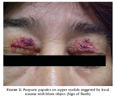
A white, 48-year-old female patient presented at the dermatology service complaining of papulous lesions with bloody content in the upper eyelids for one year. The first lesions appeared following emotional stress and regressed spontaneously within 15 days. Since then she started presenting new lesions in outbreaks, always in the same places. Such lesions worsened when there was local trauma. Concerning associated pathologies, arrhythmia (she was using a pacemaker), hypertension and fibromyalgia were informed. During the physical examination confluent papules of varied sizes were observed in the internal half of upper eyelids and more discreet in the internal corner of the lower eyelids (Figure 1). After local trauma with a blunt object, there was onset of local purpuric papules - a Sign of Barth (Figure 2).
Biopsies of the eyelid, gums and rectal mucosa lesions were performed, with the following results:
Skin: accumulation of amorphous and eosinophilic material, fragmented and globular in the papillary and reticular dermis, replacing collagen fibers; dilated and congested blood vessels (Figure 3). Congo red staining confirmed it was an amyloid substance (Figures 4 and 5).
Gums: No alterations. Absence of amyloid material.
Rectal mucosa: unspecific chronic retinitis. Absence of amyloid material.
The hemogram showed that creatinine, protein electrophoresis, VHS, iron and ferritin were within normal values. Cranium and long bones X-rays did not reveal lytic lesions. The echocardiogram was normal, from an anatomic viewpoint.
Among the abnormal tests, we found: proteinuria 570mg/24 hours; positive Bence-Jones proteinuria; and myelogram with 70% atypical plasmocytes.
Thus it was possible to make the diagnosis of multiple myeloma in the initial phase in a patient that presented only cutaneous manifestations of amyloidosis.
The patient was referred to a hematology service for multiple myeloma treatment. She died 5 months after the diagnosis.
DISCUSSION
The prevalence of amyloidosis associated with multiple myeloma varies from 13% to 26%.4
Among the patients with multiple myeloma, 15% may develop some form of amyloidosis.1, 4
The sequential analysis of aminoacids demonstrated that in systemic amyloidosis associated with multiple myeloma, fibrils are composed of an AL protein (light chain amyloidosis protein), which in turn is composed of variable immunoglobulin light chain (aminoterminal), intact light chain, or both.1
The most common cutaneous signs include: petechiae, purpuras and ecchymoses. Such signs occur spontaneously following trauma and are due to amyloid infiltration in the vascular walls.5 Other lesions may be seen, such as: plaques, papules and nodules; serous, smooth and shiny, amber or skin colored, non pruriginous, and they may be hemorrhagic. They affect mainly the eyelids, lips, tongue, oral mucosa, neck, axillae, umbilical scar and retroauricular, inguinal and anogenital regions. 1 Bullous lesions are rare.6
Some less frequent cutaneous alterations are: jaundice, paleness, hyperpigmentation, infiltrate similar to sclerodermia, alopecia areata or universal, nail dystrophies, cutis laxa and lesions similar to cutis verticis girata in the scalp.1
Actually, the amyloid material may be deposited in any tissue and/or organ, with specific manifestations.
In primary systemic amyloidosis, the deposit occurs predominantly in tissues such as in the gastrointestinal tract, smooth and skeletal muscles, carpal connection, nerves and skin. In contrast, in the secondary systemic form the amyloid deposition takes place above all in parenchymatous organs: liver, spleen, kidneys and adrenal glans.1
The amyloid deposits in the gastrointestinal tract may result in a variety of manifestations: abdominal pain, dysphagia, altered movements. Spleen and liver are present in 10% and 25% of cases, respectively.4
The tongue may be affected with macroglossia in 20% of cases, and there may be ulcerations, fissures, hemorrhages and teeth marks laterally. 4
The heart may be involved in 25% to 40% of cases and the kidneys in 30 to 50%, which are determinants of prognosis.4
Multiple myeloma is a bone marrow plasmocyte dyscrasia diagnosed when there is more than 10% plasmocytes in the bone marrow or a plasmocytoma and at least one of the following findings: 1) monoclonal serum protein; 2) monoclonal urine protein; 3) lytic bone lesions7-8 The presence of anemia, kidney insufficiency, hypercalcemia e amyloidosis were found.7
The prognosis of amyloidosis associated with multiple myeloma is unfavorable, with between 12 and 15 months mean survival rate.4 It depends on patient response to therapy and the extension of the disease. It is very important to make the earliest possible diagnosis, and always investigate multiple myeloma, especially in cases of systemic amyloidosis in patients considered suspect after the anatomic-pathological test used in this case.
Received on 28.03.2011.
Approved by the Advisory Board and accepted for publication on 11.05.2011.
Conflict of interest: None
Financial funding: None
- 1. Melo LV, Reis VMS, Criado PR, Müller H, Valente NYS. Amiloidose sistêmica associada a mieloma múltiplo: relato de caso com amiloidose cutânea exuberante. An Bras Dermatol. 1997;72:151-4.
- 2. Azulay RD, Azulay DR. Dermatologia. 2 ed. Rio de Janeiro: Guanabara Koogan; 2004. p. 346-7.
- 3. Sampaio SAP, Rivitti EA. Dermatologia. 3 ed. São Paulo: Artes Médicas; 2008. p. 895-9.
- 4. López L, González K, Navarrete G, Novales J, Guarneros A, Cortés B, et al. Multiple myeloma and systemic amyloidosis. Int J Dermatol. 2008;47:165-7.
- 5. Silverstein SR. Primary, systemic amyloidosis and the dermatologist where classic skin lesions may provide the clue for early diagnosis. Dermatol Online J. 2005;11:5.
- 6. Wang XD, Shen H, Liu ZH. Diffuse haemorrhagic bullous amyloidosis with multiple myeloma. Clin Exp Dermatol. 2007;33:94-6.
- 7. Yücel A, Akman A, Denli YG, Acar MA, Karakas M, Hazar B, et al. A case of systemic amyloidosis associated with multiple myeloma presented as macroglossia and purpura. J Eur Acad Dermatol Venereol. 2004;18:378-9.
- 8. Souza DAF, Freitas THP, Paes RAP, Müller H, Hungria VTM. Mieloma múltoplo com plasmocitomas cutâneos. An Bras Dermatol. 2004;79:581-5.
Publication Dates
-
Publication in this collection
02 Apr 2012 -
Date of issue
Feb 2012
History
-
Received
28 Mar 2011 -
Accepted
11 May 2011