Abstracts
Metastatic tuberculous abscesses or cutaneous tuberculous gummas occur mostly by reactivation of ancient cutaneous foci and is triggered mainly by pharmacological immunosuppression, diabetes mellitus, alcoholism or acquired immunodeficiency syndrome. The present case report refers to a 33-year-old male patient with polymyositis and a previous diagnosis of treated pulmonary tuberculosis. He presented cutaneous abscesses and fever months after the tuberculosis regimen. The patient was diagnosed as metastatic tuberculous abscesses associated with immunosuppression as result of polymyositis treatment.
Adrenal cortex hormones; Immunosuppression; Mycobacterium tuberculosis; Myositis; Polymyositis; Tuberculosis, cutaneous
Abscesso tuberculoso metastático ou goma tuberculosa cutânea, ocorre principalmente pela reativação de surtos cutâneos precoces desencadeados principalmente por imunodepressão farmacológica, diabetes mellitus, alcoolismo ou síndrome da imunodeficiência adquirida. O presente caso relatado refere-se a um paciente do sexo masculino de 33 anos portador de polimiosite e com diagnóstico prévio de tuberculose pulmonar tratada. Apresentou, meses depois do tratamento para tuberculose, abscessos cutâneos e febre. Após a investigação clínica foi diagnosticado como portador de tuberculose gomosa cutânea associada à imunodepressão consequente ao tratamento de polimiosite.
Corticosteróides; Imunossupressão; Miosite; Mycobacterium tuberculosis; Polimiosite; Tuberculose cutânea
INTRODUCTION
Tuberculosis is an infectious disease caused by Mycobacterium tuberculosis and may also be caused by other species of the same genus such as M. bovis, M. africanum, M. microti or M. canetti. 11. Kasper DL, Braunwald E, Fauci AS, Hauser SL, Longo DL, Jameson JC, organizadores. Harrison Medicina Interna. Volume I. 17. ed. Rio de Janeiro: McGraw Hill; 2008. Capítulo 158, Tuberculose; p. 1006-20. These infectious agents are aerobic bacilli with a thick waxy cell wall composed of mycolic acid, which renders them acid-fast.22. Cotran RS, Kumar V, Robbins ST, organizadores. Patologia. Bases patológicas das doenças. 7 ed. Rio de Janeiro: Elsevier; 2005. Cap 8, Doenças infecciosas; p. 397.
Pulmonary involvement is the most common form of the disease. Nevertheless, several organ systems may also be affected by hematogenous dissemination, causing extrapulmonary tuberculosis. The extrapulmonary tuberculosis presentations have probably been increasing as a result of AIDS and the increased use of immunosuppressive drugs.11. Kasper DL, Braunwald E, Fauci AS, Hauser SL, Longo DL, Jameson JC, organizadores. Harrison Medicina Interna. Volume I. 17. ed. Rio de Janeiro: McGraw Hill; 2008. Capítulo 158, Tuberculose; p. 1006-20.
Cutaneous tuberculosis is responsible for about 2.1% of all cases of tuberculosis. The greatest incidence occurs in people on a low income. With the advent of AIDS there was a rise in incidence probably due to a process of reactivation of latent tuberculosis in immunocompromised people by an increase in the number of bacilli that are resistant to treatment or because immunosuppression facilitates infection by less virulent strains.11. Kasper DL, Braunwald E, Fauci AS, Hauser SL, Longo DL, Jameson JC, organizadores. Harrison Medicina Interna. Volume I. 17. ed. Rio de Janeiro: McGraw Hill; 2008. Capítulo 158, Tuberculose; p. 1006-20.
CASE REPORT
A 33-year-old male patient resident of Manguinhos - Rio de Janeiro - Brazil was diagnosed 18 months ago with polymyositis and was irregularly taking methotrexate (15mg/weak) with prednisone (40 mg/day); however, he failed to attend medical followup visits regularly. One year ago, he was hospitalized with pulmonary tuberculosis. He was prescribed rifampicin, isoniazid and pyrazinamide and completed a six-month treatment period.
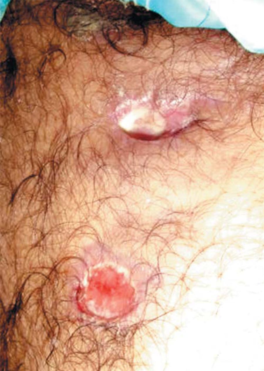
Six months later, he was admitted at the Hospital with painful warm nodules of various sizes on his body in addition to gummatous lesions developed two months earlier (Figures 1, 2 and 3). He presented intermittent fever which responded to metamizole. There was throbbing pain in his limbs, aggravated by movement and alleviated by resting. He also complained of muscle weakness in pectoral and pelvic girdle.
At admission, he had tachypnea, tachycardia and fever. Lymphadenopathy was absent and the blood pressure was normal. The laboratory tests showed leukocytosis (21,100 leukocytes), thrombocytosis (573,000 platelets) and normocytic normochromic anemia. Lactate dehydrogenase (LDH) levels were 515 U/I and creatine phosphokinase (CPK) 282 IU/l. Chest x-ray revealed no abnormalities. During hospitalization, leukocytosis, thrombocytosis and normocytic normochromic anemia persisted, as well as increased LDH levels. Creatine phosphokinase returned to normal levels.
The differential diagnosis of the nodular disease mainly considered myositis and vasculitis, methotrexate-related panniculitis, Weber-Christian disease, subcutaneous panniculitis-like T-cell lymphoma, erythema nodosum,> bacterial, fungal and mycobacterial infections.
At admission antibiotic therapy with oxacillin was initiated against infection by Staphylococcus. However, no specimen has confirmed the suspicion of diagnosis. These results hampered the investigation for presence of staphylococci bacteria and made the decision to maintain or not the antibiotics difficult. The patient's fever and skin rash persisted despite the therapy. On the seventh day of treatment, the antibiotic was changed to vancomycin at 500 mg qid intravenously for 14 days; no improvement occurred in the patient's clinical condition.
The material collected from the lesions was studied for histopathology patterns, bacterioscopy and culture studies.
Ultrasound confirmed a subcutaneous structural alteration on the posterior region of the upper third of the right leg, with 8 cm in size and without liquid. The possibility of purulent material being accumulated inside the lesion could not be discarded.
Skin histopathology studies revealed acanthosis, edema of the reticular dermis, and mild infiltrate of lymphocytes with some neutrophils around vessels and skin adnexa. Panniculitis was suggested by the presence of subcutaneous necrosis of adipocytes, deposits of fibrinoid material and hyaline eosinophilic strands. Initially, aspirated secretion from the lesions was negative for acid-fast bacilli.
One week later, a new sample showed acid-fast bacilli (+ + +). Rifampicin 600 mg qd, isoniazide 400 mg qd, pyrazinamide 2 g qd and ethambutol 1.2 g qd were initiated. Within a five-day treatment period the patient improved considerably, the fever disappeared and he was discharged. Culture studies revealed growth of Mycobacterium tuberculosis. Lesions were totally cured after 6 months.
DISCUSSION
Cutaneous tuberculosis is a rare disease with specific clinical features and natural history. Clinical manifestation and following events, may depend on multiple factors such as how bacilli accessed the skin; the strain's virulence; number of inoculated bacilli; immune state of the patient and the relation of Mycobacterium with the anatomical region affected.33. Azulay RD, Azulay DR. Dermatologia. 5. ed. Rio de Janeiro: Guanabara Koogan; 2008. p, 337.
Skin lesions can be divided according to exposure in primary and secondary lesions, by local inoculation or by distant dispersion, respectively. Tuberculids are a hypersensitivity reaction to a distant focus of infection.44. Rabello FE, Fraga S. Atlas de Dermatologia. Rio de Janeiro: Guanabara Koogan; 1970. p.176-283.
Diagnosis of cutaneous tuberculosis may be achieved by direct microscopy, PCR, culture and histopathology study of a biopsy specimen. Chest xray with pulmonary involvement, active or not, as well as positive PPD may help the diagnosis.55. Hinrichsen SL, Moura LV, Arraes LC, Reis L, Lamprea D, Gava RK. Tuberculose cutânea e AIDS: relato de um caso. An Bras Dermatol. 1996;71:511-4.
The patient had a previous diagnosis of polymyositis. It causes an autoimmune chronic inflammation of the muscles with marked pain, weakness and/or loss of muscle mass in the proximal musculature, particularly in the shoulder and pelvic girdle. The standard treatment is pharmacologic immunosuppression.
The simultaneously presentation of polymyositis and cutaneous mycobacteriosis is rare. After a literature review we have found 3 similar cases of patients with cutaneous tuberculosis plus dermatomyositis and one case of neutrophilic tuberculous panniculitis in a patient with polymyositis. The immunosuppressive treatment seems to be responsible for the presentation of cutaneous tuberculosis. Besides, because of the number of case reports, a study is necessary to evaluate the direct relationship between polymyositis and cutaneous tuberculosis.66. Kim JE, Ko JY, Bae SC, Ro YS. Tuberculous cellulitis as a manifestation of miliary tuberculosis in a patient with malignancy-associated dermatomyositis. J Am Acad Dermatol. 2011;65:450-2.
7. Fujita M, Arakawa K, Mizuno S, Wakabayashi M, Totani Y, Demura Y, Ameshima S, et al. A case of cutaneous tuberculosis under steroid & immunosuppressant therapy for dermatomyositis. Kekkaku. 2002;77:465-70.
8. Algayrès JP, Mayaudon H, Godeau B, Schill H, Valmary J, Maurel C, et al. Mycobacterium kansasii extensive subcutaneous infection in dermatomyositis. Ann Med Interne (Paris). 1990;141:633-5.-99. Langenberg A, Egbert B. Neutrophilic tuberculous panniculitis in a patient with polymyositis. J Cutan Pathol. 1993;20:177-9.
In the present case, the treatment of polymyositis may have induced the development of pulmonary tuberculosis; and one year later the onset of cutaneous disease was observed. During hospitalization, chest x-ray and CT scanning showed no evidence of tuberculosis activity. The lack of response of the tuberculin test and the absence of granuloma histopathology findings were probably due to immunosuppression of the cell-mediated immunity. An additional investigation of secretion showed acid-fast bacilli (+ + +) and growth of Mycobacterium tuberculosis in culture. The diagnosis of cutaneous metastatic tuberculous abscess, a form of tuberculosis that results from reactivation of primary foci, was established from the presence of subcutaneous, multibacillary abscesses that formed fistulae and drained spontaneously.1010. Lopes AJ, Capone D, Mogami R, Tessarollo B, da Cunha DL, Capone RB, et al. Tuberculose extrapulmonar: aspectos clínicos e de imagem. Pulmão RJ. 2006;15:253-61.
The standard RIPE regimen for six months has been shown to be effective in these forms of the disease.
REFERENCES
-
1Kasper DL, Braunwald E, Fauci AS, Hauser SL, Longo DL, Jameson JC, organizadores. Harrison Medicina Interna. Volume I. 17. ed. Rio de Janeiro: McGraw Hill; 2008. Capítulo 158, Tuberculose; p. 1006-20.
-
2Cotran RS, Kumar V, Robbins ST, organizadores. Patologia. Bases patológicas das doenças. 7 ed. Rio de Janeiro: Elsevier; 2005. Cap 8, Doenças infecciosas; p. 397.
-
3Azulay RD, Azulay DR. Dermatologia. 5. ed. Rio de Janeiro: Guanabara Koogan; 2008. p, 337.
-
4Rabello FE, Fraga S. Atlas de Dermatologia. Rio de Janeiro: Guanabara Koogan; 1970. p.176-283.
-
5Hinrichsen SL, Moura LV, Arraes LC, Reis L, Lamprea D, Gava RK. Tuberculose cutânea e AIDS: relato de um caso. An Bras Dermatol. 1996;71:511-4.
-
6Kim JE, Ko JY, Bae SC, Ro YS. Tuberculous cellulitis as a manifestation of miliary tuberculosis in a patient with malignancy-associated dermatomyositis. J Am Acad Dermatol. 2011;65:450-2.
-
7Fujita M, Arakawa K, Mizuno S, Wakabayashi M, Totani Y, Demura Y, Ameshima S, et al. A case of cutaneous tuberculosis under steroid & immunosuppressant therapy for dermatomyositis. Kekkaku. 2002;77:465-70.
-
8Algayrès JP, Mayaudon H, Godeau B, Schill H, Valmary J, Maurel C, et al. Mycobacterium kansasii extensive subcutaneous infection in dermatomyositis. Ann Med Interne (Paris). 1990;141:633-5.
-
9Langenberg A, Egbert B. Neutrophilic tuberculous panniculitis in a patient with polymyositis. J Cutan Pathol. 1993;20:177-9.
-
10Lopes AJ, Capone D, Mogami R, Tessarollo B, da Cunha DL, Capone RB, et al. Tuberculose extrapulmonar: aspectos clínicos e de imagem. Pulmão RJ. 2006;15:253-61.
-
* Study carried out at the Teaching Hospital, Federal University of Rio de Janeiro (Hospital Universitário Gaffrée e Guinle. Universidade Federal do Estado do Rio de Janeiro - HUGG-UNIRIO) - Rio de Janeiro (RJ), Brazil.
Publication Dates
-
Publication in this collection
Feb 2013
History
-
Received
17 Jan 2011 -
Accepted
15 Jan 2012