Abstracts
The authors report a case of ectopic cutaneous schistosomiasis in a 35 year-old female who presented clustered reddish macules and papules on the left buttock. The diagnosis was not suspected during clinical evaluation and required visualization of Schistosoma mansoni eggs on sections of tissue.
Schistosoma mansoni; Schistosomiasis mansoni; Skin
Os autores relatam um caso de esquistossomose cutânea ectópica em uma paciente de 35 anos que apresentou máculas e pápulas eritematosas agrupadas na nádega esquerda. O diagnostico não foi suspeitado durante a avaliação clínica, tendo sido obtido através da visualização dos ovos no exame histopatológico.
Esquistossomose mansoni; Pele; Schistosoma mansoni
CASE REPORT
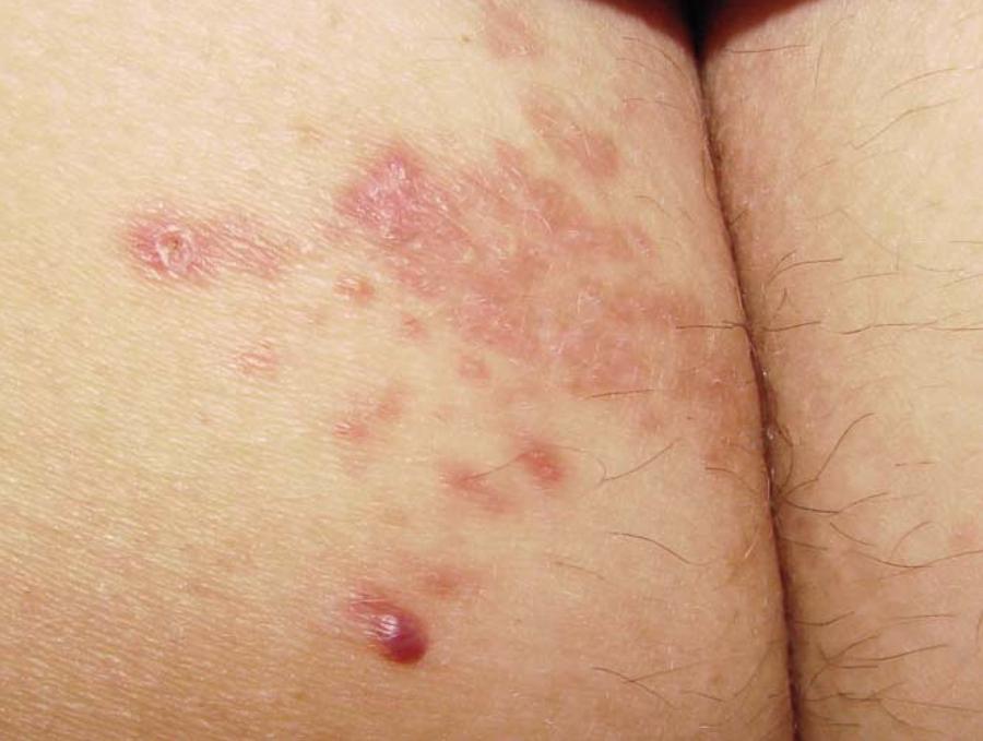
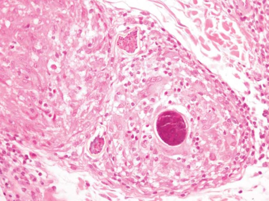
This was a thirty-five year-old, white, female patient born in Paraíba and resident in Jacarepaguá/RJ for the last 34 years. She presented with a cluster of reddish-coppery asymptomatic macules and papules on the left buttock, arising 30 days after a waterfall bathing at the countryside of the state (Figures 1 and 2). The diagnostic hypotheses were larva migrans, residual herpes simplex and sarcoidosis. Histopathological examination showed epithelioid granulomas surrounding eggs with an insinuation of a lateral spine, containing miracidia (Figure 3 and 4). Complete examination of gastrointestinal system and parasitological stool analysis did not show any alterations.
Reddish-coppery macules and papules, clustered and with a smooth surface. One of the lesions is eroded
Medium dermis with an epithelioid granuloma surrounding three Schistosoma mansoni eggs. HE, 100X
Detail of the three Schistosoma mansoni eggs containing viable miracidia. Notice that the in the eggs on the left side, it is possible to identify the characteristic lateral spine. HE, 400x
DISCUSSION
Ectopic cutaneous schistosomiasis happens when the trematodes' eggs are deposited in the
dermis, instead of being eliminated via stools (S. japonicum and
S. mansoni) or urine (S. hematobium).11. Andrade Filho Jde S, Lopes MS, Corgozinho Filho AA, Pena GP. Ectopic
cutaneous schistosomiasis: report of two cases and a review of the literature. Rev
Inst Med Trop São Paulo. 1998;40:253-7.
,
22. Lopes R, Sousa MAJ, Jeunon T. Tropical troves: what is your
diagnosis? Dermatopathol Pract Conc. 2010 [cited 2012 Sep 9];16:3. Available from:
http://www.derm101.com.
http://www.derm101.com...
The only species reported in Brazil is S. mansoni, whose
eggs are identified by the presence of a thick lateral spine.33. Nunes FC, Costa MCE, Filhote MIF, Sharapinn M. Perfil Epidemiológico
da esquistossomose mansônica no bairro Alto da Boa Vista, no Rio de Janeiro.
Epidemiological profile o mansonic schistosomiasis at Alto da Boa Vista neighborhood
in Rio de Janeiro. Cad Saúde Colet (Rio J). 2005;13:605-16. Lesions are typically located at the periumbilical region, torso,
superior dorsal region, buttocks, and genital area. They appear as skin-colored papules,
sometimes with a reddish-brown coloration, and may spontaneously regress or evolve to
lichenified, vegetant or even tumoral lesions. Ulceration and fistulization may also
occur.33. Nunes FC, Costa MCE, Filhote MIF, Sharapinn M. Perfil Epidemiológico
da esquistossomose mansônica no bairro Alto da Boa Vista, no Rio de Janeiro.
Epidemiological profile o mansonic schistosomiasis at Alto da Boa Vista neighborhood
in Rio de Janeiro. Cad Saúde Colet (Rio J). 2005;13:605-16.
,
44. Carmo EH, Bina JC, Barreto ML. Schistosomiasis related morbidity in
Brazil; Spatial distribution, clinical features evolution and medical services
assessment. In: Simpósio internacional sobre esquistossomose, Recife-PE.
1997;6:166.
,
55. Costa IM, Moreira RR, Moraes MA. Esquistossomose mansônica cutânea
ectópica. Ectopic cutaneous mansonic schistosomiasis. An Bras Dermatol.
1989;64:183-4.
There were previous case reports of schistosomiasis in the region of Jacarepaguá. We believe the patient had already contracted the disease previously to this last waterfall bathing, because the acute phase usually presents with hepatoesplenomegalia and the period of egg laying starts 40 days after the infection.33. Nunes FC, Costa MCE, Filhote MIF, Sharapinn M. Perfil Epidemiológico da esquistossomose mansônica no bairro Alto da Boa Vista, no Rio de Janeiro. Epidemiological profile o mansonic schistosomiasis at Alto da Boa Vista neighborhood in Rio de Janeiro. Cad Saúde Colet (Rio J). 2005;13:605-16. , 66. Ferreira LF, Naveira JB, Silva JR. Fase toxêmica da esquistossomose mansoni. Toxemic phase of mansonic schistosomiasis. Rev Inst Med Trop Sao Paulo. 1960;2:112-20. , 77. Domingues AL, Medeiros TB, Lopes EP. Ultrasound versus biological markers in the evaluation of periportal fibrosis in human Schistosoma mansoni. Mem Inst Oswaldo Cruz. 2011;106:802-7. In the chronic phase of schistosomiasis, the process of egg laying is diminished, and it becomes difficult to detect them on stool samples. Only 2 to 7% of (chronic) patients develop portal hypertension in Brazil44. Carmo EH, Bina JC, Barreto ML. Schistosomiasis related morbidity in Brazil; Spatial distribution, clinical features evolution and medical services assessment. In: Simpósio internacional sobre esquistossomose, Recife-PE. 1997;6:166. , 66. Ferreira LF, Naveira JB, Silva JR. Fase toxêmica da esquistossomose mansoni. Toxemic phase of mansonic schistosomiasis. Rev Inst Med Trop Sao Paulo. 1960;2:112-20. , 77. Domingues AL, Medeiros TB, Lopes EP. Ultrasound versus biological markers in the evaluation of periportal fibrosis in human Schistosoma mansoni. Mem Inst Oswaldo Cruz. 2011;106:802-7.
REFERENCES
-
1Andrade Filho Jde S, Lopes MS, Corgozinho Filho AA, Pena GP. Ectopic cutaneous schistosomiasis: report of two cases and a review of the literature. Rev Inst Med Trop São Paulo. 1998;40:253-7.
-
2Lopes R, Sousa MAJ, Jeunon T. Tropical troves: what is your diagnosis? Dermatopathol Pract Conc. 2010 [cited 2012 Sep 9];16:3. Available from: http://www.derm101.com.
» http://www.derm101.com -
3Nunes FC, Costa MCE, Filhote MIF, Sharapinn M. Perfil Epidemiológico da esquistossomose mansônica no bairro Alto da Boa Vista, no Rio de Janeiro. Epidemiological profile o mansonic schistosomiasis at Alto da Boa Vista neighborhood in Rio de Janeiro. Cad Saúde Colet (Rio J). 2005;13:605-16.
-
4Carmo EH, Bina JC, Barreto ML. Schistosomiasis related morbidity in Brazil; Spatial distribution, clinical features evolution and medical services assessment. In: Simpósio internacional sobre esquistossomose, Recife-PE. 1997;6:166.
-
5Costa IM, Moreira RR, Moraes MA. Esquistossomose mansônica cutânea ectópica. Ectopic cutaneous mansonic schistosomiasis. An Bras Dermatol. 1989;64:183-4.
-
6Ferreira LF, Naveira JB, Silva JR. Fase toxêmica da esquistossomose mansoni. Toxemic phase of mansonic schistosomiasis. Rev Inst Med Trop Sao Paulo. 1960;2:112-20.
-
7Domingues AL, Medeiros TB, Lopes EP. Ultrasound versus biological markers in the evaluation of periportal fibrosis in human Schistosoma mansoni. Mem Inst Oswaldo Cruz. 2011;106:802-7.
-
Financial funding: None
-
*
Work performed at the Laboratory of Dermatology Investigation (ID)- Rio de Janeiro (RJ), Brazil.
Publication Dates
-
Publication in this collection
Oct 2013
History
-
Received
04 Sept 2012 -
Accepted
22 Oct 2012