Abstract
Superficial Acral Fibromyxoma is a rare tumor of soft tissues. It is a relatively new entity described in 2001 by Fetsch et al. It probably represents a fibrohistiocytic tumor with less than 170 described cases. We bring a new case of SAF on the 5th toe of the right foot, in a 43-year-old woman. After surgical excision with safety margins which included the nail apparatus, it has not recurred (22 months of follow up). We carried out a review of the location of all SAF published up to the present day.
Nails; Neoplasm recurrence, local; Neoplasms; Recurrence
INTRODUCTION
Superficial Acral Fibromyxoma (SAF) is a rare tumor of soft tissues with slow growth and acral location. It has a benign behavior, but it may persist or recur if not properly treated.
CASE REPORT
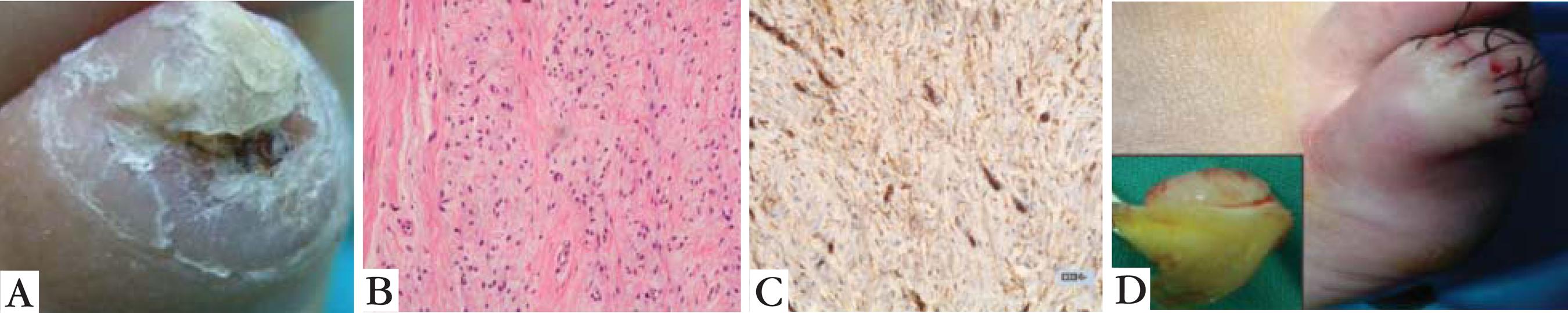
We present a 43-year-old woman, without any known allergies whose personal history reports beta thalassemia. She referred having had cutaneous changes not associated with any trauma for 8 years, consisting of swelling, partial nail loss and distal ulcerations with occasional bleeding on the 5th toe of the right foot. When the patient wore open shoes it was painless; however, it hurt and bled when she wore closed shoes. Upon examination, the distal end of the 5th toe presented a central ulcer with blood remains and partial onycholysis (Figure 1A).
A: Loss of the distal end of the 5th toe of the right foot and partial onycholysis; B: Fusiform cell proliferation in a myxoid stroma (HE, x100); C: Fusiform cells show cytoplasmic positivity to CD34 (PAP, x200); D: Image after excision. The macroscopic part of the tumor along with nail apparatus can be seen
Antero-posterior and oblique X-rays were requested of both feet, which showed subluxation of the distal phalanges of the 5th toes without signs of bone infiltration and a diagnostic biopsy was performed.
Histological results showed neoplastic dermal proliferation of fusiform cells without any relevant atypia, immersed in a myxoid stroma with collagenized areas and a prominent vascular weave (Figure 1B). Immunohistochemical studies reported positive results for CD34 and negative for S100, AME and AML (Figure 1C). The proliferation index, valued with Ki67 was low (less than 1%). These findings led to the diagnosis of Superficial Acral Fibromyxoma.
The subsequent therapeutic approach included complete removal of the tumor as well as the nail in order to avoid recurrence.
Histological examination of the surgical piece was similar to the previously described. The tumor was extirpated with wide margins, including the nail matrix, respecting the distal phalanx (Figure 1D). Resection margins were reported as tumor-free.
After a 22-month follow-up there was no recurrence of the tumor.
DISCUSSION
Superficial Acral Fibromyxoma (SAF) is a rare tumor of soft tissues, with slow growth
and located in the subungual or periungual region of the hands and feet11. Ashby-Richardson H, Rogers GS, Stadecker MJ. Superficial acral
fibromyxoma: an overview. Arch Pathol Lab Med. 2011;135:1064-6.
2. Wakabayashi Y, Nakai N, Takenaka H, Katoh N. Superficial acral
fibromyxoma of the great toe: case report and mini-review of the literature. Acta
Dermatovenerol Croat. 2012;20:263-6.
3. Messeguer F, Nagore E, Agustí-Mejias A, Traves V. Fibromixoma acral
superficial, un tumor periungueal CD34 positivo. Actas Dermosifiliogr.
2012;103:67-9.
4. Fetsch JF, Laskin WB, Miettinen M. Superficial acral fibromyxoma: a
clinicopathologic and immunohistochemical analysis of 37 cases of a distinctive soft
tissue tumor with a predilection for the fingers and toes. Hum Pathol.
2001;32:704-14.
5. Wei C, Fleegler EJ. Superficial acral fibromyxoma of the thumb.
Eplasty. 2013;13:ic13
6. Prescott RJ, Husain EA, Abdellaoui A, Al-Mahmoud RM, Khan M, Salman
WD, et al. Superficial acral fibromyxoma: a clinicopathological study of 41 new cases
from the U.K.: should myxoma (NOS) and fibroma (NOS) continue as part of 21stcentury
reporting? Br J Dermatol. 2008;159:1315-21-77. Al-Daraji WI, Miettinen M.. Superficial acral fibromyxoma: a
clinicopathological analysis of 32 tumors including 4 in the heel. J Cutan Pathol.
2008 Nov;35(11):1020-6. (Table 1). However, the heel, palm
and ankle can also be affected.55. Wei C, Fleegler EJ. Superficial acral fibromyxoma of the thumb.
Eplasty. 2013;13:ic13 It affects young
adults (mean age 43 years old), with higher frequency in men than in women in a 2:1
proportion. It is a relatively new entity described in 2001 by Fetsch et
al.44. Fetsch JF, Laskin WB, Miettinen M. Superficial acral fibromyxoma: a
clinicopathologic and immunohistochemical analysis of 37 cases of a distinctive soft
tissue tumor with a predilection for the fingers and toes. Hum Pathol.
2001;32:704-14. It probably represents a
fibrohistiocytic tumor with less than 170 described cases (SAF series and isolated case
reports).22. Wakabayashi Y, Nakai N, Takenaka H, Katoh N. Superficial acral
fibromyxoma of the great toe: case report and mini-review of the literature. Acta
Dermatovenerol Croat. 2012;20:263-6.
Pain is not usually mentioned. Ungual involvement may be present. Only one case has been associated with previous trauma. X-rays rarely show bone alterations.11. Ashby-Richardson H, Rogers GS, Stadecker MJ. Superficial acral fibromyxoma: an overview. Arch Pathol Lab Med. 2011;135:1064-6.,88. Goo J, Jung YJ, Kim JH, Lee SY, Ahn SK. A Case of Recurrent Superficial Acral Fibromyxoma. Ann Dermatol. 2010;22:110-3.
Histologically, it is a well delimited, non-encapsulated dermal tumor that may extend
towards the hypodermis. It is composed of a proliferation of cells from a fibroblastic
line usually accompanied by many mast cells. The presence of a myxoid stroma with a rich
vascular weave is very noticeable. Epidermis hyperplasia with hyperkeratosis is also
frequent. CD34 positivity is characteristic but CD10, CD99, EMA, and nestin
immunoreactivity are also common. Negative results for neural and muscular
differentiation markers (S-100, HMB-45, SMA, desmin, actin), cytokeratin and
apolipoprotein D are expected.11. Ashby-Richardson H, Rogers GS, Stadecker MJ. Superficial acral
fibromyxoma: an overview. Arch Pathol Lab Med. 2011;135:1064-6.
2. Wakabayashi Y, Nakai N, Takenaka H, Katoh N. Superficial acral
fibromyxoma of the great toe: case report and mini-review of the literature. Acta
Dermatovenerol Croat. 2012;20:263-6.-33. Messeguer F, Nagore E, Agustí-Mejias A, Traves V. Fibromixoma acral
superficial, un tumor periungueal CD34 positivo. Actas Dermosifiliogr.
2012;103:67-9.,
99. Quaba O, Evans A, Al-Nafussi AA, Nassan A. Superficial acral
fibromyxoma. Br J Plast Surg. 2005;58:561-4.,1010. Frierson HF, Cooper PH. Myxoid variant of dermatofibrosarcoma
protuberans. Am J Surg Pathol. 1983;7:445-50.
Although it is an infrequent event, it must be included in differential diagnosis of
tumors present on the fingers and toes.11. Ashby-Richardson H, Rogers GS, Stadecker MJ. Superficial acral
fibromyxoma: an overview. Arch Pathol Lab Med. 2011;135:1064-6.
2. Wakabayashi Y, Nakai N, Takenaka H, Katoh N. Superficial acral
fibromyxoma of the great toe: case report and mini-review of the literature. Acta
Dermatovenerol Croat. 2012;20:263-6.
3. Messeguer F, Nagore E, Agustí-Mejias A, Traves V. Fibromixoma acral
superficial, un tumor periungueal CD34 positivo. Actas Dermosifiliogr.
2012;103:67-9.
4. Fetsch JF, Laskin WB, Miettinen M. Superficial acral fibromyxoma: a
clinicopathologic and immunohistochemical analysis of 37 cases of a distinctive soft
tissue tumor with a predilection for the fingers and toes. Hum Pathol.
2001;32:704-14.
5. Wei C, Fleegler EJ. Superficial acral fibromyxoma of the thumb.
Eplasty. 2013;13:ic13
6. Prescott RJ, Husain EA, Abdellaoui A, Al-Mahmoud RM, Khan M, Salman
WD, et al. Superficial acral fibromyxoma: a clinicopathological study of 41 new cases
from the U.K.: should myxoma (NOS) and fibroma (NOS) continue as part of 21stcentury
reporting? Br J Dermatol. 2008;159:1315-21-77. Al-Daraji WI, Miettinen M.. Superficial acral fibromyxoma: a
clinicopathological analysis of 32 tumors including 4 in the heel. J Cutan Pathol.
2008 Nov;35(11):1020-6. Differential diagnosis
considerations are summarized in chart 1.
SAF has a benign behavior but may persist or recur if not properly treated.77. Al-Daraji WI, Miettinen M.. Superficial acral fibromyxoma: a clinicopathological analysis of 32 tumors including 4 in the heel. J Cutan Pathol. 2008 Nov;35(11):1020-6.,88. Goo J, Jung YJ, Kim JH, Lee SY, Ahn SK. A Case of Recurrent Superficial Acral Fibromyxoma. Ann Dermatol. 2010;22:110-3. Thus complete removal and follow-up is recommended. Up to this date, malignization has not been described.
In conclusion, this is the description of a rare case of Superficial Acral Fibromyxoma on the nail apparatus of a 43-year-old woman. There are less than 170 published cases. It is a benign tumor with slow growth and, although rare, it should be considered in differential diagnosis of acral lesions. Surgery is curative but requires adequate margins due to the high risk of recurrence. Malignization has never been described.
REFERENCES
-
1Ashby-Richardson H, Rogers GS, Stadecker MJ. Superficial acral fibromyxoma: an overview. Arch Pathol Lab Med. 2011;135:1064-6.
-
2Wakabayashi Y, Nakai N, Takenaka H, Katoh N. Superficial acral fibromyxoma of the great toe: case report and mini-review of the literature. Acta Dermatovenerol Croat. 2012;20:263-6.
-
3Messeguer F, Nagore E, Agustí-Mejias A, Traves V. Fibromixoma acral superficial, un tumor periungueal CD34 positivo. Actas Dermosifiliogr. 2012;103:67-9.
-
4Fetsch JF, Laskin WB, Miettinen M. Superficial acral fibromyxoma: a clinicopathologic and immunohistochemical analysis of 37 cases of a distinctive soft tissue tumor with a predilection for the fingers and toes. Hum Pathol. 2001;32:704-14.
-
5Wei C, Fleegler EJ. Superficial acral fibromyxoma of the thumb. Eplasty. 2013;13:ic13
-
6Prescott RJ, Husain EA, Abdellaoui A, Al-Mahmoud RM, Khan M, Salman WD, et al. Superficial acral fibromyxoma: a clinicopathological study of 41 new cases from the U.K.: should myxoma (NOS) and fibroma (NOS) continue as part of 21stcentury reporting? Br J Dermatol. 2008;159:1315-21
-
7Al-Daraji WI, Miettinen M.. Superficial acral fibromyxoma: a clinicopathological analysis of 32 tumors including 4 in the heel. J Cutan Pathol. 2008 Nov;35(11):1020-6.
-
8Goo J, Jung YJ, Kim JH, Lee SY, Ahn SK. A Case of Recurrent Superficial Acral Fibromyxoma. Ann Dermatol. 2010;22:110-3.
-
9Quaba O, Evans A, Al-Nafussi AA, Nassan A. Superficial acral fibromyxoma. Br J Plast Surg. 2005;58:561-4.
-
10Frierson HF, Cooper PH. Myxoid variant of dermatofibrosarcoma protuberans. Am J Surg Pathol. 1983;7:445-50.
-
*
Work performed at the Hospital Universitario Virgen Macarena - Sevilla, Spain.
-
Financial Support: none
Publication Dates
-
Publication in this collection
Jan-Feb 2014
History
-
Received
04 Apr 2013 -
Accepted
17 July 2013