Abstract
We report a case of Tinea nigra in an adolescent living in Itapema, Santa Catarina, Brazil, who presented a hyperchromic macule on the palm of the left hand, close to another erythematous macule caused by a rabbit bite. The patient received guidance on accidents and animal bites and evolved well treated with topical butenafine for the dermatomycosis. The authors also highlight the efficacy of the dermoscopic exam in diagnosing Tinea nigra with animal bite lesions and other traumas.
Bites and stings; Dermatomycoses; Dermoscopy; Mycoses; Tinea
Tinea nigra (TN) manifests as an asymptomatic hyperchromic macule in the
palmar region, and eventually the plantar or other atypical areas.11. Zaitz C, Campbell I, Marques AS, Ruiz LRB, Framil VMS. Dermatomicoses
por fungos filamentosos septados demácios. In: Zaitz C, Campbell I, Marques SA, Ruiz
LRB, Framil VMS. Compêndio de Micologia Médica. 2. ed. Rio de Janeiro: Guanabara
Koogan, 2010. p. 180-6.
2. Rossetto AL, Cruz RC, Haddad Junior V. Double-blind study with the
topical isoconazole and terbinafine for the treatment of one patient bilateral Tinea
nigra plantaris and suggestions for new differential diagnoses. Rev Inst Med Trop Sao
Paulo. 2013;55:125-8.
3. Rossetto AL, Cruz RCB. Tinea nigra in geographical forms of "heart"
and "parrot beak". An Bras Dermatol. 2011;86:389-90.
4. Rossetto AL, Cruz RC. Tinea nigra: successful treatment with topical
butenafine. An Bras Dermatol. 2012;87:939-41.-55. Rossetto AL, Cruz RCB. Spontaneous cure in a case of tinea nigra. An
Bras Dermatol. 2012;87:160-2. Dermoscopy exam
reveals its unique characteristic pattern, but aetiology is established by the mycologic
examination.66. Bonifaz A, Badali H, de Hoog GS, Cruz M, Araiza J, Cruz MA, et al.
Tinea nigra by Hortaea werneckii, a report of 22 cases from Mexico. Stud Mycol.
2008;61:77-82.
7. Xavier MH, Ribeiro LH, Duarte H, Saraça G, Souza AC. Dermatoscopy in
the diagnosis of tinea nigra. Dermatol Online J. 2008;14:15.
8. Paschoal FM, de Barros JA, de Barros DP, de Barros JC, Filho CD.
Study of the Dermatoscopic Pattern of Tinea Nigra: Report of 6 Cases. Skinmed.
2010;8:319-21.-99. Piliouras P, Allison S, Rosendahl C, Buettner PG, Weedon D.
Dermoscopy improves diagnosis of tinea nigra: A study of 50 cases. Australas J
Dermatol. 2011;52:191-4. Treatment response is good to topical antifungals with reports of
spontaneous cure.22. Rossetto AL, Cruz RC, Haddad Junior V. Double-blind study with the
topical isoconazole and terbinafine for the treatment of one patient bilateral Tinea
nigra plantaris and suggestions for new differential diagnoses. Rev Inst Med Trop Sao
Paulo. 2013;55:125-8.
3. Rossetto AL, Cruz RCB. Tinea nigra in geographical forms of "heart"
and "parrot beak". An Bras Dermatol. 2011;86:389-90.
4. Rossetto AL, Cruz RC. Tinea nigra: successful treatment with topical
butenafine. An Bras Dermatol. 2012;87:939-41.
5. Rossetto AL, Cruz RCB. Spontaneous cure in a case of tinea nigra. An
Bras Dermatol. 2012;87:160-2.-66. Bonifaz A, Badali H, de Hoog GS, Cruz M, Araiza J, Cruz MA, et al.
Tinea nigra by Hortaea werneckii, a report of 22 cases from Mexico. Stud Mycol.
2008;61:77-82.
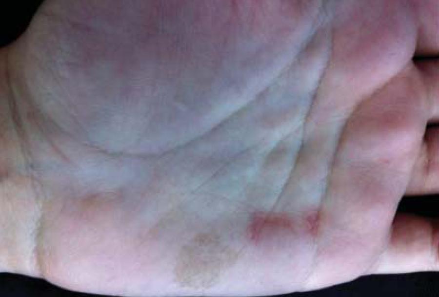
The patient was a thirteen year old female with two asymptomatic marks measuring 1.0 x 2.0 cm in the left palmar region, one black spotted and the other erythematous, of geographic shape and scaly border, caused by a European rabbit (Oryctolagus cuniculus) bite while playing with the animal (Figure 1).
Detail of two macules, both sized approximately 1.0 x 2.0 cm and located on the internal edge of the left palmar region, one black spotted and not scaly, and one erythematous of geographic shape, poorly delimited borders, and with discrete scaling on the distal border
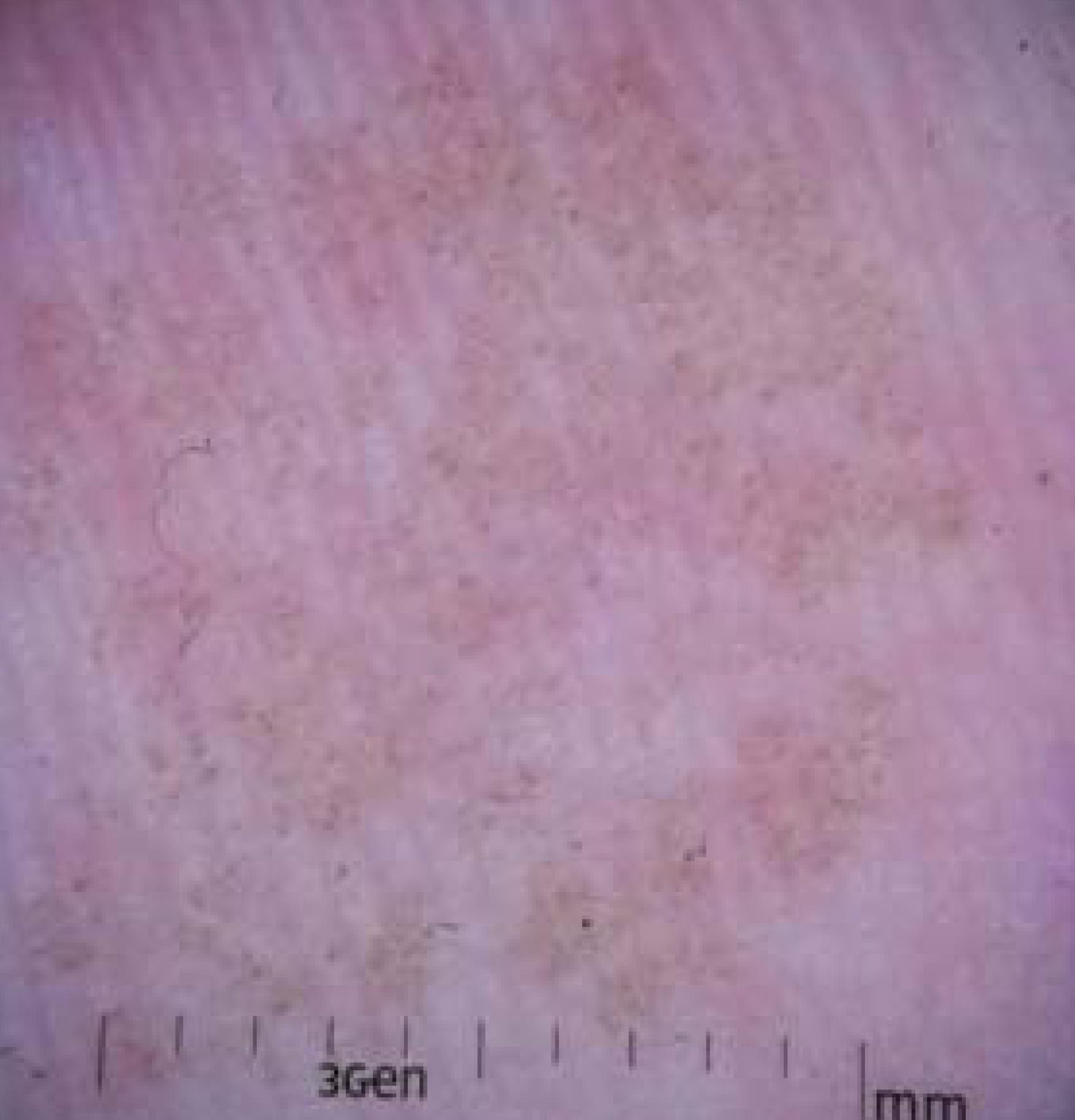
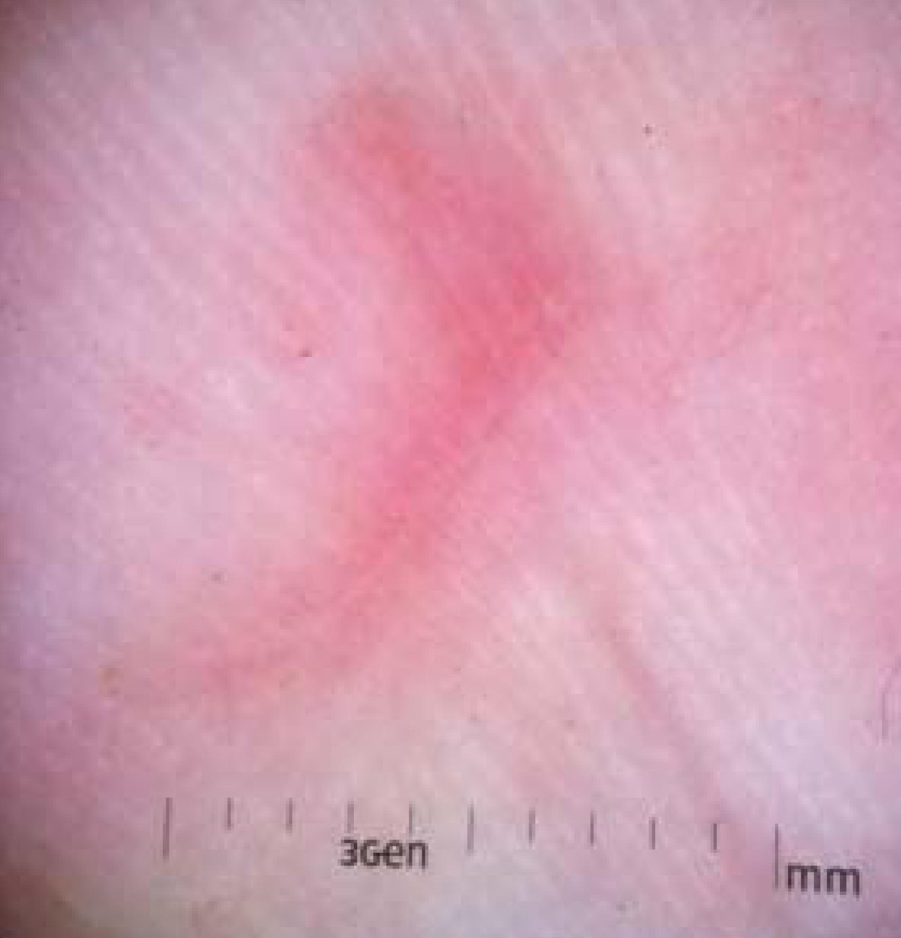
Dermoscopy of the hyperchromic macule showed darkened irregular puntiform pigmentation, a pattern typical to TN (Figure 2). The erythematous macule had a red homogenous colour without red spots or crown (Figure 3). In both there was an absence of typical pigmented melanocyte characteristics such as pigment network, globules, and stripes. The images were captured with a DermLite DL1 3Gen connected in a 5 megapixels iPhone 4S camera.
Dermoscopy of the hyperchromic macule showed darkened pigmentation, distributed in an irregular spotty shape without the standard characteristics of melanocytic pigmented lesions such as pigment network, globules, and stripes. (Magnification 10x)
Dermoscopy of the erythematous macule showing an area of reddened homogenous regularly distributed colouration with imprecise limits, an absence of red spots crown. (Magnification 10x)
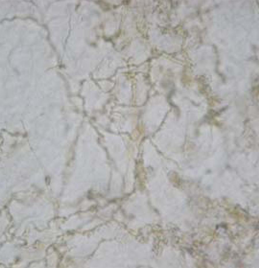
Mycological exam of the hyperchromic macule revealed septate ramified dematiaceous hyphae (Figure 4). In Sabouraud Agar culture, wrinkled pigmented colonies were grown and H. werneckii was identified in microculture. The macule regressed after application of topical butenafine twice a day for four weeks with no recurrence in six months.
Mycological exam cleared with 20% potassium hydroxide, showing irregular septate and ramified dematiaceous hyphae.
Dermascopy in TN, initially described in 1997, is a simple conclusive examination which was
effective in this case.66. Bonifaz A, Badali H, de Hoog GS, Cruz M, Araiza J, Cruz MA, et al.
Tinea nigra by Hortaea werneckii, a report of 22 cases from Mexico. Stud Mycol.
2008;61:77-82.
7. Xavier MH, Ribeiro LH, Duarte H, Saraça G, Souza AC. Dermatoscopy in
the diagnosis of tinea nigra. Dermatol Online J. 2008;14:15.
8. Paschoal FM, de Barros JA, de Barros DP, de Barros JC, Filho CD.
Study of the Dermatoscopic Pattern of Tinea Nigra: Report of 6 Cases. Skinmed.
2010;8:319-21.
9. Piliouras P, Allison S, Rosendahl C, Buettner PG, Weedon D.
Dermoscopy improves diagnosis of tinea nigra: A study of 50 cases. Australas J
Dermatol. 2011;52:191-4.-1010. Gupta G, Burden AD, Shankland GS, Fallowfield ME, Richardson MD.
Tinea nigra secondary to Exophiala werneckii responding to itraconazole. Br J
Dermatol. 1997;137:483-4. The growing habit of keeping animals such as rabbits
can occasionally lead to accidental bites and the resulting cutaneous lesion could be
confused with TN.
REFERENCES
-
1Zaitz C, Campbell I, Marques AS, Ruiz LRB, Framil VMS. Dermatomicoses por fungos filamentosos septados demácios. In: Zaitz C, Campbell I, Marques SA, Ruiz LRB, Framil VMS. Compêndio de Micologia Médica. 2. ed. Rio de Janeiro: Guanabara Koogan, 2010. p. 180-6.
-
2Rossetto AL, Cruz RC, Haddad Junior V. Double-blind study with the topical isoconazole and terbinafine for the treatment of one patient bilateral Tinea nigra plantaris and suggestions for new differential diagnoses. Rev Inst Med Trop Sao Paulo. 2013;55:125-8.
-
3Rossetto AL, Cruz RCB. Tinea nigra in geographical forms of "heart" and "parrot beak". An Bras Dermatol. 2011;86:389-90.
-
4Rossetto AL, Cruz RC. Tinea nigra: successful treatment with topical butenafine. An Bras Dermatol. 2012;87:939-41.
-
5Rossetto AL, Cruz RCB. Spontaneous cure in a case of tinea nigra. An Bras Dermatol. 2012;87:160-2.
-
6Bonifaz A, Badali H, de Hoog GS, Cruz M, Araiza J, Cruz MA, et al. Tinea nigra by Hortaea werneckii, a report of 22 cases from Mexico. Stud Mycol. 2008;61:77-82.
-
7Xavier MH, Ribeiro LH, Duarte H, Saraça G, Souza AC. Dermatoscopy in the diagnosis of tinea nigra. Dermatol Online J. 2008;14:15.
-
8Paschoal FM, de Barros JA, de Barros DP, de Barros JC, Filho CD. Study of the Dermatoscopic Pattern of Tinea Nigra: Report of 6 Cases. Skinmed. 2010;8:319-21.
-
9Piliouras P, Allison S, Rosendahl C, Buettner PG, Weedon D. Dermoscopy improves diagnosis of tinea nigra: A study of 50 cases. Australas J Dermatol. 2011;52:191-4.
-
10Gupta G, Burden AD, Shankland GS, Fallowfield ME, Richardson MD. Tinea nigra secondary to Exophiala werneckii responding to itraconazole. Br J Dermatol. 1997;137:483-4.
-
*
Work performed at the Dermatology Outpatient Clinic, Vale do Itajaí University (Univali) - Itajaí, SC, Brazil.
-
Financial funding: None
Publication Dates
-
Publication in this collection
Jan-Feb 2014
History
-
Received
13 Feb 2013 -
Accepted
15 Apr 2013