Abstract:
Dermoscopy is a non-invasive technique widely used to aid in the characterization and diagnosis of pigmented skin lesions. Recently, it has also been employed for the evaluation of non-pigmented skin tumours, and inflammatory and infectious cutaneous diseases. Although the diagnosis of cutaneous leishmaniasis is confirmed by the demonstration of amastigotes in infected skin or by the growth of promastigotes in culture medium, dermoscopy could be useful as a further diagnostic test. We report a patient with a nodular lesion located on the right cheek for almost two years. The lesion was clinically suggestive of cutaneous leishmaniasis. Dermoscopy showed yellow tears, erythema and vascular structures. The diagnosis was confirmed by the demonstration of amastigotes in a skin scraping sample.
Keywords:
Leishmaniasis, Cutaneous; Dermoscopy; Diagnosis
INTRODUCTION
Leishmaniasis includes a wild spectrum of chronic infections caused by more than 20 species of an obligated intracellular flagellated protozoan of the Leishmania genus. Infection is transmitted by the bite of a female sandfly infected with Phlebotomus (Old World) and Lutzomya (New World) genuses. Vectors are dogs and rodents.11 Goto H, Lauletta Lindoso JA. Cutaneous and mucocutaneous leishmaniasis. Infect Dis Clin North Am. 2012;26:293-307.
Leishmaniasis has a worldwide distribution, and is endemic in the Mediterranean area, North Africa, South America and parts of Asia.22 Moore EM, Lockwood DN. Leishmaniasis. Clin Med (Lond). 2011;11:492-7. On the Mediterranean coast, the main responsible agent of both visceral and cutaneous disease is Leishmania infantum. The cutaneous form from the Old World is more frequent in children.11 Goto H, Lauletta Lindoso JA. Cutaneous and mucocutaneous leishmaniasis. Infect Dis Clin North Am. 2012;26:293-307.,22 Moore EM, Lockwood DN. Leishmaniasis. Clin Med (Lond). 2011;11:492-7.
Lesions are usually isolated and located in exposed areas (face, neck, arms, legs) at the site of the bite. Clinically, the lesion appears as a slow-growing erythematous infiltrated papule, which develop into a nodule or plaque with central ulcer and crusting within a few weeks. The suspected clinical diagnosis is confirmed by the demonstration of amastigotes in infected skin biopsy or by the growth of promastigotes in culture medium. The lesion usually heals in 3-6 months, leaving an atrophic scar. It sometimes continues active with positive smears during a 24-month period (called non-curable chronic cutaneous leishmaniasis).
Dermoscopy is a non-invasive technique for the in vivo observation of the skin, which allows a magnified view of lesions and submacroscopic structures located in the epidermis and upper dermis. This technique is highly effective in the diagnosis and characterisation of pigmented lesions. More recently, it has been gaining relevance as a complementary procedure in an number of non-pigmented lesions (inflammatory, infectious, hair and nail diseases, etc.).33 Micali G, Lacarrubba F, Massimino D, Schwartz RA. Dermatoscopy: Alternative uses in daily clinical practice. J Am Acad Dermatol. 2011;64:1135-46.,44 Lallas A, Zalaudek I, Argenziano G, Longo C, Moscarella E, Di Lernia V, et al. Dermoscopy in general dermatology. Dermatol Clin. 2013;31:679-94. Dermoscopy is a great aid in the study of Leishmaniasis, not only for diagnosis, but also in the evaluation of the course and treatment response.55 Llambrich A, Zaballos P, Terrasa F, Torne I, Puig S, Malvehy J. Dermoscopy of cutaneous leishmaniasis. Br J Dermatol. 2009;160:756-61.,66 Gómez Moyano E, Hidalgo Gamero A, Martín González T, Fernández Ballesteros MD. Hallazgos dermatoscópicos en la leishmaniasis cutánea. Piel. 2012 ;27:50-1.
CASE REPORT
A 2-year-old female presented with a nodular lesion on her right cheek, which she had had since she was 2 months old. Medical history was unremarkable. Physical examination revealed an erythematous nodule with a central hyperkeratotic crust (Figure 1).
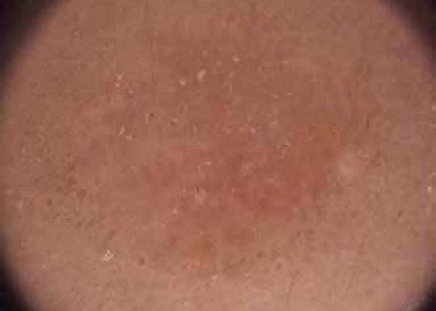
The main suspected diagnosis was cutaneous leishmaniasis. Leishmania investigation was performed, which confirmed the infection. The patient was treated with oral rifampicin for 10 weeks. Digital dermoscopy (Fotofinder dermoscope) at 6 weeks showed diffuse erythema with an erythemato-orangish central area with yellow tears and peripheral irregular vascular structures (Figure 2). At the end of the treatment, the lesion had healed, leaving only a small depressed scar (Figure 3). At that time dermoscopy showed less erythema and vessels, no yellow tears and a white central scar (Figure 4).
Dermoscopy at 6 weeks with treatment. Diffuse erythema, yellow tears and vascular structures. (X20)
Dermoscopy after treatment. Decreasing of erythema and vascular structures. Central white scar. (X20)
DISCUSSION
Differential diagnosis of cutaneous leishmaniasis includes furuncles, tuberculosis, sarcoidosis, atypical micobacteriosis, pyogenic granuloma and skin cancer.55 Llambrich A, Zaballos P, Terrasa F, Torne I, Puig S, Malvehy J. Dermoscopy of cutaneous leishmaniasis. Br J Dermatol. 2009;160:756-61. Diagnosis is performed by cytological and histological studies, with the aim of demonstrating amastigotes in dermal macrophages (Leishman-Donovan bodies), or by the visualization of promastigotes in culture medium. Polymerase chain reaction may be useful, but serology has poor specificity.77 García-Almagro D. Leishmaniasis cutánea. Actas Dermosifilogr. 2005;96:1-24.
Pentavalent antimonial drugs are used as first-line treatment. Isolated lesions have a satisfactory response to cryotherapy. Rifampicin is a classic treatment, with less adverse effects.88 Ruiz-Villaverde R, Blasco J, Linares J, Burkhardt P, Naranjo R. Leishmaniasis cutánea crónica: respuesta a N-metil glucamina intralesional tras fracaso con paramomicina tópica. Actas Dermosifiliogr. 2002;93:263-6.
The polymorphous clinical spectrum of cutaneous leishmaniasis often makes its clinical diagnosis difficult. In this sense, dermoscopy plays an important role as a complementary non-invasive method. The most common dermoscopic findings (almost 100%), described by Lambrich A et al. (2011),55 Llambrich A, Zaballos P, Terrasa F, Torne I, Puig S, Malvehy J. Dermoscopy of cutaneous leishmaniasis. Br J Dermatol. 2009;160:756-61. include generalized diffuse erythema and vascular structures, which correspond to dilated vessels (Table 1). Other signs are hyperkeratosis, central erosion or ulceration, "yellow tears", and "white starburst-like patterns". "Yellow tears" are follicular plugs due to the lateral compression of follicular ostiums caused by the growing of the lesion, whereas a "white starburst-like pattern" is attributed to the presence of parakeratotic hyperkeratosis located around the erosion. These authors describe two main dermoscopic patterns related with the evolution of the lesions: pattern 1 includes initial lesions (<6 months) and is characterised by papular lesions with vascular structures and "yellow tears"; pattern 2 is consistent with advanced lesions (>6 months) and encloses tumours with central erosion or ulceration and hyperkeratosis, a "white starburst-like pattern", and peripheral vascular structures. Some mixed or incomplete forms may also appear.
Recently, Taheri AR et al. (2013)99 Taheri AR, Pishgooei N, Maleki M, Goyonlo VM, Kiafar B, Banihashemi M, et al. Dermoscopic features of cutaneous leishmaniasis. Int J Dermatol. 2013;52:1361-6. have expanded these previously described findings to include a yellowish hue and milia-like cysts (Table 1). In this study, generalized erythema, hyperkeratosis and a "white starburst-like pattern" are more common in advanced, clinically nodular and ulcerated lesions. Moreover, the vascular pattern is correlated with the clinical presentation, location and duration of the lesion. Therefore, dotted vessels and comma-shaped vessels are more frequent in ulcerated lesions, whereas linear irregular vessels and hairpin vessels present a higher prevalence in nodular and advanced lesions. By contrast, glomerular-like vessels are more prevalent in lesions located in the lower extremities. However, dotted vessels are the most common vascular structure in all lesions. Milia-like cysts are related to lesions found in the head and neck. A yellowish hue is appreciated in papular lesions, with short period of evolution. This is similar to the structures already defined in sarcoidosis and tuberculosis lesions; hence, it is suggestive of granulomatous disease.
To the known dermoscopic structures (Table 1), Yücel A et al. (2013)1010 Yücel A, Günasti S, Denli Y, Uzun S. Cutaneous leishmaniasis: new dermoscopic findings. Int J Dermatol. 2013;52:831-7. added the salmon-colored ovoid structures, possibly related to granulomatous disease. These are more frequent in plaques and nodules, as well as in advanced lesions. Another new finding is the perilesional hypopigmented halo, which is more common in papular forms.
Our patient came to our department with an advanced lesion (>6 months), although dermoscopic imagery proved to be more compatible with pattern 1, as described by Lambrich A et al. ("yellow tears" and vascular structures), which has been described in initial lesions (<6 months). In addition, the lesion's time of evolution (almost 2 years) and the early age of the patient are both remarkable findings.
Dermoscopic differential diagnosis includes Spitz nevi, amelanotic melanoma, nodular basal cell carcinoma, squamous cell carcinoma, keratoacanthoma and pyogenic granuloma.55 Llambrich A, Zaballos P, Terrasa F, Torne I, Puig S, Malvehy J. Dermoscopy of cutaneous leishmaniasis. Br J Dermatol. 2009;160:756-61.,99 Taheri AR, Pishgooei N, Maleki M, Goyonlo VM, Kiafar B, Banihashemi M, et al. Dermoscopic features of cutaneous leishmaniasis. Int J Dermatol. 2013;52:1361-6.
In conclusion, dermoscopy is as a non-invasive procedure that provides us relevant data about cutaneous leishmaniasis, helping in both the diagnosis and evaluation of the treatment response. It would be interesting to introduce dermoscopy in our daily clinical practice as a complementary test in the study of cutaneous leishmaniasis. This would expand the available data with the aim of the standardisation of dermoscopy in the evaluation of infectious lesions like cutaneous leishmaniasis.
-
*
Work performed at the Dermatology Department of the University Hospital San Cecilio, Granada, Spain.
-
Financial support: none.
REFERENCES
-
1Goto H, Lauletta Lindoso JA. Cutaneous and mucocutaneous leishmaniasis. Infect Dis Clin North Am. 2012;26:293-307.
-
2Moore EM, Lockwood DN. Leishmaniasis. Clin Med (Lond). 2011;11:492-7.
-
3Micali G, Lacarrubba F, Massimino D, Schwartz RA. Dermatoscopy: Alternative uses in daily clinical practice. J Am Acad Dermatol. 2011;64:1135-46.
-
4Lallas A, Zalaudek I, Argenziano G, Longo C, Moscarella E, Di Lernia V, et al. Dermoscopy in general dermatology. Dermatol Clin. 2013;31:679-94.
-
5Llambrich A, Zaballos P, Terrasa F, Torne I, Puig S, Malvehy J. Dermoscopy of cutaneous leishmaniasis. Br J Dermatol. 2009;160:756-61.
-
6Gómez Moyano E, Hidalgo Gamero A, Martín González T, Fernández Ballesteros MD. Hallazgos dermatoscópicos en la leishmaniasis cutánea. Piel. 2012 ;27:50-1.
-
7García-Almagro D. Leishmaniasis cutánea. Actas Dermosifilogr. 2005;96:1-24.
-
8Ruiz-Villaverde R, Blasco J, Linares J, Burkhardt P, Naranjo R. Leishmaniasis cutánea crónica: respuesta a N-metil glucamina intralesional tras fracaso con paramomicina tópica. Actas Dermosifiliogr. 2002;93:263-6.
-
9Taheri AR, Pishgooei N, Maleki M, Goyonlo VM, Kiafar B, Banihashemi M, et al. Dermoscopic features of cutaneous leishmaniasis. Int J Dermatol. 2013;52:1361-6.
-
10Yücel A, Günasti S, Denli Y, Uzun S. Cutaneous leishmaniasis: new dermoscopic findings. Int J Dermatol. 2013;52:831-7.
Publication Dates
-
Publication in this collection
Nov-Dec 2017
History
-
Received
16 Feb 2015 -
Accepted
04 May 2015