Abstracts
OBJECTIVES: To compare the electrical activation of the rectus femoris (RF), long head of the biceps femoris (BF) and semitendinosus (ST) and the resistance torque (T R) of the hip extension (HE) movement performed on the Pilates Cadillac with the attachable spring in two different positions. METHODS: Twelve subjects performed five hip extensions with the attachable spring in two positions (high and low). Electromyography (EMG) and electrogoniometry data were colleted simultaneously. The root mean square (RMS) was calculated and normalized based on the maximal voluntary contraction. A free-body diagram (FBD) and movement equations were used to calculate T R. One-way ANOVA was used to investigate EMG differences between spring positions (p<0.05). RESULTS: When the spring was in the high position, T R was classified as descending and occurred in the "direction" of flexion over most of the range of motion (ROM). In the low position, T R descended until 60º of hip flexion, in the direction of flexion, and from there it took on an ascending pattern in the direction of extension. CONCLUSIONS: The EMG analysis seemed to follow the T R, with higher values for the RF in the low position and higher activation values for the BF and ST in the high position, where the external demand was greater. EMG and T R data supply complementary information for prescribing Pilates exercises.
torque; electromyography; Pilates; hip extension
OBJETIVOS: Comparar a ativação elétrica do reto femoral (RF), do bíceps femoral cabeça longa (BF) e semitendíneo (ST) e o torque de resistência (T R) do movimento de extensão de quadril (EQ) realizado com a mola fixada em duas posições distintas no Cadillac. MÉTODOS: 12 sujeitos realizaram 5 repetições de EQ com a mola fixada em duas posições (alta e baixa). Dados de eletromiografia (EMG) e eletrogoniometria foram coletados simultaneamente. O root mean square foi calculado e normalizado com base na contração voluntária máxima. Para o cálculo do T R, foram usados diagramas de corpo livre (DCL) e equações de movimento. ANOVA one-way foi usada para verificar as diferenças para EMG entre as posições de mola (p<0,05). RESULTADOS: Com a mola fixa na posição alta, o T R foi classificado como decrescente e ocorreu no "sentido" de flexão na maior parte da amplitude de movimento (ADM). Para posição baixa, o T R foi descrescente até 60º de flexão de quadril no sentido de flexão e, a partir daí, assumiu um comportamento crescente no sentido da extensão. CONCLUSÕES: A análise EMG pareceu acompanhar o T R, apresentando valores maiores para o RF na posição baixa e maiores valores de ativação para o BF e ST na posição alta, onde a demanda externa foi maior. Dados de EMG e T R fornecem informações complementares para prescrição de exercícios no Pilates.
torque; eletromiografia; Pilates; extensão do quadril
ORIGINAL ARTICLE
Analysis of the external resistance and electromyographic activity of hip extension performed according to the Pilates method
Silva YO; Melo MO; Gomes LE; Bonezi A; Loss JF
Laboratory of Physical Exercise Research (LAPEX), Universidade Federal do Rio Grande do Sul (UFRGS), Porto Alegre (RS), Brazil
Correspondence to
ABSTRACT
OBJECTIVES: To compare the electrical activation of the rectus femoris (RF), long head of the biceps femoris (BF) and semitendinosus (ST) and the resistance torque (TR) of the hip extension (HE) movement performed on the Pilates Cadillac with the attachable spring in two different positions.
METHODS: Twelve subjects performed five hip extensions with the attachable spring in two positions (high and low). Electromyography (EMG) and electrogoniometry data were colleted simultaneously. The root mean square (RMS) was calculated and normalized based on the maximal voluntary contraction. A free-body diagram (FBD) and movement equations were used to calculate TR. One-way ANOVA was used to investigate EMG differences between spring positions (p<0.05).
RESULTS: When the spring was in the high position, TR was classified as descending and occurred in the "direction" of flexion over most of the range of motion (ROM). In the low position, TR descended until 60º of hip flexion, in the direction of flexion, and from there it took on an ascending pattern in the direction of extension.
CONCLUSIONS: The EMG analysis seemed to follow the TR, with higher values for the RF in the low position and higher activation values for the BF and ST in the high position, where the external demand was greater. EMG and TR data supply complementary information for prescribing Pilates exercises.
Key Words: torque; electromyography; Pilates; hip extension.
Introduction
The Pilates method was originally developed by Joseph Pilates during the First World War and brought over to the USA in 19231-3. The initial concept blended elements from gymnas-tics, martial arts, yoga and dance, focusing on the relationship between the body and mental discipline3-5. Working with the Pilates method includes the use of special apparatus to place the external overload (external load) on the musculoskeletal structure with the aid of springs3. More recently, new elements were incorporated into this method, which aims at both physi-cal conditioning6-8 and rehabilitation programs9-11.
In physical therapy, Pilates exercises have been used for therapeutic purposes, neuromuscular reeducation, functional activity, and lumbopelvic stabilization3,12,13. However, the criteria for choosing the variables that modulate the overload of the Pilates exercises, i.e. position of the individual and spring position, are still based on subjective evaluations. In contrast, the knowledge of the resistance torque (TR) offered by a given exercise, alongside the information concerning the muscle activation, should also be regarded as criteria for selecting the exercises. Nevertheless, in spite of the great popularity of Pilates in clinical practice, there is a distinct lack of scientific studies on its applications to physical therapy and studies with kinesiologic, physiological, and/or biomechanical approaches12,14,15. Outside the realm of Pilates, studies with free weights, machine weights and elastic bands have demonstrated, among other things, that the TR analysis can show whether a given external load is heavier at the beginning or the end of a range of motion (ROM) and whether it is compatible with the acting muscles' ability to produce strength and, thus, support the exercise selection16-18. Moreover, other studies have quantified and compared the electromyographic (EMG) activity and the external loads applied to the muscles being studied during typical strength-training exercises and suggested that biomechanical data (EMG and external load) should be taken into account when a rehabilitation program is being developed18,19.
In the physical therapy intervention, Pilates apparatus has been commonly used for hip extension focusing on the activation of specific muscles, such as the hip extensors and the gluteus maximus, to stabilize the lumbopelvic region12,20,21. One such piece of equipment that allows several movement patterns and postures is the traditional Cadillac. When the hip extension (HE) movement is performed slowly and constantly, as indicated in the Pilates method, the TR offered by that apparatus depends on the ratio of factors such as a) the deformation coefficient of the spring (K), b) the position of the spring c) the weight of the moving human segment, and d) the perpendicular distances of the forces involved (of the spring and of the segment weight) to the joint axis in the center of the hip joint. Despite its complexity, some techniques in biomechanics research, such as the representation of the forces involved by means of a free-body diagram (FBD) and equations of movement, may help objectively classify the type of resistance in a given exercise. The knowledge of the TR behavior and of the EMG muscles during the Pilates exercises may be regarded as a tool to indicate the overload on the muscle-tendon system and complement the choice of Pilates exercises during a rehabilitation program. Thus, the objectives of the present study were to compare the electrical activation of the rectus femoris (RF), the long head of the biceps femoris (BF), and the semitendinosus (ST), and the resistance torque of the HE movement performed with the spring attached in two different positions.
Methods
Sample
Twelve participants of both sexes took part in this study, all of whom were Pilates practitioners. The mean age was 34.25±11.48 years; mean height 163.75±11.48cm and mean body mass 62.14±13.96Kg. The criteria for inclusion were that the individuals had to be healthy, with no history of musculoskeletal injury and that they had attended at least 30 Pilates sessions. All signed a consent form and were informed of the right to drop out of the data collection at any time. During the evaluation protocol, there was no record of sample loss. This study was approved by the Ethics Committee of Universidade Federal do Rio Grande do Sul, where it was carried out under protocol number 2007903.
Data acquisition
Evaluation protocol
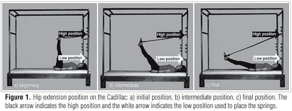
The evaluation protocol was performed on a Pilates Cadillac machine, which has two key features that allow a large number of exercises, i.e. a) options for height adjustment and placement of the springs, and b) the use of springs with different deformation coefficients and color-coded according to the level of resistance (Figure 1). Firstly, the participants were positioned in dorsal decubitus on the Cadillac. Each one of them performed five HE movements, starting from 90º hip flexion to 0º hip flexion, in two different positions: 1) with the springs in the high position (86cm in relation to the level of the participant), and 2) with the springs in the low position (20cm in relation to the level of the participant). A one-minute interval was allowed between the positions. The springs color-coded in blue by the manufacturer were previously calibrated, and the elastic constant (K) was 0.013 kg/cm.
Electromyography and goniometry
EMG and electrogoniometry data were collected simultaneously by means of the Miotec Data Acquisition System (Miotec Equipamentos Biomédicos Ltda, Porto Alegre, Brazil). The angle positions of the hip joint during the evaluation protocol of the exercises were registered by means of an electronic goniometer (Miotec Equipamentos Biomédicos Ltda, Porto Alegre, Brazil). The sampling rate for both the EMG and the electrogoniometry was 2000Hz. In order to capture the EMG signal, all the procedures recommended by the International Society of Electrophysiology and Kinesiology (ISEK) were followed, such as shaving, sanitizing with alcohol, placement of electrodes, and impedance check (accepted when inferior to 5KΩ). The reference electrode was positioned on the right knee, over the styloid process of fibula. The muscles monitored were RF, BF, and ST. Pairs of electrodes with disposable surfaces were used (Kendall Meditrace 100; Ag/AgCl; 2.2cm in diameter with a fixing adhesive, in the bipolar configuration), for each muscle. The electrodes were placed onto the muscle belly, parallel to the muscle fibers and 2cm apart. For the RF, the electrodes were attached at 50% on the line between the anterior superior iliac spine and the upper part of the patella and, for the BF and the ST, the electrodes were placed at 50% on the line between the ischiatic tuberosity and the lateral and medial epicondyle, respectively22. For the purpose of comparison, the EMG signals were normalized based on the maximal isometric activation of the hip extensors. To achieve that, all participants underwent a maximal voluntary isometric contraction (MVIC) test before the aforementioned evaluation protocol began. This consisted of performing two MVICs, lasting five seconds each, with a 2-minute interval between the MVICs, with the hip positioned at a 90° flexion. The greatest MVIC was used as a reference for normalization.
Data analysis
To analyze the data, the EMG and electrogoniometry signals were submitted to a process of digital filtering with the aid of the SAD32 software, version 2.61.07mp. For the EMG signal, a third-order Butterworth band-pass filter was used, with cutoff frequencies between 20 and 500Hz, whereas for the electrogoniometry signal a moving average filter with a 3Hz cutoff frequency was used. In order to keep the focus of the analysis on the HE movement, the EMG curves were firstly divided according to the start and finish of each repetition performed, based on the angle positions recorded. For a more detailed evaluation, the HE movement was divided into two parts, which corresponded to 50% of the extension movement: first half (H1) and second half (H2). The RMS value for each section cut off from each repetition and for each half of the extension was calculated, and afterwards the mean of those values was computed, normalized and used for the statistical analysis.
The TR consists of the weight torque (TW) of the segment and the spring strength torque (TSP), which is generated by the strength of the spring and its respective perpendicular distance in relation to the center of the hip rotation. For the calculation of the TR, FBDs of the thigh-leg-foot moving segment were drawn. Based on those and on Euler's equilibrium equation (Equation 1), it was possible to establish a relationship between the torques of the exercise. To estimate the TR, the clockwise torques were considered positive, whereas the counterclockwise ones were considered negative. Due to the low speed of execution of the exercise, the situation was considered "almost-static", and every inertial effect involved was ignored.

In which:
TM=muscle torque
TW=segment weight torque
TSP=spring strength torque
The muscle torque is representative of the activity of the muscles which cover the hip joint, and it is the net expression of the recruitment of both the extensor and the flexor muscles. The TR of the exercise, which opposes the muscle activity, is given by the sum of the other torques. For the representation of the TR, equation 1 can be reformulated (equation 2).

The right-hand side of equation 2 represents the resistance imposed on the segment involved. Under this approach, three situations are possible: (1) TSP>TW, characterizing a TR in the direction of the hip flexion, and consequently an extension muscle torque; (2) TSP<TW, characterizing a TR in the direction of the hip extension, therefore a flexion muscle torque; and (3) TSP=TW, characterizing zero TR, thus resulting in zero muscle torque. Given the dynamic characteristic of the exercise, in which the torques of the spring and of the weight of the segment vary constantly, situation (3), if it occurred, would take place for a very short lapse of time, and therefore may be disregarded. The perpendicular distances of the strength of the spring and the weight of the segment were obtained from trigonometric relationships, by directly measuring the hip angle throughout the range of motion, and, in the beginning and at the end of the movement, by measuring the angle between the spring and the human segment and the corresponding distances. For the spring strength evaluation along the entire arch of motion, we used trigonometric relationships associated to the deformation coefficient determined by the calibration23. The inertial parameters used (weight, position of the center of mass, and of the segments involved, i.e. thigh-leg-foot) were estimated based on the anthropometric tables obtained in the literature24. The calculations necessary for the quantification of the TR were developed on Excel (version 1997, Microsoft Windows).
For the graphic representation of the simultaneous behavior of the EMG with the TR during the evaluated exercise, the filtered and normalized EMG signal underwent a softening process in which the RMS value was calculated in 1-second Hamming windows.
Statistics
The data were used by means of the SPSS 10.0 software. Initially, the variance equivalence was checked (Levene's test), and then the data normality (Shapiro-Wilk). Once the adherence to the normal model was confirmed, one-way ANOVA was applied to check the differences between the EMG activity of each muscle (RF and BF) as follows:
1) Comparison of the EMG of the muscles tested during the full HE movement, between positions 1 and 2; 2) comparison of the EMG of each muscle recorded in the first half of the HE movement in position 1, with the first half of the HE movement in position 2; 3) comparison of the EMG of each muscle recorded in the second half of the HE movement in position 1, with the second half of the HE movement in position 2; 4) comparison of the EMG of each muscle recorded in the first half of the movement with the second half of the HE movement with the spring in position 1; 5) comparison of the EMG of each muscle recorded in the first half of the movement with the second half of the HE movement with the spring in position 2. The level of significance adopted for all tests was p<0.05.
Results
The TR had a very similar behavior for all individuals, with small variations due to the inertial and anthropometric parameters of each participant, but which did not alter the line of the TR curve in the exercise being evaluated (Figures 2 and 3). Thus, with the spring in the high position, the TR was classified as descending and occurred in the "direction" of flexion, and with the spring in the low position, the TR was classified as descending until approximately 60º of hip flexion in the "direction" of the flexion, and after that, ascending in the "direction" of extension.
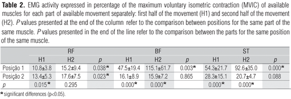
Figures 2 and 3 show a behavior typical of the TR simultaneously with the EMG response obtained during the HE for one individual. In general, the EMG had higher percentages of activation for the BF and ST when compared to the RF in the high position (Figure 2), and greater RF values when compared to the BF and ST in the low position (Figure 3). By visually comparing the halves of the movement in each spring position, it is possible to identify, in Figure 2, a greater ascending EMG activation of all evaluated muscles in the second half of the movement, during which the TR is descending. Figure 3, however, shows that the RF activation is greater than that of the extensors, particularly in the second half of the movement, in which the TR occurred in the direction of extension. These results were confirmed with significance through statistical treatment and are displayed in Tables 1, 2, and 3. Table 1 shows the total EMG results for the muscle evaluated during the HE movement and the result of the comparison between positions 1 and 2, separately for each muscle. Table 2 shows the partial EMG results divided in sections (H1 and H2) for the muscles evaluated during the HE movement. In this table, the results of the comparisons between the positions for the same section of the same muscle are shown in the last line, while the results of the comparison between sections for the same position of the same muscle are shown in the intermediate columns.
Discussion
Based on the differences in the TR behavior observed between the two spring positions evaluated in this study, the muscles are expected to generate different neuromuscular strategies to overcome the resistance variation imposed on them and still provide stability to the joints. In that sense, when comparing the EMG signal of the muscles tested between positions 1 and 2, significant differences were detected only for the group that comprises the hip extensors, and the EMG activity of the BF and of the ST in the high position was greater than the EMG activity found for the BF and for the ST in the low position (p<0.05) (Table 1). These differences in the EMG activity may have occurred due to the reduced need to recruit motor units due to a lower external demand of the extensor muscles evaluated, considering that, for most of the ROM with the spring in the low position, the torque of resistance was in the direction of extension.
When comparing the EMG signal of the muscles evaluated between the positions, the differences found for the extensor group were maintained in relation to the full movement (Table 2). Nevertheless, that did not happen with the RF, which had higher activation values for the spring in the low position, when the first half of the movement was analyzed. It is speculated that the greater RF recruitment in this section of the movement, in which it is not a primary motor of the extension, is due to a greater need for control and stabilization of the movement in response to the descending behavior of TR, which reaches zero in the first half of the movement.
Also concerning the analysis of the response of the EMG signal during the exercise in position 1 and contrary to the theoretical expectation that there might be a greater recruitment of motor units during the highest TR, i.e. in H1, the hip extensor group had larger EMG activity values in H2 (p<0.05) (Figure 1A) (Table 2). Considering that the exercise was performed in an extremely slow and controlled fashion, the greater EMG activity of the extensor muscles in this section may be explained by the greater need to perform posterior tilt of the hip and support the lumbopelvic region as the thigh-leg-foot segment came closer to the floor. In this situation, it is very likely that other muscles, such as the gluteus and the abdomen, contribute in synergism to prevent the pelvis from moving in anterior tilt and the spine from executing a hyperextension. In a rehabilitation program focusing on the stabilization of the lumbopelvic region, where the posterior hip muscles need to be strengthened, this exercise in the high position may be an option for the initial phase of the treatment. With the spring in the high position, the TR of the exercise was descending, meaning that the physical therapist can vary the external load on the muscles during ROM and demand more when the muscles are more stretched out, i.e. in a more favorable physiological situation for the production of muscle strength (strength-length ratio)25. Furthermore, based on the results of the EMG activity, the high position at the outset of a given treatment allows the physical therapist to gradually induce the recruitment of motor units as the patient executes the full extension of the hip, and this result is obtained without increasing the external overload on the muscles and tendons (Figure 2). Thus, the treated muscles are submitted to a safe and gradual neural adaptation to the exercise, which is also an advantage when the main goal is to treat an injury to the passive components of the posterior muscles of the hip.
When the spring was set in position 2, the TR occurred in the direction of flexion, and then changed to the direction of extension (Figure 3). Hence, it is understood that the extensor TR was balanced by the TM generated, among other structures, by the hip flexor muscle group. This mechanical characteristic tends to produce a neuromuscular response different from that observed in position 1, at least in respect to the levels of activation of the hip extensor muscle group, which are not primary motors of hip flexion. In this situation, the hip flexor group is likely to be contributing eccentrically as primary motors, whereas the extensors increasingly act as stabilizers of the hip joint. Although this study only include the EMG of a single hip flexor muscle, the findings tend to confirm this hypothesis, because the highest levels of EMG activity of the RF muscle were found in H2 (p<0.05) (Table 2). It is interesting to note that there were no differences in the activation of the BF and ST muscles for position 2, which may indicate that, even if the RF muscle contributes eccentrically as the primary motor of the movement (preventing the mobile segment from "falling"), the activation of the extensors takes over the task of maintaining the desired joint stability for the exercise. These results corroborate the principles defended by the Pilates method that allows a combination of the activation of all the muscles involved in a given exercise in order to achieve the best motor strategy to stabilize the joints20,21. As a clinical application, the exercise performed with the spring in position 2 may be recommended for the rehabilitation of muscle-tendon injuries in runners, such as tendinitis26. The most noticeable characteristic of the exercise in the low position is the fact that the TR has a low external demand, changing its direction along the ROM. This may be an interesting strategy for the intermediate phase of a rehabilitation program for eccentric strengthening of the hip flexors. In this exercise, in the section where the TR is greater, the flexors are stretched out (greater capacity to generate strength)25, and the levels of EMG activity of the RF are higher throughout the ROM (Figure 3). This indicates that there is a greater contribution of the passive and active components in the production of strength, a mechanism that is recommended for the rehabilitation of this type of injury26,27.
The findings of the present study suggest that certain changes in spring height during the hip extension exercise on the Cadillac may be more suitable for a particular clinical objective than for another. In other words, changing the spring placement, as is commonly done in the clinical use of Pilates, does not only change the "intensity" of the exercise from weak to moderate, or from moderate to intense, it also defines whether the muscles should be considered as primary muscles or otherwise in the performance of a given movement. Thus it interferes with the importance of the contribution of passive and/or active structures to generate strength during the exercise.
Conclusion
At this stage, it can be concluded that, in the general comparisons, the EMG analysis followed the TR, showing greater values for the RF in the low position, and greater activation values for the BF and ST in the high position, where the external demand was increased. However, in the partial comparisons with the high spring, EMG activity was greater in the part of the movement where there was less TR. With the spring in the low position, even when the TR shifted its direction along the ROM, the EMG activity levels were constant. The present study may be the first step towards setting objective criteria (TR and EMG) for the development of rehabilitation programs using Pilates exercises.
References
- 1. Muscolino JE, Cipriani S. Pilates and the "powerhouse". J Bodyw Mov Ther. 2004;8(1):15-24.
- 2. Latey P. The Pilates method: history and philosophy. J Bodyw Mov Ther. 2001;5(4):275-82.
- 3. Rydeard R, Leger A, Smith D. Pilates-based therapeutic exercise: effect on subjects with nonspecific chronic low back pain and functional disability: a randomize controlled trial. J Orthop Sports Phys Ther. 2006;36(7):472-84.
- 4. Self BP, Bagley AM, Triplett TL, Paulos LE. Functional biomechanical analysis of the Pilates-based reformer during demi-plié movements. J Appl Biomech. 1996;12(3):326-37.
- 5. Latey P. Updating the principles of the Pilates method Part 2. J Bodyw Mov Ther. 2002;6(2): 94-101.
- 6. Jago R, Jonker ML, Missaghian M, Baranowski T. Effect of 4 weeks of Pilates on the body composition of young girls. Prev Med. 2006;42(3):177-80.
- 7. Segal NA, Hein J, Basford JR. The effects of Pilates training on flexibility and body composition: an obserrvational study. Arch Phys Med Rehabil. 2004;85(12):1977-81.
- 8. Bertolla F, Baroni BM, Leal Junior ECP, Oltramar JD. Efeito de um programa de treinamento utilizando o método Pilates® na flexibilidade de atletas juvenis de futsal. Rev Bras Med Esporte. 2007;13(4):222-6.
- 9. Donzelli S, Di Domenica E, Cova AM, Galletti R, Giunta N. Two different techniques in the rehabilitation treatment of low back pain: a randomized controlled trial. Eura Medicophys. 2006;42(3):205-10.
- 10. Blum CL. Chiropractic and pilates thepapy for the treatment of adult scoliosis. J Manipulative Phisiol Ther. 2002;25(4):E3.
- 11. Kolyniak IEGG, Cavalcanti SMB, Aoki MS. Avaliação isocinética da musculatura envolvida na flexão e extenção do tronco: efeito do método Pilates. Rev Bras Med Esporte. 2004;10(6):487-90.
- 12. Bernardo LM. The effectiveness of Pilates training in healthy adults: An appraisal of the research literature. J Bodyw Mov Ther. 2007;11(2):106-10.
- 13. Bryan M, Hawson S. The benefits of pilates exercise in orthopaedic rehabilitation. Tech in Orthop. 2003;18(1):126-9.
- 14. Souza MS, Vieira CB. Who are the people looking for the Pilates method? J Bodyw Mov Ther. 2006;10(4):328-34.
- 15. Touche RL, Escalante K, Linares MT. Treating non-specific chronic low back pain through the Pilates method. J Bodyw Mov Ther. 2008; 12(4):364-70.
- 16. Kulig K, Andrews JG, Hay JG. Human strength curves. Exerc Sport Sci Rev. 1984;12:417-66.
- 17. Toledo JM, Ribeiro DC, Loss JF. Critérios mecânicos para progressão de exercícios de rotação interna e externa do ombro no plano sagital. Rev Bras Fisioter. 2007;11(1):49-56.
- 18. Ribeiro DC, Loss JF, Cañeiro JPT, Lima CS, Martinez FG. Electromyographical analysis of the quadríceps during knee extension at different speed. Acta Ortop Bras. 2005;3(4):189-93.
- 19. Matheson JW, Kernozek TW, Fater DC, Davies GJ. Electromyographic activity and applied load during seated quadriceps exercises. Med Sci Sports Exerc. 2001;33(10):1713-25.
- 20. Rydeard R, Leger A, Smith D. Pilates-based therapeutic exercise: effect on subjects with nonspefic chronic low back pain and functional disability: a randomized controlled trial. J Orthop Sports Phys Ther. 2006;36(7):472-84.
- 21. Herrington L, Davies R. The influence of Pilates training on the ability to contract the transversus abdominis muscle in asymptomatic individuals. J Bodyw Mov Ther. 2005;9(1):52-7.
-
22SENIAM. [home page na Internet]. Netherlands; c2006-2008 [atualizada em 2008 may 21; acesso em 2008 may 21]. Disponível em: < http://www.seniam.org>
» link - 23. Loss JF, Koetz AP, Soares DP, Scarrone FF, Hennemann V, Sacharuk VZ. Quantificação da resistência oferecida por bandas elásticas. Rev Bras Cienc Esporte. 2002;24(1):61-72.
- 24. Winter DA. Biomechanics and Motor Control of Human Movement. 3Ş ed. New York: John Wiley Professio; 2004.
- 25. Rassier DE, MacIntosh BR, Herzog W. Length dependence of active force production in skelectal muscle. J Appl Physiol. 1999;86(5):1445-57.
- 26. Paluska SA. An overview of hip injuries in running. Sports Med. 2005;35(11):991-1014.
- 27. Fredericson M, Moore W, Guillet M, Beaulieu C. High hamstring tendinopathy in runners: meeting the challenges of diagnosis, treatment and rehabilitation. Phys Sportsmed. 2005;33(5):32-43.
Correspondência para:
Publication Dates
-
Publication in this collection
20 Feb 2009 -
Date of issue
Feb 2009
History
-
Accepted
17 Oct 2008 -
Reviewed
26 Sept 2008 -
Received
17 June 2008