Abstracts
A experimental study with 40 rats of the Lewis type was done focusing the influence of sodic risedronate on fractures consolidation in the animals. They were submitted to a protein nutrition diet to a non-protein one, divided randomly in four groups, having 10 animals in each group. Like this: group 1, with a protein nutrition diet, without risedronate (control group); group II, protein nutrition diet t with risedronate , group III, non-protein diet, without risedronate; group IV, non-protein diet with risedronate. The rats were submitted to similar fractures, on the 15º day and to the euthanasia on the 43º of the experiment. The variability analyzed included the ponderous evaluation, radiographic evaluation, the bone densitometry, histomorphometric bone callus evaluation, blood dosage of calcium, phosphorus, alkaline phosphatase, total proteins, albumin and osteocalcin. It was found out the risedronate had positive influence on the fractures consolidation process in nourished rats and malnourished rats and the mineral bone density increased. The risedronate caused the formation of the best nutrition and morphology of the ripen bone tissue.
Protein malnutrition; Risedronate; Fracture healing; Rats
Realizou-se um estudo experimental com 40 ratos da raça Lewis visando-se avaliar a influência do risedronato sódico na consolidação de fraturas em animais submetidos à dieta aprotéica e dieta protéica, divididos aleatoriamente em quatro grupos, com 10 animais em cada grupo, assim constituídos: grupo I, com dieta protéica, sem risedronato (grupo controle); grupo II, dieta protéica, com risedronato; grupo III, dieta aprotéica, sem risedrionato; grupo IV, dieta aprotéica, com risedronato. Os ratos foram submetidos a fraturas semelhantes, no 15º dia e à eutanásia no 43º dia do experimento. As variáveis analisadas incluíram a evolução ponderal, avaliação radiográfica, densitometria óssea, avaliação histomorfométrica do calo ósseo, dosagens sanguíneas de cálcio, fósforo, fosfatase alcalina, proteínas totais, albumina e osteocalcina. Concluiu-se que o risedronato exerceu influência positiva no processo de consolidação de fraturas em ratos nutridos e desnutridos, e aumentou a densidade mineral óssea. O risedronato ocasionou a formação de tecido ósseo maduro de melhor qualidade e morfologia.
Desnutrição protéica; Risedronato; Consolidação de fraturas; Ratos
ORIGINAL ARTICLE
The evaluation of the Sodic Risedronate effect in the fractures consolidation: experimental study with rats
Luiz Antonio Alcântara de OliveiraI; Roberto GuarnieroII; Consuelo Junqueira RodriguesII; Paulo José de SantanaI; Marco Antonio BatistaIII
IAssociate Phd Professor at FMUEL
IIFull Professor at FMUEL
IIIAssistant Professor at FMUEL
Correspondence Correspondence to Rua José Evanildo Clóvis de Souza, 180 Jardim Monte Belo - Londrina, Paraná. CEP 86041-590 Telefones: (43) 3342-7993; 9991-4312 email: alcantar@sercomtel.com.br
SUMMARY
A experimental study with 40 rats of the Lewis type was done focusing the influence of sodic risedronate on fractures consolidation in the animals. They were submitted to a protein nutrition diet to a non-protein one, divided randomly in four groups, having 10 animals in each group. Like this: group 1, with a protein nutrition diet, without risedronate (control group); group II, protein nutrition diet t with risedronate , group III, non-protein diet, without risedronate; group IV, non-protein diet with risedronate. The rats were submitted to similar fractures, on the 15º day and to the euthanasia on the 43º of the experiment. The variability analyzed included the ponderous evaluation, radiographic evaluation, the bone densitometry, histomorphometric bone callus evaluation, blood dosage of calcium, phosphorus, alkaline phosphatase, total proteins, albumin and osteocalcin. It was found out the risedronate had positive influence on the fractures consolidation process in nourished rats and malnourished rats and the mineral bone density increased. The risedronate caused the formation of the best nutrition and morphology of the ripen bone tissue.
Key words: Protein malnutrition; Risedronate; Fracture healing; Rats.
INTRODUCTION
The fractures consolidation is an organism specialized response where the regeneration of the bone leads to the restoration and integrity of the skeleton. Although many of the fractures consolidate without problems, some factors such as, protein malnutrition, can interfere negatively in the fractures consolidation(9).
There is not a drug in the pharmaceutical medicaments which helps in the fast fracture consolidation. Even with studies that have been done in order to find a result(3,12). Therefore; the sodic risedronate, a biphosphanates a bone body builder were used, wanting to observe its effectiveness in fractures consolidation, by the experimental study in nourished and non-nourished rats.
With the statistics analyses, the biochemical behaviour of the calcium, phosphorus and alkaline phosphatase, furthermore the total proteins and albumin, and the biochemical marker of the bone formation, the osteocalcin were analyzed. The obtained data by ponderate, radiography, densitometry, histological and histhomorphometrical evaluations were also analyzed.
The purpose of this work is to evaluate the sodic residronate influence, experimentally, in the fractures process consolidation in adult male rats, nourished or non-nourished, in which were done the right tibial fractures.
This work was approved by the Ethical committee of Research at the Clinicas Hospital in São Paulo University under the protocol 669/00.
MATERIAL AND METHOD
40 rats of the Lewis type, male, adult, isogenic, with the ideal weight ranging from 250 to 342 grams were used. The animals were divided randomly into four groups with 10 animals in each group, distributed as the following: group 1 with protein diet, without risedronate (control group); group II, with protein diet, with disedronate; group III, with protein diet, without risedronate; group IV, with non-protein diet, with disedronate, kept in cages, with five animals in each of them, made of opaque polyethylene and close with a stainless lid in grade shape, covered with sawdust and cleaned three times a week. In the cages where the animals were kept, the light period was of twelve hours, with temperature, humidity and noise level remaining stable
The non-protein diet, given to groups III and IV, is standardized and used to the malnutrition experimental in laboratory animals.
To the biochemical analyses, blood collection were done with cardiac puncture on the 1º, 15º and 43º day of the experiment.
On the 15º day, the rats were submitted to a close fracture of the median third of the right tibias, done manually by forced flexion, trying to have similarity to the local traced and the peristoneous mobilization, having the animals under anesthetics, by intraperiosteal injection of a cetamine solution of 40 mg/kg and xilasine of a dosage of 5 mg/kg(7,14) of body weight dissolved in 1 ml of saline solution. After doing the fractures, the animals of group II and IV started having daily doses of 1mg/kg/day of sodic risedronate, by the gavage using a catheter of polyethylene especially adapted, measuring 5cm length and 1 nm diameter, preparing the solution immediately before each oral administration. The contention of the animal was done by a capable technician, and, separating the jaw with a spatula, administrated the drug to groups II and IV, with no anesthetics. The administration of the sodic Risedronate was done with an only daily dose, after the fracture for 28 days.
The euthanasia of the animals occurred 28 days after the fractures, on the 43º of the experiment, considering a protocol of administration of the lethal dose with intraperitoneal of barbituric injectable anesthetics after anesthesia induction with halothane, finishing with lethal dose of chloride of potassium After this procedure, the heaviness and the disarticulation of the right femoral thigh of the animals to the bone callus study.
The evaluation of the studied material was done according with the ponderous evolution of the animals and serial doses of calcium, phosphorus, alkaline phosphatase, total proteins, albumin and osteocalcin 1º, 15º and 43º days, and the radiographic study, planimetry, densitometry, histology and histomorphometry of the bone callus at the end of the study.
To the statistics studies, the variance analyses technique was used in order to the outlining plan entirely casual, and mascarating the double-blind type, in the factorial scheme of the treatment, with the unbent of the liberty degrees of the effect of interaction between the diet and the medication. The obtained data were analyzed in significance of 5 %, that is, the tests were significantly considered when p <0,005.
RESULTS
Next, the tables with the averages and the standard variable factor measured and its respective variance analyses.


 ]
]


DISCUSSION
The comparative and systematic clinical evaluation of the bone callus formation shows many difficulties, due to a lot of individual differences about the nature and the fracture place, the course and consolidation. However, it is particularly difficult the therapeutics methods studies to be able to influence the bone callus formation. Therefore, it is natural the approach of this experiment problem. Furthermore, in developing countries like Brazil, the nutrition must be considered as an important factor of the fractures consolidation, because it depends on it the skeleton integrity restoration.
In this experiment, the rat was the animal used, due to be a practical and common model. The fractures have fast consolidation, not need internal or external mobilization, and without important complications. The model also enables the blood collection to the Biochemistry studies. The bone alterations related to the bone callus evolution can be easily seen in these animals model(10). The model also allows the evaluation of the bone callus formation in situations such as, malnutrition(9), or the effect of the substances as calcitonin(8), sodic alendronate(12), sodium fluoride(2), bone ossific hydroxyapathitis(3).
The fractures were obtained by manual osteoclasis(13), and showed satisfactory similarities, with small local differences, however with no need for creating a mechanical device to do it. The comparison of experimental fractures consolidation demands similarity, and what was tried to be found in this experiment. The animals were not mobilized, letting them free for strolling, the mobilization, besides being unnecessary, could create the possibility of interference due to the presence of the synthesis material(8).
The peritoneal via, to the anesthetics, is the most used for rats. There were no complications during the anesthetics act, what allows us assure the effectiveness of this action.
The non-protein to the experimental malnutrition(11) led to the protein malnutrition, checked in the values analyses of the total proteins (p<0,0001) and the albumin (p=0,0153), the protein malnutrition therefore obtained had direct interference in the fractures consolidation, which goes with obtained observations of other studies(8,9).
The medication used in this experiment in order to analyzed its possible influence as supporter in the fracture consolidation, the sodic risedronate, it is a third generation pyridine bisphosphonates, together with other bisphosphonates, inhibit the bone reabsorption mediated for the osteoclast(5).
The formation of a bridge between the fragments of a fractured bone is the most important step in its consolidation; the quantity of bone callus is the most visible area in the radiographies(1). The experimental fractures in the tibias of rats reach the ossification phase around the fourth week evolution, when already has shown evidences in the consolidation radiographies. Based on these data, the euthanasia of the animals was determinated 28 days after the fractures action, to the radiography, densitometry and histoly evaluation of bone callus.
The malnutrition of the animals of group III and IV was proved because of the significant weight loss (p<0,0001), what could have been seen on the 15º of the experiment, when the fractures were done, the weight loss continuing until the end of the experiment.
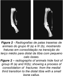
To the planimetric evaluation were done radiographies (Figures 1 and 2) after the knee disarticulation. In the radiography evaluation, the difference between the average results of the bone callus was statistically significant (p<0,0001), having smaller area of the bone callus in groups which consumed the diet without proteins, which is in accordance with what was found in other studies(9) that showed to have more abundant callus number in fractures in nourished rats when compared to the non-nourished ones.
The densitometry analyses showed difference statistically significant when the administration of risedronate happened. The bisphosphonates adheres to the bone minerals, acting on the osteoclasts, has highly selective location and retention in the bones, and can increase the bone bosy(5). This confirms the mechanism of action of the risedronate as an inhibitor of the bone reabsorption mediate by the osteoclasts.
Comparing the animals with non-protein diet with or without risedronate, the histological studies does not show significant difference to the quality of the bone formed, however it was observed the in the non-protein diet group with risedronate, there was more rape bone tissue, showing the calcification to the anatomic-pathologic exam, when compared to the non-protein diet group without medication. Thus, the risedronate contributed qualitatively with the rape bone tissue formation, best quality and morphology , although has not offered significant difference quantitatively to the histological exams in samples of groups submitted to protein and non-protein diet (Figures 3 and 6). This difference in the quality of the bone tissue was also observed in groups with protein diet, comparing them to the groups with or without risedronate, what shows the risedronate improves the trabecular architecture(6,12).


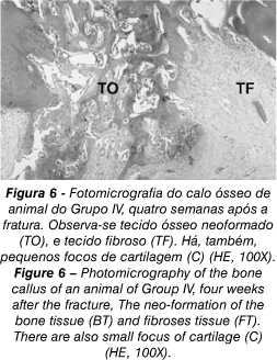
In the evaluation of the tissues components of the different samples, when evaluating the percentage of the fibroses tissues, and the neo-formed bone, it was seen there was significative statistics in groups with different diets, with or without the risedronate administration. However, when evaluated the percentage of the cartilaginous tissue, the statistically significance difference (p<0,05), with the increase of it, when the risedronate was administrated to the group of non-protein diet. This is significative for being likely the cartilage evolutes to the endochondral ossification. However, there is no guarantee of it, considering it is possible the evolution for the consolidation retard or even for the pseudartroses.
In the laboratorial evaluation of the level of calcium, a significative different was observed in the serial of calcium level (p<0,05) in the groups which receive risedronate in both the protein and non-protein diet, confirming the hipocalcemiant effect of the bisphosphonates(5).
The analyses of the level of the serial alkaline phosphates showed differences statistically significant in the groups with different diets, being smaller in groups with non-protein diet what can be explained with the malnutrition; however, the adding of risedronate did not cause difference statistically significant in different groups.
The analyses of the varieties of the serial levels of the total proteins, enable to observe the difference statistically significant when the analyzed diet (p<0,001), confirming the protein malnutrition. The adding of risedronate did not cause significative difference in the groups which received the medication.
In relation of the analyses of the serial level of albumin, there was difference statistically significant, what suggests maybe this medication interferes in the metabolism of this protein, drawing to more discussion about this subject.
The osteocalcin, a protein of low molecular weight, synthesized by the osteoblasts, and specify to the bones study, has its seric level increased in the bone formation(4). In this experiment, the serial levels of this protein were increased in the group where the risedronate was administrated to the animals that consumed the protein diet, showing difference statistically significant (p=0,0024) in the 43 days. Also showed difference statistically significant (p=0,0498) in the group which received protein diet. The osteocalcin is produced and liberate by the osteoblastos during the bone formation, and the serial increase of this bone metabolism marker in this experiment, mainly when the risedronate administration, shows a high osteoblastic activity which translate bone production, that is, anabolism.
The risedronate had significative influence in several variances, such as, relative amount of cartilage, that is the place to the enchondral ossification, present in the bone callus, in the bone densitometry; in the marker of the bone formation, the osteocalcin, being its action mainly in malnutrition of the animals. This can have an important clinical application, mainly when referring to patients who suffered from fractures being in situation of nutrition careless, as patients in bed for long time, elder, or in another situation of disadvantage, what certainly challenge the new researches in this area.
CONCLUSIONS
The sodic risedronate administrated by oral via in dose of 1 mg/kg of body weight had positive influence in the fractures consolidation process in nourished or non-nourished rats.
The sodic risedronate showed signicant statistics in the evaluation of the bone mineral density of the nourished and non-nourished rats.
In the biochemical evaluation, the osteocalcin showed the disedronate contributed positively to the bone formation of the animals.
REFRÊNCIAS BIBLIOGRÁFICAS
Work performed at the Orthopedics and Traumatology Institute of Clinicas Hospital of the Medicine School of the State of São Paulo (FMUSP) and in the Medicine State School in Londrina City (FMUEL).
- 1. Aro, H., Eerola, E., Aho, A. J.: Determination of callus quantity in 4-week-old fractures of the rat tibia. J. Orthop. Res. 3: 101-108, 1985.
- 2. Batista, M. A.: Avaliação do efeito do fluoreto de sódio na consolidação das fraturas: estudo experimental em ratos. Londrina, 2000. 67 p. Dissertação (mestrado). Universidade Estadual de Londrina.
- 3. Campos, W. G.: Avaliação do efeito do complexo osseína-hidroxiapatita na consolidação das fraturas na desnutrição protéica: estudo experimental em ratos. Acta Ortop. Bras. 9: 21-25, 2001.
- 4. Delmas, P. D.: Clinical use of biochemical markers of bone remodelling in osteoporosis. Bone 13: 17-21, 1992.
- 5. Fleisch, H.: Mechanisms of action of the bisphosphonates. Medicina, Buenos Aires 57 (suppl) 1: 65-75, 1997.
- 6. Goa, K. L., Balfour, J. A.: Risedronate. Drugs Aging: 13: 83-91, 1998.
- 7. Goss-Sampson, M. A., Kriss, A.: Effects of pentobarbital and ketamine-xylazine anaesthesia on somatosensory, brainstem auditory and peripheral sensory-motor responses in the rat. Laboratory animals: 25:360-66, 1991.
- 8. Guarniero, R., Barros, T. E. P., Zerbini, C. A. F., Rodrigues C. J. J., Pedrinelli Corsato, M., Reis, P. R.: Estudo da consolidação de fraturas na desnutrição protéica: trabalho experimental com o uso da calcitonina em ratos desnutridos. São Paulo, 1979. 45p. (Monografia) Departamento de Ortopedia e Traumatologia da Faculdade de Medicina da Universidade de São Paulo.
- 9. Guarniero, R., Barros Fº, T. E. P,; Tannuri, U., Rodrigues, C. J., Rossi J. D. M. B. A.: Study of fracture healing in protein malnutrition. Rev. Paul. Med., 110: 63-68, 1992.
- 10. Maeda, H., Kimmel, D. B., Lane, N., Raab, D.: The musculoskeletal response to immobilization and recovery. Bone 14: 153-159, 1993.
- 11. Modolin, M. L. A., Bevilacqua, R. G., Margarido, N. F., Lima-Gonçalves, E.: Cicatrização das feridas abertas na desnutrição com hipoproteinemia. Rev. Hosp. Clin. Fac. Med. S. Paulo 37: 275-278, 1982.
- 12. Santana, P. J.: Estudo da consolidação de fraturas na desnutrição protéica: trabalho experimental com o uso de alendronato em ratos. São Paulo, 1999. 122p. Tese (doutorado). Faculdade de Medicina, Universidade de São Paulo.
- 13. Urist, M. R., McLean, F. C.: Bone repair in rats with multiple fractures. Am. J. of Surgery 15: 685-695, 1950.
- 14. Wixson, S. K., White, W. J., Hughes, Jr, H. C., Lang, C. M., Marshall, W. K.: The effect of pentobarbital, fentanyl-droperidol, ketamine-xylazine and ketamine-diazepam on core and surface body temperature regulation in adult male rats. Laboratory Animal Science 37: 743-749.
Publication Dates
-
Publication in this collection
16 June 2004 -
Date of issue
June 2004
History
-
Accepted
06 Apr 2004 -
Received
14 Apr 2003