Abstracts
This study intended to examine the effects of radiofrequency shrinkage (RF) on patellar ligament (PL) and posterior cruciate ligaments (PCL) of fresh human cadavers, measuring stiffness and maximum deformation. Eleven PCLs and 14 PLs were studied with traction tests being performed with the aid of a Kratos® K5002 machine. The structures were reduced by 15-20%, after the shrinkage. However, this reduction was partially lost after the traction test. Conclusion: RF was successful in reducing the length of the structures studied, in spite of the statistically significant stiffness loss. Then, RF was not fully successful in maintaining the reduction of ligament length under the traction forces of the test.
Posterior Cruciate Ligament; Patellar Tendon; Biomechanics; Catheter Ablation
Este trabalho visou estudar os efeitos da radiofreqüência sobre os ligamentos patelares (LP) e ligamentos cruzados posteriores (LCP) de cadáveres, levando em conta as características de rigidez e deformação máxima. Foram utilizados 11 LCP e 14 LP, sendo feitas as aferições com o aparelho Kratos® K5002 . Foi realizada a termoabrasão das estruturas, com encurtamento obtido entre 15 e 20% do comprimento inicial. Observou-se que essas deformações (encurtamento) não se mantiveram no ensaio pós RF. Conclusão: A radiofreqüência permite o encurtamento do LP e LCP. O encurtamento obtido não se mantém completamente quando os ligamentos são submetidos a cargas tensionais padronizadas neste ensaio biomecânico. O uso de radiofrequência causa redução da rigidez do tecido (LP e LCP).
Ligamento cruzado posterior; Ligamento patelar; Biomecânica; Ablação por Cateter
ORIGINAL ARTICLE
Study of the mechanical properties of the posterior cruciate ligament and patellar tendon on fresh human cadavers after radiofrequency shrinkage
Aline Fazzolari DotaI; Maurício Rodrigues ZenaideI; Marco Kawamura DemangeII; Gilberto Luis CamanhoIII; Arnaldo José HernandezIV
IResident Doctors (R3), Orthopaedics and Traumatology Institute, Hospital das Clínicas, Medical College, University of São Paulo
IIPreceptor and Post-graduation Student, Orthopaedics and Traumatology Institute, Hospital das Clínicas, Medical College, University of São Paulo
IIIAssociate Professor, Department of Orthopaedics, Medical College, University of São Paulo
IVAssociate Professor, Department of Orthopaedics, Medical College, University of São Paulo
Correspondences to Correspondences to: Rua Dr. Ovídio Pires de Campos, 333 3º. Andar - Cerqueira César São Paulo Capital CEP: 05403-010 Tel: 11-55431538/91634615
SUMMARY
This study intended to examine the effects of radiofrequency shrinkage (RF) on patellar ligament (PL) and posterior cruciate ligaments (PCL) of fresh human cadavers, measuring stiffness and maximum deformation. Eleven PCLs and 14 PLs were studied with traction tests being performed with the aid of a Kratos® K5002 machine. The structures were reduced by 15-20%, after the shrinkage. However, this reduction was partially lost after the traction test. Conclusion: RF was successful in reducing the length of the structures studied, in spite of the statistically significant stiffness loss. Then, RF was not fully successful in maintaining the reduction of ligament length under the traction forces of the test.
Keywords: Posterior Cruciate Ligament; Patellar Tendon; Biomechanics; Catheter Ablation.
INTRODUCTION
Anterior cruciate ligament (ACL) and posterior cruciate ligament (PCL) reconstruction corresponds to an increasingly common procedure, where techniques improvements are ongoing. This reconstruction aims to restore knee stability and function after rupture/ failure of these ligaments.
In clinical practice, we can find partial and total cruciate ligaments rupture cases, as well as ligament laxity or non-functional neoligaments/ stretched grafts conditions. Those with partial rupture or stretching without continuity loss can be treated conservatively or by means of surgical anterior cruciate ligament reconstruction.(1)
The mechanical properties of ligaments have been studied, as well as those of its potential replacements. Tendons and ligaments are predominantly constituted of collagen, a triple-helix-shaped structure with a variable ability to retain fluids as a result of mechanical demand (state of deformity resulting from magnitude, duration and range of applied strengths). Deformation of a purely elastic tissue is directly proportional to the strength applied, while the properties of a viscous tissue are dependent on time and speed of the strength applied. Biological tissues present elastic and viscous properties, providing them with a viscoelastic behavior.(2) This behavior determines that locomotive apparatus structures are more adjusted to high-speed mechanical demands, or impacts continuously occurring in lifetime.
Today, we can see a growing number of surgeries for ACL reconstruction review, a significant number of times being resultant from this ligaments stretching, as previously mentioned (3) , a fact that is also seen with the posterior cruciate ligament. Thus, it is regarded as of interest the development of techniques enabling ligament repair or its correspondent replacement grafts allowing for knee stability, not requiring ligament reconstruction surgery.
Radiofrequency equipment enables shrinkage of collagen fibers (4,5) without cutting them. When turned on, the tips convey an alternate current from its end to the tissue. Tissue ions follow the alternate current direction generating rubbing heat. Several radiofrequency equipment models exist, including those adjusted to surgical arthroscopic use with the presence of fluids (sunk in saline solution). These instruments are used in orthopaedic and arthroscopic surgeries for resection, ablation, soft tissues excision, blood vessels homeostasis, and soft tissues coagulation.(6)
The use of electro thermal shrinkage techniques by radiofrequency (RF) targeting the shrinkage of collagen fibers may have clinical importance. Its use in shoulder arthroscopy for capsule shrinkage, the so-called thermal capsuloplasty, consists of one of the clinical uses and research lines with radiofrequency instruments in orthopaedic surgeries performed through arthroscopy.(7,8,9) These instruments started being used in knee surgeries(1,3,4), also in attempts of eliminating anterior cruciate ligament laxity. (1)
The methods using radiofrequency for this purpose are regarded as relatively easy, and can be performed by most of surgeons experienced with arthroscopy, showing a low level of complications.
OBJECTIVE
To assess the mechanical properties of posterior cruciate ligament (PCL) and patellar ligament (PL) following radiofrequency, comparing them with the original biomechanical properties of these structures.
MATERIALS AND METHODS
The mechanical characteristics stiffness and maximum deformation were assessed. For this, the central third of the patellar ligament (PL) and the posterior cruciate ligament (PCL) of cadavers knees were used. Fourteen knees were assessed (11 PCL and 14 PL). Specimens indicating previous consumptive diseases or knee changes such as deformities, surgical scars or evidences of previous injuries were not included, targeting the study of knees regarded as normal.
Anatomical pieces collection was limited to those cadavers in which captivation < 7 days after death was allowed and which had been previously stored at the DES (death examination service) at a temperature < 4ºC.
The knees were removed from anterior approach, following the criteria described by the Death Examination Service of São Paulo city. After removed, the anatomical pieces were stored into plastic bags, with an ID number provided by DES and placed into freezers at 20ºC, until mechanical assays could be performed, for a period not exceeding 2 months, although they could be kept at that temperature for up to 3 months with no interference on the result.(10,11)
Patellar ligament and posterior cruciate ligament were dissected and prepared for radiofrequency and mechanical assays. For this, the anatomical pieces were allowed to unfreeze during 4 hours at room temperature and soaked into 0.9% sodium chloride solution.
Patellar ligament was prepared with two bone blocks (2.0 x 1.0 x 1.0 cm) each at one end, with 1-cm large tendon, similarly to the method employed for surgical anterior cruciate ligament reconstruction, using its medial third. (12)
The posterior cruciate ligament was prepared by making small incisions on the femur and on the tibia, so that the piece adjusted to circular jaws and remained axially aligned on the machine.
For fixating test bodies of patellar ligament, jaws were used (which fixed bone blocks) composed by two rectangular metal plates, with a sinusoidal threaded profile on its inner surface and with intermediate devices in one of its ends, enabling them to be mounted on the machine where assays were performed. (13)
For fixing the posterior cruciate ligament, a pair of circular jaws was used, fixing each bone block on their equator, keeping ligament alignment, without tension.
Jaws were compressed against each other with the same controlled torque.
We used the radiofrequency machine on ligaments fixed to an instrument, with saline solution, simulating a knee arthroscopy environment and developed in order to minimize the effect of ligaments dehydration, thus avoiding bias.(14) The compartment was constituted of an acrylic box, similar to an aquarium, approximately 10-cm high, long and wide, and with a removable cap. The box, with capacity to 01 liter of solution, was mounted with a rubber seal cap and locks at the four edges in order to minimize fluid leakage. On the cap, a central hole allowed for fixating the tendon with an external hook.
In opposite walls of the box, two holes were made for radiofrequency instruments insertion (hole with silicone membrane), allowing for a symmetric approach to studied structures.
Ligaments length at baseline was measured with a pachymeter.
Mechanical assays were performed at the Biomechanics Laboratory LIM 41 of the Orthopaedics and Traumatology Institute of the Medical College, University of São Paulo, in a universal mechanical assay machine Kratos® K 5002 , with a built-in electronic load cell CCI of 100kgf (Figure 1). The accuracy of that load system is 10g. The assays were accompanied by a graphic register monitored by an IBM-PC-compatible microcomputer.
A universal brace, normalized for traction assays, was used aiming to centralize and homogenize tensions between structures, offsetting occasional alignment variations between ligaments and tendons fibers or bundles.
The jaws and ligaments sets were mounted at an axial plane on the mechanical assays machine.
Two traction assays were performed, one before RF and the other afterwards. The assay started with a free single axis traction, which was increased to a rate of 20 mm/min up to a 150N strength, which was considered as peak strength (MAXS). This strength was established intending to maintain structures integrity for subsequent RF use and new assay. Such strength had already been reported by literature for performing repeated assays.(13)
Then, the upper brace of the machine was lowered to a range of 15-20% of the initial length, then applying radiofrequency to the structure by means of the tip of the PCEs Vulcan machine (Figure 2). This device was used to control the amount of shrinkage, since the machine was able to detect the moment when the structure was tensioned again. Tip temperature was kept around 75º C.
The shortened structure was then submitted again to a traction assay, obtaining the biomechanical parameters after RF use.
RESULTS
Biomechanical properties parameters were measured before and after RF use.
Stiffness was obtained by means of strength (N) vs. deformation (mm) graphs, representing the tangent of the bending angle of the ascending phase diagram until MAXS, pre-determined at 150 N, was reached.
By separately taking the assays as 2 groups pre- and post-RF a graph of individual profiles was built for both structures studied PL and PCL. (Graphs 1 and 2)
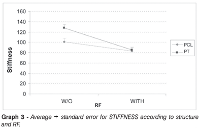
A statistical analysis was performed between both structures behaviors, being observed in both a reduced level of stiffness after RF use, with such reduction being greater for PL. (Graph 3)
For PCL, a reduced stiffness was noticed, reaching, in average, 18.1±8.6 between pre-RF and post-RF groups, which was shown to be statistically significant (p=0.048).
Similarly, for PL, a mean stiffness reduction of 42.6±7.7 was seen between both groups, which was shown to be statistically significant (p<0.001).
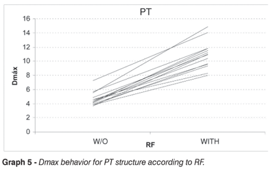
Maximum deformation experienced by the structure during tests was also studied, by an analysis in 2 groups, pre- and post-RF. Below, a graph is shown for individual profiles, showing a consistent increase of this deformation after RF. (Graphs 4 and 5)
Subsequently, the statistical analysis was performed by measuring the averages, standard deviation, median, minimum measure and maximum measure, characterizing the following graph. (Graph 6)
According to the analysis, for PCL, a mean increment of 4.6±0.6mm was seen on the maximum deformation from pre- to post-RF, being considered as statistically significant (p<0.0001). For PL, a mean increment of 6.3±0.6mm was reported, also statistically significant (p<0.0001).
DISCUSSION
We studied the PL and the PCL here, because these are structures that are frequently considered in knee ligaments pathologies and reconstructions.
Studies addressing PCL and its injury-associated biomechanical behavior are scarce when compared to studies addressing the ACL. Considering that cruciate ligaments have similar proportionality and maximum strength constants between each other (5), this study was aimed to enable these results to be applied also to the ACL, due to their comparable biomechanical behavior (15) .
The central portion of the patellar ligament (PL) has been used by many orthopaedic surgeons for anterior cruciate ligament reconstruction. Thus, we chose to study this ligament as a comparative element to cruciate ligaments behavior.
The use of radiofrequency is a new technique that has recently been employed for knee surgeries (1,6,16), as well as for shoulder procedures(7,8,9).
Studies show that tendons and ligaments submitted to radiofrequency do not show significant changes on their biomechanical properties at an interval ranging from 65° to 75°, however leading to structure rupture at temperatures above 80°.(4,17) It is also known that the maximum tolerable shrinkage would be around 15 - 20%, without significant collagen properties loss (4). Therefore, we sought to keep the temperature around 75º, and shrinkage within a 15 - 20% range.
By this study, we realize that RF is an easily-applicable and efficient method for knee structures shrinkage, such as the PL and the PCL, which is consistent to other in vitro e in vivo studies(1,6,14,18). Furthermore, we objectively evidenced that RF significantly changes a tissues mechanical properties.
We noticed that the shrinkage occurred at the expense of a significant tissue stiffness reduction. For PCL, a mean stiffness reduction of 18.1±8.6 occurred, with p=0.048, while PL presented a mean reduction of 42.6±7.7, with p<0.001. This stiffness reduction after RF use was also reported by other authors in animal experiments and in other biological structures.(19-21)
Stiffness loss is noticeable by a major increase of maximum deformity of the structure as shown by the assay. The PCL presented a mean increase of 4.6±0.6 mm, while the PL showed 6.30±0.6mm increase, both with p<0.001. Thus, we can see that the shrinkage obtained by RF can be partially lost when the structure is submitted to a given tensional load, as observed in the increased maximum deformation after a standardized traction assay.
Considering the major stiffness reduction, and the resulting deformation potential at lower tensional loads, it may be suggested that after its use in knee surgeries protection may be necessary for the shortened structure intending to maintain the outcome achieved, as previously employed by some other authors.(3,16)
CONCLUSION
Shrinkage by thermo-abrasion provided by RF is an effective method for providing shrinkage of the studied structures (PCL and PL). After RF use, the mechanical properties of the structures are significantly changed, with a major reduction of baseline stiffness. Therefore, under tension, a higher amount of deformation occurs on the structure, as well as a baseline shrinkage loss.
REFERENCES
Received in: 09/20/06, Approved in: 10/25/06
Study developed at the Department of Orthopaedics and Traumatology, Orthopaedics and Traumatology Institute, Hospital das Clínicas, University of São Paulo (IOT/HC/FMUSP)
- 01 - Carter T, Bailie D, Edinger S. Radiofrequency electrothermal shrinkage of the anterior cruciate ligament. Am J Sports Med.2002; 30:221-6.
- 02 - Taylor DC, Dal Ton JD, Seaber AV, Garrett WE. Viscoelastic properties of mus cletendon units. Am J Sports Med. 1990;18:300-9.
- 03 - Spahn G, Schindler S. Tighthnig elonganted ALC grafts by application of bipolar eletromagnetic energy (ligament shrinkage). Knee Surg Sports Traumatol Arthrosc. 2002; 10:66-71.
- 04 - Vangsness CT Jr, Mitchell W, Nimni M, Erlich M, Saadat V, Schmotzer H Collagen shortening. An experimental approach with heat. Clin Orthop Relat Res.1997; 337:267-71.
- 05 - Wall MS, Deng XH, Torzilli PA, Doty SB, O'Brien SJ, Warren RF.Thermal modification of collagen. Shoulder Elbow Surg.1999; 8:339-44.
- 06 - Roach RA. Roberts SNJ, Rees DA. The potential benefit of thermal shrinkage for lax anterior cruciate ligaments. Acta Orthop. Belg. 2004, 70 247-52.
- 07 - Anderson K, Warren RF, Altchek DW, Craig EV, OBrien SJ. Risk factors for early failure after thermal capsulorrhaphy. Am J Sports Med. 2002; 30:103-7.
- 08 - Levy O, Wilson M, Williams H. "Thermal capsular shrinkage for shoulder instability: Mid-term longitudinal outcome study. J Bone Joint Surg Br. 2001; 83:640-5.
- 09 - Mishra DK, Fanton GS. Two-year outcome of arthroscopic bankart repair and electrothermal-assisted capsulorrhaphy for recurrent traumatic anterior shoulder instability. Arthroscopy. 2001; 17:844-9.
- 10 - Viidik A, Lewin T. Changes in tensile strength characteristics and histology of rabbit ligaments induced by different modes of postmortal storage. Acta Orthop Scand.1966; 36: 141-55.
- 11 - Woo S, Orland C. Effects of posmortem storage by freezing on ligament tensile behavior. J Biomech.1986; 19:399-404.
- 12 - Noyes FR, Butler DL, Grood ES, Zernicke RF, Hefzy MS. Biomechanical analysis of human ligament grafts used in knee-ligament repairs and reconstructions. J Bone Joint Surg Am. 1984; 66:344-52.
- 13 - Gorios C. Estudo do relaxamento à tensão e da rigidez do ligamento cruzado anterior do joelho e dos enxertos para sua reconstrução com o ligamento patelar e com os tendões dos músculos semitendíneo e grácil [dissertação]. São Paulo: Faculdade de Medicina, Universidade de São Paulo; 2000.
- 14 - Chimich D, Shrive N, Frank C, Marchuk L, Bray R. Water content alters viscoelastic behaviour of the normal adolescent rabbit medial collateralligament. J Biomech.1992; 25: 831-7.
- 15 - Hernandez AJ. Correlação das propriedades biomecânicas dos ligamentos do joelho com seus parâmetros antropométricos [tese]. São Paulo: Faculdade de Medicina, Universidade de São Paulo; 1994.
- 16 - Indelli PF. "Monopolar thermal treatment of symptomatic anterior cruciate ligament instability." Clin Orthop Relat Res. 2003; 407:139-47.
- 17 - Hayashi K, Thabit G, Massa KL. The effect of thermal heating on the lenght and histologic proprieties of the glenohumeral joint capsule. Am J Sports Med.1997; 25:107-12
- 18 - Thabit G. The arthroscopic monopolar radiofrequency treatment of chronic anterior cruciate ligament instability. Oper Tech Sports Med.1998; 6:157-60.
- 19 - Potzl W, Heusner T, Kumpers P, Marquardt B, Steinbeck J. Does immobilization after radiofrequency-induced shrinkage influence the biomechanical properties of collagenous tissue? An in vivo rabbit study. Am J Sports Med. 2004; 32:681-7.
- 20 - Potzl W, Kumpers P, Szuwart T, Filler T, Marquardt B, Steinbeck J. Neuronal regeneration after application of radiofrequency energy to collagenous tissue is affected by limb immobilization: an in vivo animal study. J Orthop Res. 2004; 22:1345-50.
- 21 - Potzl W, Kumpers P, Szuwart T, Gotze C, Marquardt B, Steinbeck J. Immobilisation after radiofrequency-induced shrinkage of tendon. A histological study in rabbits. J Bone Surg Br. 2004; 86:752-8.
Correspondences to:
Publication Dates
-
Publication in this collection
04 Sept 2007 -
Date of issue
2007
History
-
Accepted
25 Oct 2006 -
Received
30 Aug 2006


