Abstract
Objectives:
This study aims to investigate the antimicrobial and the anti-biofilm activities of Lactobacillus plantarum extract (LPE) against a panel of oral Staphylococcus aureus (n = 9) and S. aureus ATCC 25923. The in vitro ability of LPE to modulate bacterial resistance to tetracycline, benzalchonium chloride, and chlorhexidine were tested also.
Methods:
The minimum inhibitory concentrations (MICs) and the minimal bactericidal concentrations of Lactobacillus plantarum extract, tetracycline, benzalchonium chloride and clohrhexidine were determined in absence and in presence of a sub-MIC doses of LPE (1/2 MIC). In addition, the LPE potential to inhibit biofilm formation was assessed by microtiter plate and atomic force microscopy assays. Statistical analysis was performed on SPSS v. 17.0 software using Friedman test and Wilcoxon signed ranks test. These tests were used to assess inter-group difference (p < 0.05).
Results:
Our results revealed that LPE exhibited a significant antimicrobial and anti-biofilm activities against the tested strains. A synergistic effect of LPEs and drug susceptibility was observed with a 2–8-fold reduction.
Conclusion:
LPE may be considered to have resistance-modifying activity. A more detailed investigation is necessary to determine the active compound responsible for therapeutic and disinfectant modulation.
Keywords:
Antibacterial; Staphylococcus aureus; Biofilm; Antibiotics; Synergetic; Lactobacillus
Introduction
Staphylococci are important causes of infections associated with various devices. Staphylococcus aureus is one of the most frequent human pathogen associated to medical implant.11 Huang H-L, Chang Y-Y, Lai M-C, Lin C-R, Lai C-H, Shieh T-M. Antibacterial TaN-Ag coatings on titanium dental implants. Surf Coat Technol. 2010;205:1636-41. It has been isolated from parotitis,22 Smith A, Jackson M, Bagg J. The ecology of Staphylococcus species in the oral cavity. J Med Microbiol. 2001;50:940-6. gingival pockets,33 Colombo APV, Teles RP, Torres MC, et al. Subgingival microbiota of Brazilian subjects with untreated chronic periodontitis. J Periodontol. 2002;73:360-9. periodontitis,44 Souto R, Andrade AFBD, Uzeda M, Colombo APV. Prevalence of non-oral pathogenic bacteria in subgingival biofilm of subjects with chronic periodontitis. Braz J Microbiol. 2006;37:208-15. carious lesions,55 Kouidhi B, Zmantar T, Hentati H, Bakhrouf A. Cell surface hydrophobicity, biofilm formation, adhesives properties and molecular detection of adhesins genes in Staphylococcus aureus associated to dental caries. Microb Pathog. 2010;49:14-22. and gingivitis.66 Koukos G, Sakellari D, Arsenakis M, Tsalikis L, Slini T, Konstantinidis A. Prevalence of Staphylococcus aureus and methicillin resistant Staphylococcus aureus (MRSA) in the oral cavity. Arch Oral Biol. 2015;60:1410-5. Orthodontic and other oral appliances may act as reservoir of resistant opportunistic S. aureus.77 Merghni A, Nejma MB, Dallel I, et al. High potential of adhesion to biotic and abiotic surfaces by opportunistic Staphylococcus aureus strains isolated from orthodontic appliances. Microb Pathog. 2016;91:61-7.
S. aureus has the capacity to adhere to various medical devices and form biofilm.88 Proctor RA. Toward an understanding of biomaterial infections: a complex interplay between the host and bacteria. J Lab Clin Med. 2000;135:14-5. Biofilm is associated to the ica gene encoding for polysaccharide intercellular adhesion (PIA)99 Mckenney D, Pouliot KL, Wang Y, et al. Broadly protective vaccine for Staphylococcus aureus based on an in vivo-expressed antigen. Science. 1999;284:1523-7. which leads to increased bacterial drug resistance compared to planktonic cells.1010 Carpentier B, Cerf O. Biofilms and their consequences, with particular reference to hygiene in the food industry. J Appl Bacteriol. 1993;75:499-511.,1111 Costerton JW, Lewandowski Z, Caldwell DE, Korber DR, Lappin-Scott HM. Microbial biofilms. Annu Rev Microbiol. 1995;49:711-45.
S. aureus is able to acquire resistance to antibiotics and to antiseptic agents via efflux pumps (TetK, msrA transporters) systems, which export certain tetracyclines and macrolides molecules outside the cells. On the other hand, some multidrug resistance (MDR) proteins like NorA and QacA confer resistance to a wide range of structurally unrelated antiseptics.1212 Marshall NJ, Piddock LJ. Antibacterial efflux systems. Microbiologia. 1997;13:285-300. In clinical practice, chlorhexidine (CHX) and quaternary ammonium compounds (QAC) are the most frequently used disinfectants to reduce and prevent the spread of pathogens. Multidrug resistance pumps have been recognized as mediators of a number of commonly used ammonium compounds and detergents.1313 Littlejohn TG, Paulsen IT, Gillespie MT, et al. Substrate specificity and energetics of antiseptic and disinfectant resistance in Staphylococcus aureus. FEMS Microbiol Lett. 1992;74:259-65. However, the use of these disinfectants in hospitals may contribute to the rise of disinfectant-resistant bacteria1414 Reverdy ME, Bes M, Brun Y, Fleurette J. Evolution of resistance to antibiotics and antiseptics of hospital Staphylococcus aureus strains isolated from 1980 to 1991. Pathol Biol (Paris). 1993;41:897-904.,1515 Russell AD. Do biocides select for antibiotic resistance. J Pharm Pharmacol. 2000;52:227-33. due to QAC-resistant genes (qacA, qacB, and qacC), which have been identified in several staphylococcal species.1616 Anthonisen I-L, Sunde M, Steinum T, Sidhu M, Sørum H. Organization of the antiseptic resistance gene qacA and Tn552-related β-lactamase genes in multidrug-resistant Staphylococcus haemolyticus strains of animal and human origins. Antimicrob Agents Chemother. 2002;46:3606-12.
17 Bjorland J, Steinum T, Kvitle B, Waage S, Sunde M, Heir E. Widespread distribution of disinfectant resistance genes among staphylococci of bovine and caprine origin in Norway. J Clin Microbiol. 2005;43:4363-8.-1818 Zmantar T, Kouidhi B, Miladi H, Bakhrouf A. Detection of macrolide and disinfectant resistance genes in clinical Staphylococcus aureus and coagulase-negative staphylococci. BMC Res Notes. 2011;4:453.
In this study, the antibacterial activity of Lactobacillus plantarum extract (LPE) was investigated. In addition, the ability of LPE to modulate the susceptibility of S. aureus, isolated from the oral cavity of Tunisian children, to tetracycline (TET), benzalchonium chloride (BC) and clohrhexidine (CHX) was tested. Secondly, the effects of disinfectant and antibiotic associated with LPE were tested for biofilm inhibition of S. aureus to polystyrene and glass using microtiter plate and atomic force microscopy assays, respectively.
Material and methods
Antibacterial activity of L. plantarum
L. plantarum strain was isolated from Tunisian traditional fermented milk (ricotta cheese), identified with a conventional method using Api-50 CHL system (BioMerieux, Marcy-l'Étoile, France) and by polymerase chain reaction (PCR) technique using L. plantarum-specific primers: forward IDL04F: 5′-AGGGTGAAGTCGTAACAAGTAGCC-3′ and reverse IDL62R 5′-CTAGTGGTAACAGTTGATTAAAACTGC-3′, giving a product size of about 428 bp as described previously.1919 Kwon HS, Yang EH, Yeon SW, Kang BH, Kim TY. Rapid identification of probiotic Lactobacillus species by multiplex PCR using species-specific primers based on the region extending from 16S rRNA through 23S rRNA. FEMS Microbiol Lett. 2004;239:267-75.
Cell-free supernatant was obtained by centrifuging (10,000 × g for 10 min at 4 °C) L. plantarum culture, grown in MRS broth for 16 h at 30° C. The supernatant was adjusted at neutral pH and filter-sterilized (0.22 µm, Millipore). The obtained L. plantarum extract (LPE) was conserved at 4 °C until use.
Antibacterial activity of LPE was tested against nine strains (Table 1) isolated from the oral cavity of Tunisian children and S. aureus ATCC 25923 using the broth microdilution method.2020 Bagamboula CF, Uyttendaele M, Debevere J. Inhibitory effects of spices and herbs towards Shigella sonnei and S. flexneri. Meded Rijksuniv Gent Fak Landbouwkd Toegep Biol Wet. 2001;66:523-30.,2121 Erdemoglu N, Kupeli E, Yesilada E. Anti-inflammatory and antinociceptive activity assessment of plants used as remedy in Turkish folk medicine. J Ethnopharmacol. 2003;89:123-9. The LPE was tested alone or in combination with antibiotics.
Minimum inhibitory and minimum bactericidal concentrations in µg/mL of Lactobacillus plantarum extract alone or in combination with benzalchonium chloride, chlorhexidine, and tetracycline.
Microorganisms
The S. aureus strains (n = 9) used in this study were isolated from the oral cavity of Tunisian children from the dental clinic of dentistry (Monastir, Center of Tunisia).
The criteria for inclusion were: no antibiotic treatment four weeks prior to sampling, no use of mouth rinses or any other preventive measure that might involve exposure to antimicrobial agents, and no systemic disease. A sterile swab was used for sample collection from the oral cavity of each patient. After incubation (24 h at 37 °C), swabs were plated on blood agar plates supplemented with 5% sheep blood (24 h at 37 °C) and bacterial identification was achieved using conventional methods.
Minimal inhibitory concentration determination
The broth microdilution method was used to determine the minimum inhibitory concentration (MIC) of LPE (5–90%, v/v), BC (Acros organics, USA) (0–1024 µg/mL), CHX (Sigma–Aldrich, USA) (0–1024 µg/mL) and tetracycline (TET) (0–1024 µg/mL) according to the Clinical and Laboratory Standards Institute (2006) guidelines.2222 CLSI Performance standards for antimicrobial susceptibility testing. Clinical and Laboratory Standards Institute. Wayne, PA: Clinical and Laboratory Standards Institute; 2006. Supplement M100-S16.
After 24 h of incubation, bacterial growth was evaluated by the presence of turbidity and a pellet on the well bottom. MIC was defined as the lowest concentration of the compound that had no macroscopically visible growth. All experiments were carried out three times.
Minimal bactericidal concentration determination (MBC)
To determine the MBC values, 10 µL of each well medium, with no visible growth was removed and inoculated in Muller Hinton agar plates. MBC was defined as the lowest concentration at which 99% of the bacteria were killed. Each experiment was repeated at least twice.2323 Magina M, Dalmarco E, Wisniewski A, et al. Chemical composition and antibacterial activity of essential oils of Eugenia species. J Nat Med. 2009;63:345-50.
Modulation of S. aureus susceptibility to BC, CHX and TET by LPE
To determine the potential effect of LPE to modulate drug resistance of S. aureus, MICs of BC, CHX and TET (ranging from 0.5 to 2048 µg/mL) were determined alone and combined with a sub-MIC of LPE (1/2 MIC, v/v) using the microtiter plates assay.2424 Kouidhi B, Zmantar T, Jrah H, et al. Antibacterial and resistance-modifying activities of thymoquinone against oral pathogens. Ann Clin Microbiol Antimicrob. 2011;10:29.
Determination of anti-biofilms activity by microtiter plates assay
The anti-adhesion properties of LPE (ranging from 10% to 90%, v/v) to S. aureus strains were tested as previously described by Merritt et al.2525 Merritt JH, Kadouri DE, O'toole GA. Growing and analyzing static biofilms. Curr Protoc Microbiol. 2005. [chapter 1]: Unit 1B 1 Briefly, the bacterial culture was grown in Tryptone soy broth (TSB) at 37 °C for 24 h. Two microliters were disposed into each well of 96-well plates in the presence of 198 µL of the medium supplemented with 2% glucose (w/v) containing LPE. Plates were incubated in aerobic conditions at 37 °C for 24 h. The anti-biofilm activity of TET, CHX, and BC ranging from 2 to 1024 µg/mL were determined against S. aureus strains with or without sub-MIC of the LPE (1/2 MIC, v/v), using the microtiter plates assay. After incubation, the plate was washed with phosphate buffer saline, stained with 100 µL of 1% (w/v) crystal violet and incubated at room temperature for 15 min. Then, the absorbance at 590 nm was determined using a microplate reader (D.E.E.D reader, Bio-Rad instrument). The mean absorbance (OD590nm) of the sample was determined, and the percentage of inhibition obtained for each concentration of LPE and drugs was determined by the following formula:
Evaluation of LPE biofilm inhibition by atomic force microscopy (AFM)
To evaluate biofilm inhibition potency of LPE, S. aureus ATCC 25923 was grown in Tryptone soy broth (TSB) at 37 °C for 24 h and then diluted into the TSB medium (105 cells/mL) supplemented with 2% glucose (w/v). For each concentration of the tested drugs (2–1024 µg/mL) and LPE (5–90%), 10 mL of bacterial suspension was incubated in a 6-well plate containing round glass cover slips for 24 h at 37 °C. After adhesion, the non-adhering bacteria were removed by rinsing the substrate four times with sterile PBS. Then the substrate was air dried and observed using AFM. All AFM images were then processed to analyze the biofilm formation behavior.
Statistical analysis
Data were analyzed using SPSS v. 17.0 software. The Friedman test, followed by the Wilcoxon signed ranks test were used to assess inter-group differences. In addition, statistical significance was set at p-value <0.05.
Results
Antimicrobial activity of LPE
As presented in Table 1, LPE demonstrated selective antimicrobial properties. Seven S. aureus strains (S3, S4, S5, S6, S1, S8 and S9) and S. aureus ATCC 25923 have a MICs value ranged from 20% to 50% (v/v). The remaining two strains (S2, S7) were more susceptible (MIC 10%). In addition, a variable MBCs (10–90%) (v/v) was observed according to the tested strain (Table 1). On the other hand, out of the nine oral S. aureus strains, eight (Table 1) were considered resistant to BC (MICs between 4 and 128 µg/mL) and only one strain (S4) was sensitive (MICs ≤ 2 µg/mL); all strains were resistant to CHX (MICs > 2 µg/mL). Moreover, only one strain (S4) was susceptible to TET (MIC < 2 µg/mL).
Modulation of drug resistance by LPE
Data presented in Table 1 showed that the presence of LPE at 1/2-MIC (v/v) in combination with BC resulted in a 4-fold reduction of MICs and MBCs of S. aureus ATCC 25923 and one oral strain (S6). Concerning the synergistic effect of CHX and LPE, there was an important diminution of MIC and MBC of CHX (2–8-fold potentiation) in six strains (Table 1). Furthermore, a 2-8-fold reduction of MIC (µg/mL) of TET against seven strains when combined with LPE was noticed (Table 1).
Biofilm inhibition by LPE
Biofilm formation of oral S. aureus strains was evaluated in 96 microtiter wells plate with LPE (ranging from 5% to 90%, v/v). Results were expressed as minimum biofilm inhibition concentration (BIC50 and BIC90). As presented in Table 2, BIC90 of LPE was reached at 54% LPE supplementation, for two oral strains (S4 and S7). While, BIC50 ranged from 28 to 77% (v/v) LPE supplementation (Table 2).
Anti-biofilm effect of Lactobacillus plantarum extract alone or in combination with benzalchonium chloride, chlorhexidine, and tetracycline against oral staphylococci.
In addition, BC showed various effects on the development of S. aureus biofilms with BIC50 values ranging from 12 to 112 µg/mL for the tested strains. Moreover, the combination of BC and 1/2 MIC LPE resulted in a reduction of BIC50 against eight strains (Table 2). In the same way BIC90 was also reduced with the combination of BC and LPE.
Concerning the effect of CHX alone and in combination with LPE, BIC50 values ranged from 34–355 µg/mL for CHX for the tested strains (Table 2) whereas the combination of CHX and 1/2 MIC of LPE % (v/v) exhibited a reduction of BIC50 and BIC90 against all the tested strains (Table 2).
Our results revealed that TET may reduce S. aureus attachment to polystyrene (BIC90 reach 30 µg/mL for S4). In addition, the combination of TET and 1/2 MIC of LPE exhibited a reduction of BIC50 and BIC90 against nine strains (Table 2).
Evaluation of LPE biofilm inhibition by AFM
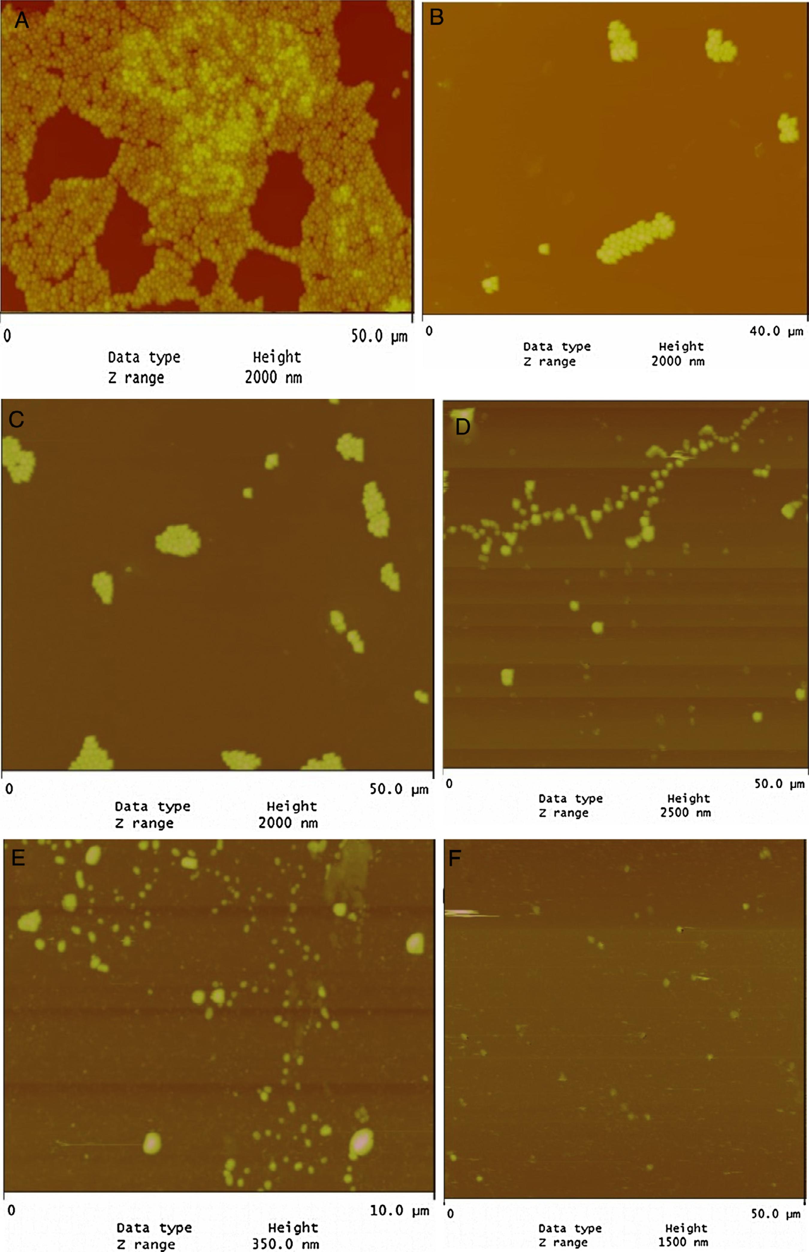
AFM revealed that the tested strains were strongly biofilm positive and had the potency to adhere to glass surface (Fig. 1A). On the other hand, LPE decreased significantly the adherence to glass (Fig. 1D). In addition, the combination of LEP with TET, BC and CHX decreased significantly the biofilm formation (Fig. 1B–E). As presented in Fig. 1F, almost total biofilm inhibition was observed with 16 µg/mL of BC combined with 1/2 MIC of LPE % (v/v).
AFM images of biofilm formed by S. aureus ATCC 25923 to glass surface: (A) TSB alone; (B) TSB with 4 µg/mL of BC + 1/2 MIC LPE; (C) TSB with 8 µg/mL of CHX + 1/2 MIC LPE; (D) TSB with + 40% LPE (v/v); (E) TSB with 8 µg/mL TET + 1/2 MIC LPE; (F) TSB with 16 µg BC + 1/2 MIC LPE.
Discussion
A wide ranges of L. plantarum produce anti-microbial peptides with important effects against Gram-positive bacteria.2626 Eijsink VG, Axelsson L, Diep DB, Havarstein LS, Holo H, Nes IF. Production of class II bacteriocins by lactic acid bacteria; an example of biological warfare and communication. Antonie Van Leeuwenhoek. 2002;81:639-54. Lactic acid bacteria (LAB) are known to have bactericidal activity against pathogenic bacteria. For that, they develop many mechanisms like hydrogen peroxide production causing denaturation of many enzymes and peroxidation of membrane leading to increased permeability.2727 Kong S, Davison AJ. The role of interactions between O2, H2O2, === Inserir caracter correspondente ao PDF === OHe- and O2-in free radical damage to biological systems. Arch Biochem Biophys. 1980;204:18-29. Also, the LAB are able to produce bacteriocins which have a bactericidal effect on other species.2828 Garneau S, Martin NI, Vederas JC. Two-peptide bacteriocins produced by lactic acid bacteria. Biochimie. 2002;84:577-92. In order to find a natural source able to inhibit pathogenic bacteria and prevent oral biofilm formation, we tested in the effect of LEP on biofilm formation of nine S. aureus isolated from the oral cavity. In addition, we tested the potential effect of LPE to modulate susceptibility of some drugs and disinfectants of 10 S. aureus.
In our study, neutralized LPE supplementation (MIC values 10–50%, v/v) had potential bactericidal effect on oral S. aureus (Table 1). As presented in Table 1, the extract is very effective against S2 and S7 and may constitute a valuable agent to counter the growth of biofilms and undesirable microorganisms on surfaces. Similar results have been found concerning the antibacterial activity of LPE against Vibrio sp.2929 Lamari F, Sadok K, Bakhrouf A, Gatesoupe F-J. Selection of lactic acid bacteria as candidate probiotics and in vivo test on Artemia nauplii. Aquacult Int. 2014;22:699-709. and Listeria. monocytogenes.3030 Ben Slama R, Kouidhi B, Zmantar T, Chaieb K, Bakhrouf A. Anti-listerial and anti-biofilm activities of potential probiotic Lactobacillus strains isolated from Tunisian traditional fermented food. J Food Saf. 2013;33:8-16. Even with sub-inhibitory concentrations (1/2 MIC) LPE showed a potential role to modulate susceptibility of the tested S. aureus. Since BC, TET, and CHX resistance is modulated by efflux pump systems, we suggest that LPE may potentiate, partially, the role of the disinfectant by blocking the respective efflux pump. The efflux pump is the one of major antibiotic resistance mechanisms utilized by bacterial cells to develop resistance. Thus, this resistance mechanism eliminates several classes of antibiotics such as fluoroquinolones, TET, and other compounds such as BC and CHX.
We also noted that the MIC of BC (2–4-fold reduction), CHX (2–8-fold reduction), and TET (2–8-fold reduction) was reduced when the medium was supplemented with LPE at a sub-inhibitory concentration (Table 1). The results showed a statistically significant difference between the antibacterial effects of drugs with and without LPE supplementation (p < 0.05). Thus, this finding confirms our hypothesis that the ability to modify susceptibility tested in this study (Table 1) is related to components that have a potential role to inhibit S. aureus efflux pump.
Biofilm formation is an important virulence factor of oral bacteria.55 Kouidhi B, Zmantar T, Hentati H, Bakhrouf A. Cell surface hydrophobicity, biofilm formation, adhesives properties and molecular detection of adhesins genes in Staphylococcus aureus associated to dental caries. Microb Pathog. 2010;49:14-22. Our results demonstrate that LPE exhibited a good anti-bioflm activity for all tested strains (Table 2). Similar results were found concerning the anti-biofilm activity of LPE against L. monoctogenes.3030 Ben Slama R, Kouidhi B, Zmantar T, Chaieb K, Bakhrouf A. Anti-listerial and anti-biofilm activities of potential probiotic Lactobacillus strains isolated from Tunisian traditional fermented food. J Food Saf. 2013;33:8-16. Previous studies have shown that the cells in a biofilm were more resistant to antimicrobial agents compared to free-floating cells.3131 Frank JF, Koffi RA. Surface-adherent growth of Listeria monocytogenes is associated with increased resistance to surfactant sanitizers and heat. J Food Protect. 1990;53:550-4.,3232 Krysinski E, Brown L, Marchisello T. Effect of cleaners and sanitizers on Listeria monocytogenes attached to product contact surfaces. J Food Protect. 1992;55:246-51. On the other hand, the anti-biofilm activity of disinfectants and antibiotic (BC, CHX and TET) to polystyrene and glass was enhanced when the medium was supplemented with LPE at different percentages (Table 2 and Fig. 1). The statistical analysis showed a correlation between the percentage inhibition of biofilm of different drugs alone or combined with a sub-MIC of LPE % (1/2 MIC, v/v) (p < 0.05). As shown in Fig. 1, AFM demonstrates that S. aureus biofilm were strongly affected by the supplementation of LPE. The identification of anti-biofilm constituents will be essential to be included as alternatives in the control of bacterial biofilms.
The MICs modifying ability of LPE tested in this study may be related to components that have a potential role to inhibit S. aureus efflux pump. Further studies are required to assess their composition and potential clinical relevance.
References
-
1Huang H-L, Chang Y-Y, Lai M-C, Lin C-R, Lai C-H, Shieh T-M. Antibacterial TaN-Ag coatings on titanium dental implants. Surf Coat Technol. 2010;205:1636-41.
-
2Smith A, Jackson M, Bagg J. The ecology of Staphylococcus species in the oral cavity. J Med Microbiol. 2001;50:940-6.
-
3Colombo APV, Teles RP, Torres MC, et al. Subgingival microbiota of Brazilian subjects with untreated chronic periodontitis. J Periodontol. 2002;73:360-9.
-
4Souto R, Andrade AFBD, Uzeda M, Colombo APV. Prevalence of non-oral pathogenic bacteria in subgingival biofilm of subjects with chronic periodontitis. Braz J Microbiol. 2006;37:208-15.
-
5Kouidhi B, Zmantar T, Hentati H, Bakhrouf A. Cell surface hydrophobicity, biofilm formation, adhesives properties and molecular detection of adhesins genes in Staphylococcus aureus associated to dental caries. Microb Pathog. 2010;49:14-22.
-
6Koukos G, Sakellari D, Arsenakis M, Tsalikis L, Slini T, Konstantinidis A. Prevalence of Staphylococcus aureus and methicillin resistant Staphylococcus aureus (MRSA) in the oral cavity. Arch Oral Biol. 2015;60:1410-5.
-
7Merghni A, Nejma MB, Dallel I, et al. High potential of adhesion to biotic and abiotic surfaces by opportunistic Staphylococcus aureus strains isolated from orthodontic appliances. Microb Pathog. 2016;91:61-7.
-
8Proctor RA. Toward an understanding of biomaterial infections: a complex interplay between the host and bacteria. J Lab Clin Med. 2000;135:14-5.
-
9Mckenney D, Pouliot KL, Wang Y, et al. Broadly protective vaccine for Staphylococcus aureus based on an in vivo-expressed antigen. Science. 1999;284:1523-7.
-
10Carpentier B, Cerf O. Biofilms and their consequences, with particular reference to hygiene in the food industry. J Appl Bacteriol. 1993;75:499-511.
-
11Costerton JW, Lewandowski Z, Caldwell DE, Korber DR, Lappin-Scott HM. Microbial biofilms. Annu Rev Microbiol. 1995;49:711-45.
-
12Marshall NJ, Piddock LJ. Antibacterial efflux systems. Microbiologia. 1997;13:285-300.
-
13Littlejohn TG, Paulsen IT, Gillespie MT, et al. Substrate specificity and energetics of antiseptic and disinfectant resistance in Staphylococcus aureus FEMS Microbiol Lett. 1992;74:259-65.
-
14Reverdy ME, Bes M, Brun Y, Fleurette J. Evolution of resistance to antibiotics and antiseptics of hospital Staphylococcus aureus strains isolated from 1980 to 1991. Pathol Biol (Paris). 1993;41:897-904.
-
15Russell AD. Do biocides select for antibiotic resistance. J Pharm Pharmacol. 2000;52:227-33.
-
16Anthonisen I-L, Sunde M, Steinum T, Sidhu M, Sørum H. Organization of the antiseptic resistance gene qacA and Tn552-related β-lactamase genes in multidrug-resistant Staphylococcus haemolyticus strains of animal and human origins. Antimicrob Agents Chemother. 2002;46:3606-12.
-
17Bjorland J, Steinum T, Kvitle B, Waage S, Sunde M, Heir E. Widespread distribution of disinfectant resistance genes among staphylococci of bovine and caprine origin in Norway. J Clin Microbiol. 2005;43:4363-8.
-
18Zmantar T, Kouidhi B, Miladi H, Bakhrouf A. Detection of macrolide and disinfectant resistance genes in clinical Staphylococcus aureus and coagulase-negative staphylococci. BMC Res Notes. 2011;4:453.
-
19Kwon HS, Yang EH, Yeon SW, Kang BH, Kim TY. Rapid identification of probiotic Lactobacillus species by multiplex PCR using species-specific primers based on the region extending from 16S rRNA through 23S rRNA. FEMS Microbiol Lett. 2004;239:267-75.
-
20Bagamboula CF, Uyttendaele M, Debevere J. Inhibitory effects of spices and herbs towards Shigella sonnei and S. flexneri Meded Rijksuniv Gent Fak Landbouwkd Toegep Biol Wet. 2001;66:523-30.
-
21Erdemoglu N, Kupeli E, Yesilada E. Anti-inflammatory and antinociceptive activity assessment of plants used as remedy in Turkish folk medicine. J Ethnopharmacol. 2003;89:123-9.
-
22CLSI Performance standards for antimicrobial susceptibility testing. Clinical and Laboratory Standards Institute. Wayne, PA: Clinical and Laboratory Standards Institute; 2006. Supplement M100-S16.
-
23Magina M, Dalmarco E, Wisniewski A, et al. Chemical composition and antibacterial activity of essential oils of Eugenia species. J Nat Med. 2009;63:345-50.
-
24Kouidhi B, Zmantar T, Jrah H, et al. Antibacterial and resistance-modifying activities of thymoquinone against oral pathogens. Ann Clin Microbiol Antimicrob. 2011;10:29.
-
25Merritt JH, Kadouri DE, O'toole GA. Growing and analyzing static biofilms. Curr Protoc Microbiol. 2005. [chapter 1]: Unit 1B 1
-
26Eijsink VG, Axelsson L, Diep DB, Havarstein LS, Holo H, Nes IF. Production of class II bacteriocins by lactic acid bacteria; an example of biological warfare and communication. Antonie Van Leeuwenhoek. 2002;81:639-54.
-
27Kong S, Davison AJ. The role of interactions between O2, H2O2, === Inserir caracter correspondente ao PDF === OHe- and O2-in free radical damage to biological systems. Arch Biochem Biophys. 1980;204:18-29.
-
28Garneau S, Martin NI, Vederas JC. Two-peptide bacteriocins produced by lactic acid bacteria. Biochimie. 2002;84:577-92.
-
29Lamari F, Sadok K, Bakhrouf A, Gatesoupe F-J. Selection of lactic acid bacteria as candidate probiotics and in vivo test on Artemia nauplii Aquacult Int. 2014;22:699-709.
-
30Ben Slama R, Kouidhi B, Zmantar T, Chaieb K, Bakhrouf A. Anti-listerial and anti-biofilm activities of potential probiotic Lactobacillus strains isolated from Tunisian traditional fermented food. J Food Saf. 2013;33:8-16.
-
31Frank JF, Koffi RA. Surface-adherent growth of Listeria monocytogenes is associated with increased resistance to surfactant sanitizers and heat. J Food Protect. 1990;53:550-4.
-
32Krysinski E, Brown L, Marchisello T. Effect of cleaners and sanitizers on Listeria monocytogenes attached to product contact surfaces. J Food Protect. 1992;55:246-51.
Publication Dates
-
Publication in this collection
Jan-Feb 2017
History
-
Received
24 Mar 2016 -
Accepted
26 Oct 2016