Abstracts
The clinical, gross and microscopical features of a granulosa cell tumour in the ovary of a 2-year-old cat are described. This is a rare tumour in cats, moreover because of the animal’s age. It was diagnosed clinically by ultrasound examination. The tumour, a cystic mass in the region of the left ovary, metastasized to the epiplon and lungs. Tumour cells expressed vimentin and were arranged in a diffuse sarcomatous pattern, supported by a fine fibrovascular stroma. The presence of hair loss and repeated oestrus as clinical signs are indicative of orthoendocrine paraneoplastic syndrome, due to excessive oestrogen stimulation. This tumour should be considered in the differential diagnosis of behavioural disturbances in female cats.
Granulosa cell tumour; Ovary; Cats; Immunohistochemistry; Paraneoplastic syndromes
As características clínicas, macroscópicas e microscópicas de um tumor de células da granulosa no ovário de uma gata de 2 anos de idade são descritas neste trabalho. Essa neoplasia é rara em gatos, principalmente devido à idade apresentada pelo animal. O diagnóstico foi feito clinicamente através de exame ultra-sonográfico. O tumor, uma massa cística na região do ovário esquerdo, metastatizou para o epíploo e para os pulmões. As células tumorais expressaram vimentina e arranjavam-se num padrão sarcomatoso e difuso, sustentado por fino estroma fibrovascular. A presença de sinais clínicos, como perda de pêlos e repetição de estros, é indicativa de síndrome paraneoplásica ortoendócrina, devido à excessiva estimulação estrogênica. A existência desse tipo de tumor deve ser considerada no diagnóstico diferencial de distúrbios comportamentais em gatas.
Tumor de células granulosas; Ovário; Gatos; Imunoistoquímica; Síndromes paraneoplásicas
SHORT COMMUNICATION / NOTA PRÉVIA
Granulosa cell tumour with metastasis in a cat
Tumor de células da granulosa com metástases numa gata
Márcia Regina GIACÓIA1 1 Departamento de Patologia da Faculdade de Medicina Veterinária e Zootecnia da USP - SP 2 Departamento de Reprodução Animal da Faculdade de Medicina Veterinária e Zootecnia da USP - SP ; Paulo César MAIORKA1 1 Departamento de Patologia da Faculdade de Medicina Veterinária e Zootecnia da USP - SP 2 Departamento de Reprodução Animal da Faculdade de Medicina Veterinária e Zootecnia da USP - SP ; Clair Motos OLIVEIRA2 1 Departamento de Patologia da Faculdade de Medicina Veterinária e Zootecnia da USP - SP 2 Departamento de Reprodução Animal da Faculdade de Medicina Veterinária e Zootecnia da USP - SP ; Idércio Luiz SINHORINI1 1 Departamento de Patologia da Faculdade de Medicina Veterinária e Zootecnia da USP - SP 2 Departamento de Reprodução Animal da Faculdade de Medicina Veterinária e Zootecnia da USP - SP ; Maria Lúcia Zaidan DAGLI1 1 Departamento de Patologia da Faculdade de Medicina Veterinária e Zootecnia da USP - SP 2 Departamento de Reprodução Animal da Faculdade de Medicina Veterinária e Zootecnia da USP - SP
CORRESPONDENCE TO:
Maria Lúcia Zaidan Dagli
Departamento de Patologia
Faculdade de Medicina Veterinária e Zootecnia da USP
Cidade Universitária Armando de Salles Oliveira
Av. Orlando Marques de Paiva, 87
05508-000 São Paulo SP
e-mail: mlzdagli@usp.br
SUMMARY
The clinical, gross and microscopical features of a granulosa cell tumour in the ovary of a 2-year-old cat are described. This is a rare tumour in cats, moreover because of the animals age. It was diagnosed clinically by ultrasound examination. The tumour, a cystic mass in the region of the left ovary, metastasized to the epiplon and lungs. Tumour cells expressed vimentin and were arranged in a diffuse sarcomatous pattern, supported by a fine fibrovascular stroma. The presence of hair loss and repeated oestrus as clinical signs are indicative of orthoendocrine paraneoplastic syndrome, due to excessive oestrogen stimulation. This tumour should be considered in the differential diagnosis of behavioural disturbances in female cats.
UNITERMS: Granulosa cell tumour; Ovary; Cats; Immunohistochemistry; Paraneoplastic syndromes.
This paper reports the gross and microscopical findings of a granulosa cell tumour in a 2-year-old Siamese cat. The animal was presented for clinical evaluation with the complaint of alopecic areas in the back and tail, and behavioral disturbances. The first diagnosis was psychogenic dermatitis, and an unsuccessful symptomatic treatment was carried out during 6 months. After this period, the animal was presented once to the clinician, with a history of repeated oestrus and polyuria. Physical examination revealed an enlargement of the abdomen, with a palpable mass in the left side of the hypogastric region. Ultrasound examination revealed the presence of an ecogenic formation in the region of the left ovary. Several nodules in the lungs were shown by chest radiographies. A treatment with vincristine was started, but no response was obtained, and the animal was euthanized.
At necropsy, the cadaver was cachetic. A whitish tumour mass of about 12 cm in diameter was observed in the region of the left ovary. The cut surface of the mass revealed solid, cystic and necrotic areas. Many nodules of about 2 to 3 cm of diameter were seen in the epiplon, and also in the diaphragmatic lung lobes. Representative slices of the tumour mass and other altered organs were harvested, fixed in 10% formalin and embedded in paraffin wax.
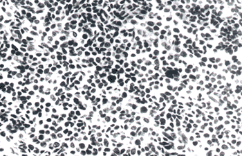
The histopathological examination of Hematoxylin-eosin (HE) stained slides revealed that the tumour mass was composed by small cells with round nuclei, similar to granulosa cells of the Graafian follicle, arranged in a diffuse sarcomatous pattern, supported by a fine fibrovascular stroma (Fig. 1). Many small blood vessels could be identified among the tumour cells. In some areas of the tumour mass a typical folliculoid pattern of tumour cells was observed. Rosette formations resembling the Call-Exner bodies were not seen. Typical and atypical mitotic Figures were frequently seen; necrotic and cystic areas were abundant.
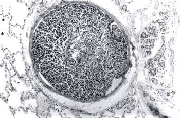
Metastatic nodules presented the same pattern of the primitive ovarian tumour. Besides the presence of metastatic nodules, lungs also presented several neoplastic emboli (Fig. 2). A cystic glandular hyperplasia was observed in the uterus (Fig. 3).
To better characterize this sex cord - gonadostromal tumour, an immunohistochemical staining was performed. Additional sections of the tumour mass were placed in silanized slides, deparaffinized, hydrated and boiled in citrate buffer pH 6.0 within a conventional microwave oven for 15 minutes. The sections were then incubated overnight at 4ºC with a dilution of 1:200 of anti-vimentin (DAKO, Denmnark) antibody, or anti-high and low molecular weight cytokeratins (AE1/AE3, Biogenex). The reactions were revealed with the streptavidin - biotin- peroxidase kit (Duet, DAKO, Denmark), with diaminobenzidine as the chromogen. Negative controls were obtained by omitting the primary antibodies. The tumour cells presented a strong cytoplasmic positivity for vimentin, but not for cytokeratins (Fig. 4).
Granulosa cell tumour is considered the most common sex cord-gonadostromal tumour in all animal species4,5. The reported mean age of incidence in cats is 11 years3, and this data was based in only 20 reported cases. In this case, the only two-year-old cat was the youngest animal diagnosed with this tumour in the literature, and this can be considered a rare diagnosis. Contraposing, the mean age of incidence of sex cord-gonadostromal tumours in bitches is five years, and some cases can be found at a slightly younger age6.
The histopathological findings in this granulosa tumour did not differ from the literature and are in accordance to the agressive behaviour described for this tumour in cats1,7.
The presence of repeated estrus, and loss of hair can represent a sign of oestrogen production by tumour cells being this considered a paraneoplastic syndrome9. The finding of a discrete enlargement of the uterus with cystic hyperplasia of the endometrium can be a consequence of this hormonal imbalance. According to CRUM2, granulosa cell tumour has clinical importance for two reasons, its potential synthesis of large amounts of estrogen and the hazard of malignancy.
The positivity for vimentin, a marker for mesenchymal cells, characterizes the sex cord stromal cells of the gonads, represented by the granulosa cells of the ovary and the Sertoli cells of the testicle8. In animals, the report of a granulosa cell tumour associated with clinical signs of repeated estrus is important as an alert to the clinician to include this pathological entity in his differential diagnosis.
ACKNOWLEDGEMENTS
The authors thank Mr. C. Arroyo, Mr.L.A. Bugalho and Mrs.M.I.S. da Silva for preparing the slides. They are also grateful to Mrs. S.M.S. Silva for the careful preparation of the photomicrographs.
RESUMO
As características clínicas, macroscópicas e microscópicas de um tumor de células da granulosa no ovário de uma gata de 2 anos de idade são descritas neste trabalho. Essa neoplasia é rara em gatos, principalmente devido à idade apresentada pelo animal. O diagnóstico foi feito clinicamente através de exame ultra-sonográfico. O tumor, uma massa cística na região do ovário esquerdo, metastatizou para o epíploo e para os pulmões. As células tumorais expressaram vimentina e arranjavam-se num padrão sarcomatoso e difuso, sustentado por fino estroma fibrovascular. A presença de sinais clínicos, como perda de pêlos e repetição de estros, é indicativa de síndrome paraneoplásica ortoendócrina, devido à excessiva estimulação estrogênica. A existência desse tipo de tumor deve ser considerada no diagnóstico diferencial de distúrbios comportamentais em gatas.
UNITERMOS: Tumor de células granulosas; Ovário; Gatos; Imunoistoquímica; Síndromes paraneoplásicas.
Received: 22/12/1997
Accepted: 23/04/1999
-
1- BAKER, E. Malignant granulosa cell tumour in a cat. Journal of American Veterinary Medical Association, p.322-4, 1956.
-
2- CRUM, C.P. Female genital tract. In: COTRAN, R.S.; KUMAR, V.; ROBBINS, S.L. Pathologic basis of disease 5.ed. Philadelphia : W.B. Saunders, 1994. P.1033-88.
-
3- HOLZWORTH, J. Diseases of the cat: medicine and surgery 1. Philadelphia : W.B. Saunders, 1987. P.520-1: tumours of the female reproductive system.
-
4- MADEWELL, B.R.; THEILEN, G.H. Veterinary cancer medicine 2.ed. Philadelphia : Lea and Febiger, 1987. P.567-82: Tumours of the urogenital tract.
-
5- NIELSEN, S.W.; KENNEDY, P.C. Tumours of the genital systems. In: MOULTON, J.E. Tumours in domestic animals 3.ed. Berkeley University of California Press, 1990. P.458-75.
-
6- NIELSEN, S.W.; MISDORP, W.; McENTEE, K. Tumours of Ovary. In: International Histological Classification of Tumours in Domestic Animals Bulletin of the World Health Organization, v.53, n.2/3, p.203-16, 1976.
-
7- NORRIS, H.J.; GARNER, F.M.; TAYLOR, H.B. Pathology of feline ovarian neoplasms. Journal of Pathology, v.97, p.138-43, 1969.
-
8- PATNAIK, A.K.; MOSTOFI, F.K. A clinicopathologic, histologic and immunohistochemical study of mixed germ cell-stromal tumours of the testis in 16 dogs. Veterinary Pathology, v.30, p.287-95, 1993.
-
9- SLAUSON, D.O.; COOPER, B.J. Mechanisms of Disease – a textbook of comparative general pathology. 2.ed. Baltimore : Williams & Wilkins, 1990. P.377-471: disorders of cell growth.
Publication Dates
-
Publication in this collection
28 Sept 2000 -
Date of issue
1999
History
-
Accepted
23 Apr 1999 -
Received
22 Dec 1997