Abstracts
This study showed a skin squamous cell carcinoma clinical case from 8-years-old female weimaranner, treated by gold-198 foil brachytherapy. The aims were to evaluate special radioactive mould performed to be used on Veterinary practioners and, it's efficiency on one case of skin tumor treatment in dog. The method showed efficacy on the skin tumor treatment and radiological protection especially for the professional team involved, low cost and better radiobiological results when it was compared with standard treatment using others radioactive elements.
Gold (198Au) foils; Skin squamous cell carcinoma; Dog
Este trabalho relata um caso clínico de carcinoma escamo celular cutâneo em um cão da raça Weimaraner, fêmea, 8 anos de idade tratado utilizando-se braquiterapia com folhas de Ouro-198. Os objetivos deste relato foram: avaliar o uso de um molde radioativo confeccionado para uso veterinário e sua eficácia no tratamento de um tumor de pele em cão. O método demonstrou ser eficaz no tratamento do tumor, mostrou ser uma prática segura para a equipe profissional envolvida, com baixos custos e resultado radiobiológico superior quando comparado com o tratamento padrão utilizado com outros elementos radioativos.
Folhas de Ouro-198; Carcinoma escamo-celular cutâneo; Cão
Gold (198Au) foils brachytherapy use on canine skin tumor
Utilização da braquiterapia com folhas de ouro (Au198) em tumor de pele de cão
Marco Antônio Rodrigues FernandesI; Alexandre Lima de AndradeII; Luciane BiazzonoII; Maria Cecília Rui LuvizottoII; Adimir dos SantosIII; Clibas CorreaI
ISanta Casa de Misericórdia de Araçatuba, Araçatuba - SP
IIDepartamento de Clínica, Cirurgia e Reprodução Animal da Unesp, Campus de Araçatuba, Araçatuba - SP
IIIInstituto de Pesquisas Energéticas e Nucleares de São Paulo, São Paulo - SP
correspondence to Endereço para correspondência MARIA CECÍLIA RUI LUVIZOTTO Departamento de Clínica, Cirurgia e Reprodução Animal Faculdade de Veterinária da UNESP Campus de Araçatuba Rua José Bonifácio, 1193 16015-050 Araçatuba - SP e-mail: mar.fer01@ig.com.br; ruimcl@fmvz.unesp.br
SUMMARY
This study showed a skin squamous cell carcinoma clinical case from 8-years-old female weimaranner, treated by gold-198 foil brachytherapy. The aims were to evaluate special radioactive mould performed to be used on Veterinary practioners and, it's efficiency on one case of skin tumor treatment in dog. The method showed efficacy on the skin tumor treatment and radiological protection especially for the professional team involved, low cost and better radiobiological results when it was compared with standard treatment using others radioactive elements.
Key-words: Gold (198Au) foils. Skin squamous cell carcinoma. Dog.
RESUMO
Este trabalho relata um caso clínico de carcinoma escamo celular cutâneo em um cão da raça Weimaraner, fêmea, 8 anos de idade tratado utilizando-se braquiterapia com folhas de Ouro-198. Os objetivos deste relato foram: avaliar o uso de um molde radioativo confeccionado para uso veterinário e sua eficácia no tratamento de um tumor de pele em cão. O método demonstrou ser eficaz no tratamento do tumor, mostrou ser uma prática segura para a equipe profissional envolvida, com baixos custos e resultado radiobiológico superior quando comparado com o tratamento padrão utilizado com outros elementos radioativos.
Palavras-chave: Folhas de Ouro-198. Carcinoma escamo-celular cutâneo. Cão.
Introduction
Canine skin squamous cell carcinoma (SCC) is frequently found on the limbs, however, the trunk, head, and neck are commonly involved 1. The majority of the dogs with skin SCC have a good prognosis after tumor surgical excision. Histological evaluation is important and most of the time, determine the prognosis2. Actually, if the tumor is well differentiated and surgical excision is complete, the rate of recurrence and metastasis are low. However, if tumor is histologically poorly differentiated, metastasis is more likely to occur3, and consequently cisplatin chemotherapy can be considered4. Thereafter, if histological residual SCC evidences exists, radiation therapy should be employed for the treatment5. The cutaneous SCC response to radiation therapy has not been well characterized. Protocols like those, recommended for oral SCC treatment, may be considered appropriated for this case 6. Radiation therapy and cisplatin chemotherapy combination may be considered adequate, when patients with SCC were both unrespectable and anaplastic8.
Radioactive elements such as Radon (222Rd) and Gold (198Au) have been employed for years in veterinary brachytherapy, generally as individual seeds in permanent implant forms8,9,10,11. However, this procedure shows difficulties, especially concerned in the seed distribution inside the lesion. In fact, this procedure compromises the radiation dose distribution because strong dose gradients, with high and low alternate points are induced12. This radiation dose heterogeneous distribution leads to recurrence of the skin tumor and to a poor prognosis. Nowadays, Iridium (192Ir) is more commonly used in a wire form and less frequently as the seed form13.
The objectives of this study were: to determine the efficacy of Gold (198Au) foils moulds in skin SCC in a dog and to evaluate the radiation dose distribution in the lesion plane.
Material and Method
Case Report
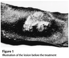
An 8-years-old female Weimaranner purebred dog was attended at the Veterinary Hospital of Universidade Estadual Paulista (Unesp) - Araçatuba city, São Paulo state - Brazil, with chronic exulceration in the right thoracic limb skin at the carpal region of five months history. The prescribed treatment prior to referral consisted of several topically administered broad-spectrum antibiotics with no improvement. Dermatological examination revealed a single and chronic exulceration, erythematous and hairless with round shape and elevated borders (Figure 1). The lesion size had about 20 cm2, and there was no evidence of other physical abnormalities. The presumptive diagnosis was skin SCC based on the history. A tissue fragment with 5 mm of diameter was extracted from the lesion border and submitted to histopathological evaluation, which confirmed skin SCC.
Treatment performed
The animal reported has been treated with teletherapy followed by brachytherapy. The dog was submitted to intravenous general anesthesia with chlorpromazine (Amplictil Rhodia) (1.0 mg/kg of body weight, IV) and thiopental sodium (Thionembutal Abbott) (12.5 mg/kg of body weight, IV) for each session. Teletherapy was performed in a cobalt (60Co) therapy unity, divided in 4 sessions. The affected area received a radiation dose of 500 cGy at 0.5 cm deepness (Cobalt-60 build'up) in each session. The first two sessions were performed in two consecutive days and the last two sessions, 7 days after in a total dose of 2,000 cGy. The radiation field reached a 6.0 cm X 8.0 cm surface area, which covered the entire tumor, and at the same time guaranteed safety margins. A material with the density equal to the human tissue (transparent bolus) and thickness to 0.5 cm was placed on the tumor 11 to allow a maximum region dose (build'up) in the tumor level (Figure 2).
Brachytherapy was performed one week after the fourth teletherapy session. A radioactive mould was manufactured with a Gold (198Au) foil in a disc shape of 2.76 cm radius which covered totally the tumor area. The mould was fixed on the tumor by an adhesive tape during the treatment period of 55 hours. The mould initial activity was 27.0 milicuries (mCi), which provides an initial dose rate of 60 cGy/h close to the center and of 17.5 cGy/h at the mold periphery. The radiation dose released under the mould level varied with the distance to the radioactive disc center and the treatment deepness (d), as shown in Table 1.
Then, the total radiation dose was composed of teletherapy and brachytherapy close to the tumor center was 4,500 cGy. The brachytherapy did not require anesthesia. The Elizabethan collar was used to prevent the patient from licking and scratching. The second biopsy was done in the treatment ending, it showed queratinized stratificated epithelium and also an inflammatory process with mononuclear cells distribution mainly around of the pilous foliculus.
After 4 months, it was observed a small lesion of circular appearance of about 1.0 cm diameter in the periphery of the irradiated field suggesting a residual lesion area. So, a second biopsy specimen was extracted from these area, and again, diagnosis of skin SCC was made. The decision was to repeat the brachytherapy procedure. However, at this time the radioactive material was distributed in two concentric distinct regions as shown in Figure 3.
The initial mould activity was 41.0 mCi, which provided an uniform initial dose rate of 58.0 cGy/h in all of the mould extension due to the activity difference between the internal and external ring. The treatment period (attachment of mold in the lesion) last seven consecutively days (uninterrupted) and the released radiation dose at 0.5 cm below the mould plane was 4,500 cGy15,16. Between the mould base and the external surface of the lesion it was placed a material (transparent bolus) at same as teletherapy.
Radioactive Mould Manufacture Procedure
Non-radioactive Gold (197Au) used in the treatment, consisted of a rectangular shape foil of 0.00125cm thickness, manufactured in the specialized Brazilian market for medical use. Manufacturer data indicate a 99.90% chemical clarity in the gold metal mass that had been confirmed by x-ray fluorescence and electronic microscopic analyses, performed by the metallurgical laboratory of Instituto de Pesquisas Energéticas e Nucleares (IPEN) in São Paulo (SP) Brazil17.
At the first brachytherapy, a disc with 5.50 cm of diameter was withdrawn from the rectangular foil. At the second brachytherapy, the foil was cut out in a ring shape as shown in Figure 3. Gold (197Au) activation was performed in the IEA-R1 reactor of IPEN. The activation homogenous was checked by measurement of the activity concentration distribution (mCi/g) using small pieces of the foil sample. For each piece it had been measured its mass and its total activity. The analysis showed the activation homogenous was inside of the desired quality. A second approach had been made by means of a gamma scanning of the Gold (198Au) foil. The spectrometry also showed that the homogenous was acceptable for brachytherapy purposes. The activation time of the gold (197Au) foils in the reactor was based on the neutron flux at the irradiation position, the required dose for the treatment, the tumor area and the foil dimension18.
The Computer System Micro Shield Version 419 was used during all the preliminary study phases of the dose distribution at the lesion level and its geometric morphology. In order to verify the ideal conditions, a radiographic film was exposed to the radioactive mould, and the obtained image was analyzed in an optical densitometer. Optical density study showed a good agreement to the desired dose distribution, which demonstrated the radioactive viability of the mould manufactured.
The radiation dose estimated for the treatment was based on brachytherapy protocols currently in use20 and the radium dosage rules recommended by the Manchester System21, which establishes the quantity of milligrammes-hour of the used radioactive element. Gold (198Au) radioactive decay and released dose attenuation were appreciated as a time function and thus, estimated the necessary treatment period for the desirable radiation dose liberation22,23.
Results, Discussion and Conclusion
The canine skin SCC is a radiosensitive tumor and responds to a wide range of dose levels, as well as occurred with mastocytomas24. The animal studied here had tolerated very well the Cobalt (60Co) teletherapy and Gold (198Au) brachytherapy. Some studies had described the use of other radioactive elements with good results1,5,7,12,13,25,26. In our knowledgement, this case report is the first one with the use of this shape of radioactive gold.
On the early periods after teletherapy, skin reaction was not observed in agreement to the literature3,20. Erythema and dry desquamation were observed three weeks after the end of teletherapy. These skin reactions, actinic dermatitis, are frequently observed in therapies with radioactive elements18,27. After mould removes, the following signs were present: hair loss and thinning epidermis were observed four weeks after the first schedule brachytherapy (Figure 4). Skin healing reactions occurred within 30 days and none other physical abnormalities were evident.
Biopsies were taken from irradiated tissue in our patient, and histological examination revealed recurrence of the skin SCC. This fact was an important procedure to indicate an additional schedule of brachytherapy with radioactive gold concentric rings. After this therapy the dog was considered clinically free of tumor. Afterwards, a new biopsy was taken of marginal lesion and reveled that marginal board was healthy.
Although others therapeutic possibilities has been already mentioned3,4,25 the proposed treatment here showed the advantage of not been invasive. The lesion area (carpals anterior face) is a local of difficult healing because it shows a great tension at wounded borders. In the majority of the cases this fact gives rise to the rupture of sutures, moreover it does not guarantee free surgical borders of tumor2. These facts confirm the advantages of the applied methods.
In fact, it was observed the skin and hair colors changing 60 days after the treatment. There was partial hair growth, leukotrichya and a skin scar remained in later periods (Figure 5).
The clinical observations after the brachytherapy showed therapeutic results similar to the ones obtained with commonly radioactive implant moulds used in the large variety of specialized centers in the small animal cancer treatment2,5,25.
The radioactive material use in concentric rings format, provided greater uniformity, in the dose distribution, better than seed implants14,17,28. The radioactive material time handling using a disc shape was lower than that observed with the use of seeds. Actually, it has been demonstrated a lower radiation exposition risk for the professionals involved in the treatment. Thereafter, the brachytherapy techniques in several cases avoid the surgical mutilations, which require a great skin area removal or even the indication of euthanasia4,5,12.
The treatment was well tolerated by the animal and the radioactive mold confection irradiated in the IEA-R1 nuclear reactor, although requiring a multidisciplinary team, can be easily performed. Finally, the proposed procedure gives rise to a good option for skin tumor treatment against recurrent lesions.
In conclusion, the development of a time-dose schedule that gives a high degree of cures, without a damaging effect upon the patient, has been the primary goal of most radiotherapists. An ideal therapy schedule should afford a time-dose relationship that delivers a destructive dose to the tumor, spares the normal tissue as much as possible, and requires a minimal number of visits by the animal patients to the therapist for treatment. Moreover, to offer the veterinary patient cancer holders an additional and reliable treatment option.
Acknowledgements
The authors thank team of Reactor IEA-R1, Instituto de Pesquisas Energéticas e Nucleares (IPEN) of Comissão Nacional de Energia Nuclear (CNEN) - São Paulo (SP) - Brazil, and Mr. Vladimir Lepki for thechinical support.
Recebido para publicação: 24/04/2002
Aprovado para publicação: 03/06/2003
-
1- BANKS, W. C.; ENGLAND, R. B. Radioactive gold in the treatment of ocular squamous cell carcinoma of cattle. Journal of the American Veterinary Medical Association, v. 163, n. 7, p. 745-748, 1973.
-
2- MELEO, K. A. Tumors of the skin and associated structures. The Veterinary Clinics of North America Small Animal Practice, radiation oncology, Philadelphia, v. 27, n. 1, 1997. p. x-y.
-
3- OGILVIE, G. K.; MOORE, A. S. Managing the veterinary cancer patient Trenton: Veterinary Learning Systems, 1995. ??? p.
-
4- SHAPIRO, W.; KITCHELL, B. E., FOSSUM, T. W.; COUTO, C. G.; THEILEN, G. Cisplatin for treatment of transitional cell and squamous cell carcinomas in dogs. Journal of the American Veterinary Medical Association, v. 193, p. 1530-1533, 1988.
-
5- RICHARD, W.; MICHAEL, W.; ALFRED, M.; JACK, H.; JOEY, S.; SANDRA, B.H. Development of brachytherapy technique for nasal tumors in dogs. American Journal of Veterinary Research, v. 51, n. 8, p. 1250-1256, 1990.
-
6- EDWARD, L. G. Veterinary radiotherapy. Journal of the American Veterinary Medical Association, v. 17, n. 11, p. 1707-1712, 1970.
-
7- BANKS, W. C.; ROYCE, R.; EARL, M.; HUSSEY, D. H. Radiotherapy techniques in veterinary medicine. Journal of the American Veterinary Medical Association, v. 160, n. 4, p. 446-50, 1972.
-
8- ALAIN, P. T.; EDWARD, C. F. Megavoltage irradiation of pituitary macrotumors in dogs with neurologic signs. Journal of the American Veterinary Medical Association, v. 213, n. 2, p. 225-231, 1998.
-
9- DAVID, A. W.; JAMES, K. B. Combined treatment of ocular squamous cell carcinoma in a horse, using radiofrequency hyperthermia and interstitial 198Au implants. Journal of the American Veterinary Medical Association, v. 196, n. 11, p. 1831-1833, 1990.
-
10- GOOSSENS-MARIELLE, M. C.; FELDMAN-EDWARD, C.; THEON-ALAIN, P.; KOBLIK-PHILIP, D. Efficacy of cobalt 60 radiotherapy in dogs with pituitary-dependent hyperadrenocorticism. Journal of the American Veterinary Medical Association, v. 212, n. 3, p. 374-376, 1998.
-
11- JANE, M. T.; ALAIN, P. T. Reirradiation of tumors in cats and dogs. Journal of the American Veterinary Medical Association, v. 193, n. 4, p. 465-469, 1988.
-
12- SWANSON, E. W.; MILLER, J. K.; CRAGLE, R. G. Effects of internal irradiation of the mammary gland with 144Cerium-144Praseodymium. Journal of Dairy Science, v. 53, n. 1, p. 46-51, 1969.
-
13- JAMES, P. T.; NORMAN, A.; JAMIE, R. B.; BRIAN, S. B.; GARY, W. E. 192Iridium brachytherapy, using an intracavitary afterload device, for treatment of intranasal neoplasm in dogs. American Journal of Veterinary Research, v. 53, n. 4, p. 617-622, 1992.
-
14- JOHNS, H. E.; CUNNINGHAM, J. R. Measurements of radiation: dosimetry, Brachytherapy - intercavitary and interstitial sources. In: ______. The physics of radiology 4. ed. Illinois: Charles C. Thomas, 1981. p. 217-269.
-
15- BENTEL, G. C. Dose Calculations in Brachyterapy. Pratical Applications of Brachyterapy Techniques. In: Radiation therapy planning 2. ed. New York: McGraw-Hill, 1996. p. 533-616.
-
16- FOWLER, J. F. The radiobiology of brachytherapy: brachytherapy HDR and LDR. In: BRACHYTHERAPY MEETING, 1989, Michigan. Procedings.. p. 121-128.
-
17- FERNANDES, M. A. R. Utilização de Moldes Radioativos Especiais de Folhas de Ouro-198 para Braquiterapia em Tumores de Pele Tese (Doutorado em...), Instituto de Pesquisas Energéticas e Nucleares, Universidade de São Paulo, São Paulo, 2000.
-
18- PEREZ, C. A.; BRADY, L. W. Principles and practice of radiation oncology, 2nd ed. Philadelphia: J.B. Lippincott, 1992. 1544 p.
-
19- NEGIN, C. A.; WORKU, G. Micro Shield Version 4: user's manual. Rockville: Grove Engineering, Maryland, 1992.
-
20- ORTON, C. G. Time-dose-factors (TDFs) in brachytherapy. British Journal of Radiology, v. 47, n. 561, p. 603-607, 1974.
-
21- MEREDITH, W. J. Radium dosage: the manchester system 2. ed. Baltimore: Williams and Wilkins, 1967. ??? p.
-
22- KUTCHER, G. J. Comprehensive QA for radiation oncology: report of AAPM Radiation Therapy Committee Task Group 40. Medical Physics, v. 21, n. 4, p. 581-618, 1994.
-
23- LANGMACK, K. A.; THOMAS, S. J. The Application of Dose-Volume Histograms to the Paris and Manchester Systems of Brachytherapy Dosimetry. The British Journal of Radiology, v. 68, p. 42-48, 1995.
-
24- SLUSHER, R.; ROENIGK, W. J.; WILSON, G. P. Effect of X-irradiation on mastocytomas in dogs. Journal of the American Veterinary Medical Association, v. 151, n. 8, p. 1049-1054, 1967.
-
25- NIKULA, K. J.; BENJAMIN, S. A.; ANGLETON, G. M.; SAUNDERS, W. J.; LEE, A. C. Ultraviolet radiation, solar dermatosis, and coetaneous neoplasia in beagle dogs. Radiation Research, v. 129, p. 11-18, 1992.
-
26- TAKEDA, M.; SHIBUYA, H.; INOUE, T. The Efficacy of Gold-198 Grain Mold therapy for mucosal carcinomas of the oral cavity. Acta Oncologica, v. 35, n. 4, p. 463-467, 1996.
-
27- MASON, K. A.; THAMES, H. D.; OCHRAN, T. G.; RUIFROK, A. C. C.; JANJAN, N. Comparison of continous and pulsed low dose rate brachytherapy: biological equivalence in vivo. International Journal of Radiation Oncology and Biological Physics, v. 28, n. 3, p. 667-671, 1994.
-
28- KHAN, F. M. A system of dosimetric irradiation. In: The physics of radiation therapy 2. ed. Baltimore: Williams & Wilkins, 1994, p. 200-225.
Publication Dates
-
Publication in this collection
06 May 2004 -
Date of issue
2003
History
-
Accepted
03 June 2003 -
Received
24 Apr 2002