
Abstracts
Syzygium cumini is a plant that has been used in popular medicine for the treatment of insulin dependent diabetes mellitus (DMID). This study verified the effect of Syzygium cumini upon the regeneration of insulin producing cells in the pancreatic duct wall. The animals were divided into four groups, control (C), treated control (TC), diabetic control (DC) and treated diabetic (TD). An aqueous extract from Syzygium cumini bark was given by gavage in a daily dose of 1g/kg of body weight. After a thirty day period the animals were euthanized and the pancreas taken to immunohistochemical analysis. In this study, it was observed the positive staining for insulin on cells of the pancreatic duct and connective tissue in the pancreas of TD and TC animals. These results indicate that Syzygium cumini bark extract stimulates development of insulin positive cells from the pancreatic duct epithelial cells.
Syzygium cumini; Alloxan; Diabetes; Immunohistochemistry
O Syzygium cumini é uma planta medicinal que tem sido utilizada popularmente para o tratamento da diabetes melito insulino dependente (DMID). Este estudo verificou o efeito do extrato da casca de Syzygium cumini sobre a regeneração de células insulino-positivas, a partir do ducto pancreático, no pâncreas de ratos normais e diabéticos. Os animais foram divididos em grupo controle (C), controle tratado (CT), diabético controle (DC) e diabético tratado (DT). Nos grupos tratados foi realizada a administração oral do extrato aquoso da casca de Syzygium cumini, na dose de 1g/kg de peso vivo. Após um período de 30 dias, os animais foram submetidos à eutanásia e o pâncreas retirado para análise imunohistoquímica. Neste estudo, foram visualizadas células insulino-positivas no ducto pancreático e no tecido conjuntivo próximo a ele, no pâncreas dos animais dos grupos DT e CT. Estes resultados indicam que o tratamento com o extrato da casca de Syzygium cumini na dose de 1g/kg estimula a formação de células insulino-positivas a partir das células epiteliais do ducto pancreático.
Syzygium cumini; Aloxano; Diabetes; Immunohistoquímica
Syzygium cumini and the regeneration of insulin positive cells from the pancreatic duct
Syzygium cumini e a regeneração de células insulino-positivas a partir do ducto pancreático
Deila Rosély C. SchosslerI; Cinthia Melazzo MazzantiII; Sônia Cristina Almeida da LuzI; Andreane FilappiIII; Danívia PrestesIII; Aron Ferreira da SilveiraI; Marcelo CecimIII
IDepartamento de Morfologia da Universidade Federal de Santa Maria, Santa Maria - RS
IIDepartamento de Bioquímica da Universidade Federal do Rio Grande do Sul, Porto Alegre - RS
IIIDepartamento de Medicina de Grandes Animais da Universidade Federal de Santa Maria, Santa Maria - RS
Correspondence Correspondence to MARCELO CECIM Departamento de Medicina de Grandes Animais - Hospital Veterinário Universidade Federal de Santa Maria 97105-900 - Santa Maria - RS mcecim@smail.ufsm.br
ABSTRACT
Syzygium cumini is a plant that has been used in popular medicine for the treatment of insulin dependent diabetes mellitus (DMID). This study verified the effect of Syzygium cumini upon the regeneration of insulin producing cells in the pancreatic duct wall. The animals were divided into four groups, control (C), treated control (TC), diabetic control (DC) and treated diabetic (TD). An aqueous extract from Syzygium cumini bark was given by gavage in a daily dose of 1g/kg of body weight. After a thirty day period the animals were euthanized and the pancreas taken to immunohistochemical analysis. In this study, it was observed the positive staining for insulin on cells of the pancreatic duct and connective tissue in the pancreas of TD and TC animals. These results indicate that Syzygium cumini bark extract stimulates development of insulin positive cells from the pancreatic duct epithelial cells.
Key-words: Syzygium cumini. Alloxan. Diabetes. Immunohistochemistry
RESUMO
O Syzygium cumini é uma planta medicinal que tem sido utilizada popularmente para o tratamento da diabetes melito insulino dependente (DMID). Este estudo verificou o efeito do extrato da casca de Syzygium cumini sobre a regeneração de células insulino-positivas, a partir do ducto pancreático, no pâncreas de ratos normais e diabéticos. Os animais foram divididos em grupo controle (C), controle tratado (CT), diabético controle (DC) e diabético tratado (DT). Nos grupos tratados foi realizada a administração oral do extrato aquoso da casca de Syzygium cumini, na dose de 1g/kg de peso vivo. Após um período de 30 dias, os animais foram submetidos à eutanásia e o pâncreas retirado para análise imunohistoquímica. Neste estudo, foram visualizadas células insulino-positivas no ducto pancreático e no tecido conjuntivo próximo a ele, no pâncreas dos animais dos grupos DT e CT. Estes resultados indicam que o tratamento com o extrato da casca de Syzygium cumini na dose de 1g/kg estimula a formação de células insulino-positivas a partir das células epiteliais do ducto pancreático.
Palavras-chave: Syzygium cumini. Aloxano. Diabetes. Immunohistoquímica.
Introduction
Diabetes mellitus is composed of a myriad of derangements on carbohydrate, lipid and protein metabolism, which are associated to an absolute or relative deficiency of insulin secretion/action.1
Syzygium cumini is one of the most used plants for the treatment of the disease. It originated in India, and today it can be found in most Brazilian States, principally on the north and north-east.2 The bark, the fruit, the seed as well as the leaves are utilized in the treatment of insulin dependant diabetes mellitus (IDDM). They are prepared as an aqueous or ethanolic extract, by infusion or as a juice of the raw plant.3
Alloxan can specifically destroy the beta (b) cells of the pancreatic islets, inducing loss of the cell turgor, nuclear picnosis, cytoplasmatic vacuolization, mitochondrial edema and fragmentation, leading to cell death.4,5
According to Waguri et al.6 the b cell can present 2 types of regeneration: differentiation of the precursors cells from the pancreatic duct, or proliferation from existing or surviving mature b cells.
The objective of the present work is to report the effect of Syzygium cumini upon the regeneration of insulin producing cells in the pancreatic duct wall.
Materials and Methods
The study used 180 adult female Wistar rats, maintained ad libitum water and standard lab chow for a 10 days adaptation period. The bark material was collected in may 2001 in Santa Maria (RS, Brazil). It was initially air dried, then placed in an oven at 50º C for 72 hours and then processed in a Wiley mill. After that the material was cold extracted with ethanol and evaporated in a low pressure rotavapor. The resulting residue was dark and viscous, yielded 10% of original bark weight and was called extract.
For the induction of IDDM 150 animals were used. After a 24 hours fasting period the animals received 150mg/kg of alloxan (Sigma-Aldrich Inc, St Louis, MO, USA, in a 2% solution, sodium citrate buffer, 0,05M, pH 4,5) intraperitoneally. Six hours after induction, animals started to receive a 10% glucose solution ad libitum to avoid hypoglycemic shock. After 15 days fasting glucose levels were determined. Only animals with values above 180mg/dl were considered diabetic and used in the experiment.
Group 1: control (C, n=11) and group 2: treated control (TC, n=9) were composed of animals not exposed to alloxan. Diabetic animals were randomly assigned to group 3: diabetic control (DC, n=10) or group 4: treated diabetic (TD, n=9). Groups 2 and 4 received a daily dose of 1g/kg of body weight of the bark extract administered by gavage. Control groups received 2ml distilled water. After 30 days of treatment, animals were euthanised, pancreas was excised and preserved in 10% formalin for 24 hours. Organs were embedded in paraffin. Serial (7m) cuts were realized and 3 sections randomly distributed were fixed in slides previously prepared with organosilene.
Sections were immuno-stained for insulin a peroxidase technique, streptoavidin-biotin method as proposed by Peinado, Pedroso and Rodrigo et al..7 The primary antibody was guinea pig antiswine insulin (Dako, Carpinteria, USA), diluition 1:500. The secundary antibody was rabbit antiguinea pig immunoglobulin conjugated with peroxidase ( Dako, Carpinteria, USA), diluition 1:200. After incubation sections were counterstained with hematoxilin. Evaluation was carried out by observation of insulin-positive cells in the duct wall and immediately around it under a 100, 400, and 1000X magnification.
Results
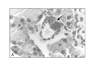
Positive staining for insulin was observed in the epithelia of the pancreatic duct (Figura 1A) and cells around the duct (Figura 1B) in 7 out of 9 animals of the TD group. In TC animals, only 2 out of 9 individuals presented the finding. Such cells were not identified in animals from the C and DC group.
Discussion and Conclusions
Positive insulin staining was found only in Syzygium cumini treated animals, either normal or diabetic. Such finding raises the possibility that Syzygium cumini can stimulate b cell regeneration by the proliferation of its precursor cells in the pancreatic duct, since practically all of its b cells were destroyed by alloxan.4,5
According to Rao, Kesavulu and Apparao1, b cell destruction leads to a fall in insulin production. In an effort to compensate that, surviving b cells increase insulin production. Such hyperactivity is followed by nuclear enlargement, making mitosis more difficult.8 However, according to Edlund9 all pancreatic cells are derived from the dorsal and ventral endoderm of the primitive gut. For this reason, pancreas and intestine precursor cells can originate any pancreatic cell, but the extrinsic factors, which may regulate this mechanism, remain unknown.
The appearance of insulin producing cells in the pancreatic duct observed in this study is in accordance with Lipsett and Finegood10, who reported b cell neoformation from precursors cells in the pancreatic duct of diabetic animals. Park and Bendayan11 also observed the phenomenon.
Waguri et al.6 concluded that b cell regeneration from the duct occurs when practically all b cells were destroyed. When there are normal b cells, regeneration occurs within the pancreatic islet from these preexisting cells. Such evidence corroborates the suggestion that Syzygium cumini stimulates precursor cell differentiation. It is important to note that in the present study, there were insulin producing cells in the pancreatic duct and adjacent connective tissue of non-diabetic animals treated with the plant (group TC). In these animals b cell population was unaffected, thus if there was regeneration, it should have been an intrapancreatic event, and not from duct cells.
Only 3 positive cells per pancreas were observed in animals from TD and TC groups. Such small number of neoformed cells may be related to the amount or efficacy (bark, instead of leaves or seeds) of the extract used. Normalization of blood glucose levels as reported in other studies, was not attained in this experiment. Also, the treatment was shorter than six weeks, as it was described in previus studies.12 Bragança2 and Pepato et al.3 stated that treatment efficiency is time related.
The present results suggest that Syzygium cumini induces the appearance of positive insulin staining cells in the epithelia of the pancreatic duct of treated animals.
Recebido para publicação: 18/06/2003
Aprovado para publicação: 18/05/2004
-
1RAO, B. K.; KESAVULU, M. M.; APPARAO, C. H. Antihyperglycemic activity of momordica cymbalaia in alloxan diabetic rats. Journal of Ethnopharmacology, v. 78, n. 1, p. 67-71, 2001.
-
2BRAGANÇA, L. A. R. Plantas medicinais antidiabéticas: Uma abordagem multidisciplinar Rio de Janeiro, Universidade Federal Fluminense, 1996. 30 p.
-
3PEPATO, M. T. et al. Lack of antidiabetic effect of Eugenia jambolana leaf decoction on rat streptozotocin diabetes. Brazilian Journal of Medical and Biological Research, v. 34, n. 1, p. 389-395, 2001.
-
4DREWS, G. et al. Contrasting effect of alloxan on islets and single mouse pancreatic beta-cells. Biochemical Journal, v. 1, n. 352, p. 398-397, 2000.
-
5MATHEUS, C. E.; LEITER, E. H. Constitutive differences in antioxidant defense status distinguish alloxan-resistant and alloxan-susceptible mice. Free Radical Biological Medical , v. 29, n. 3-4, p. 449-455, 1999.
-
6-WAGURI, M. et al. Demonstration of two different processes of b-cells regeneration in a new diabetic mouse model induced by selective perfusion of alloxan. Diabetes, v. 46, n. 5, p. 1281-1290, 1997.
-
7PEINADO, M. A.; PEDROSA, J. A.; RODRIGO, J. Avances em immunocitoquímica y técnicas relacionadas Jaén: Gráfica La Paz, 1996. 401 p.
-
8GEPTS, W.; LECOMPTE, P. M. The pancreatic islet in diabetes. The American Journal of Medicine, v. 70, n. 1, p. 105-113, 1981.
-
9EDLUND, H. Pancreas: how to get there from the gut. Current Opinion in Cell Biology, v. 70, n. 6, p. 663-668, 1999.
-
10LIPSETT, M.; FINEGOOD, D. T. Beta-cell neogenesis during prolonged hyperglycemia in rats. Diabetes, v. 51, n. 6, p. 1834-1841, 2002.
-
11PARK, I.; BENDAYAN, M. Endocrine cells in the rat pancreatic and bile duct system: alteration in diabetes. Pancreas, v. 9, n. 5, p. 566-573, 1994.
-
12PRINCE, P. S. M., MENON, V. P., PARI, L. Hypoglycaemic activity of Syzygium cumini seeds: effect on lipid peroxidation in alloxan diabetic rats. Journal of Ethnopharmacology, v. 61, n. 1, p. 1-7, 1998.
Correspondence to
Publication Dates
-
Publication in this collection
09 Aug 2005 -
Date of issue
Aug 2004
History
-
Received
18 June 2003 -
Accepted
18 May 2004

