
Abstract
In this study, biogenic SiO2 of high purity and high surface area obtained from rice husk was used for prepare a nanostructured SnO2/SiO2composite. The predominantly amorphous silica was extracted in an acidic route and then the nanocomposite was done via sol-gel route using ethylene glycol and citric acid followed by heat treatment. SiO2 content of the rice husk was determined by X-ray fluorescence (XRF) and its specific surface area determined by nitrogen adsorption. The composite nanostructured SnO2/SiO2 was structurally characterized by the techniques of X-ray diffraction (XRD), Raman and Fourier transform infrared (FT-IR) spectroscopy. The morphological characteristics were revealed by scanning electron microscope (SEM).
biogenic SiO2; rice husk; SnO2/SiO2 composite; Pechini method
1 Introduction
Agroindustry has generated a large content of residues and a necessity of utilization of these residues might reduce pollution and increase energy savings. Among these residues, rice husks have received attention due to its large volume produced and high content of amorphous silica, approximately 21%11 Umeda J, Kondoh K and Michiura Y. Process parameters optimization in preparing high-purity amorphous silica originated from rice husks. Materials Transactions. 2007; 48(12):3095-3100. http://dx.doi.org/10.2320/matertrans.MK200715.
http://dx.doi.org/10.2320/matertrans.MK2...
,22 Shen J, Liu X, Zhu S, Zhang H and Tan J. Effects of calcination parameters on the silica phase of original and leached rice husk ash. Materials Letters. 2011; 65(8):1179-1183. http://dx.doi.org/10.1016/j.matlet.2011.01.034.
http://dx.doi.org/10.1016/j.matlet.2011....
. SiO2 can be obtained by direct calcination followed by calcination chemical treatment, and by sol-gel route. Silica from rice husk is considered as an alternative to commercial silica33 Adam F, Appaturi JN, Khanam Z, Thankappan R and Nawi MAM. Utilization of tin and titanium incorporated rice husk silica nanocomposite as photocatalyst and adsorbent for the removal of methylene blue in aqueous medium. Applied Surface Science. 2013; 264:718-726. http://dx.doi.org/10.1016/j.apsusc.2012.10.106.
http://dx.doi.org/10.1016/j.apsusc.2012....
due to low cost and wide application, such as to obtain silicon carbide44 Gorthy P and Pudukottah M. Production of silicon carbide from rice husks. Journal of the American Ceramic Society. 1999; 82(6):1393-1400. http://dx.doi.org/10.1111/j.1151-2916.1999.tb01929.x.
http://dx.doi.org/10.1111/j.1151-2916.19...
, catalysts55 Adam F, Appaturi JN and Iqbal A. The utilization of rice husk silica as a catalyst: review and recent progress. Catalysis Today. 2012; 190(1):2-14. http://dx.doi.org/10.1016/j.cattod.2012.04.056.
http://dx.doi.org/10.1016/j.cattod.2012....
, adsorbents66 Adam F and Chua JH. The adsorption of palmytic acid on rice husk ash chemically modified with Al(III) ion using the sol-gel technique. Journal of Colloid and Interface Science. 2004; 280(1):55-61. http://dx.doi.org/10.1016/j.jcis.2004.07.006. PMid:15476773.
http://dx.doi.org/10.1016/j.jcis.2004.07...
, zeolites77 Gaydhankar TR, Joshi PN, Kalita P and Kumar R. Optimal synthesis parameters and application of Sn-MCM-41 as an efficient heterogeneous catalyst in solvent-free Mukaiyama-type aldol condensation. Journal of Molecular Catalysis A Chemical. 2007; 265(1-2):306-315. http://dx.doi.org/10.1016/j.molcata.2006.10.041.
http://dx.doi.org/10.1016/j.molcata.2006...
, silicates88 Naskar MK and Chatterjee M. A novel process for the synthesis of lithium aluminum silicate powders from rice husk ash and other water-based precursor materials. Materials Letters. 2005; 59(8-9):998-1003. http://dx.doi.org/10.1016/j.matlet.2004.06.075.
http://dx.doi.org/10.1016/j.matlet.2004....
and cement99 Ganesan K, Rajagopal K and Thangavel K. Rice husk ash blended cement: Assessment of optimal level of replacement for strength and permeability properties of concrete. Construction & Building Materials. 2008; 22(8):1675-1683. http://dx.doi.org/10.1016/j.conbuildmat.2007.06.011.
http://dx.doi.org/10.1016/j.conbuildmat....
.
Compared to conventional materials, nanomaterials have interesting physical and chemical properties, besides several areas of application, and relative easy production carried out by different methods such as microemulsion1010 Lakshmanan R, Okoli C, Boutonnet M, Jaras S and Rajarao GK. Microemulsion prepared magnetic nanoparticles for phosphate removal: Time efficient studies. Journal of Environmental Chemical Engineering. 2014; 2(1):185-189. http://dx.doi.org/10.1016/j.jece.2013.12.008.
http://dx.doi.org/10.1016/j.jece.2013.12...
, hydrothermal1111 Yang W, Gao Z, Wang J, Wang B and Liu L. Hydrothermal synthesis of reduced graphene sheets/Fe2O3 nanorods composites and their enhanced electrochemical performance for supercapacitors. Solid State Sciences. 2013; 20:46-53. http://dx.doi.org/10.1016/j.solidstatesciences.2013.03.011.
http://dx.doi.org/10.1016/j.solidstatesc...
and sol-gel1212 Ebrahimi A, Pirouz A, Abdi Y, Azimi S and Mohajerzadeh S. Selective deposition of CuO/SnO2 sol–gel on porous SiO2 suitable for the fabrication of MEMS-based H2S sensors. Sensors and Actuators. B, Chemical. 2012; 173:802-810. http://dx.doi.org/10.1016/j.snb.2012.07.104.
http://dx.doi.org/10.1016/j.snb.2012.07....
.
Tin oxide (SnO2) is an important semiconductor oxide in the industry1313 Manjula P, Satyanarayana L, Swarnalatha Y and Manorama SV. Raman and MASNMR studies to support the mechanism of low temperature hydrogen sensing by Pd doped mesoporous SnO2. Sensors and Actuators. B, Chemical. 2009; 138(1):28-34. http://dx.doi.org/10.1016/j.snb.2009.02.051.
http://dx.doi.org/10.1016/j.snb.2009.02....
, but despite its wide usage1414 Zhao Y, Liu J, Liu Q, Sun Y, Song D, Yang W, et al. One-step synthesis of SnO2 hollow microspheres and its gas sensing properties. Materials Letters. 2014; 136:286-288. http://dx.doi.org/10.1016/j.matlet.2014.08.073.
http://dx.doi.org/10.1016/j.matlet.2014....
15 Wang F, Song X, Yao G, Zhao M, Liu R, Xu M, et al. Carbon-coated mesoporous SnO2 nanospheres as anode material for lithium ion batteries. Scripta Materialia. 2012; 66(8):562-565. http://dx.doi.org/10.1016/j.scriptamat.2012.01.003.
http://dx.doi.org/10.1016/j.scriptamat.2...
16 Shimizu K, Katagiri M, Satokawa S and Satsuma A. Sintering-resistant and self-regenerative properties of Ag/SnO2 catalyst for soot oxidation. Applied Catalysis B: Environmental. 2011; 108–109:39-46. http://dx.doi.org/10.1016/j.apcatb.2011.08.003.
http://dx.doi.org/10.1016/j.apcatb.2011....
-1717 Dou X, Sabba D, Mathews N, Wong LH, Lam YM and Mhaisalkar S. Hydrothermal Synthesis of High Electron Mobility Zn-doped SnO 2 Nanoflowers as Photoanode Material for Efficient Dye-Sensitized Solar Cells. Chemistry of Materials. 2011; 23(17):3938-3945. http://dx.doi.org/10.1021/cm201366z.
http://dx.doi.org/10.1021/cm201366z...
has low thermal stability and tendency to aggregation1818 Feng YS, Zhou SM, Li Y and Zhang LD. Preparation of the SnO2/SiO2 xerogel with a large specific surface area. Materials Letters. 2003; 57(16-17):2409-2412. http://dx.doi.org/10.1016/S0167-577X(02)01245-4.
http://dx.doi.org/10.1016/S0167-577X(02)...
, that can be overcome by incorporating the amorphous SiO2 to SnO2[1919 Granger G, Restoin C, Roy P, Jamier R, Rougier S, Lecomte A, et al. Nanostructured optical fibers in the SiO2/SnO2 system by the sol-gel method. Materials Letters. 2014; 120:292-294. http://dx.doi.org/10.1016/j.matlet.2014.01.104.
http://dx.doi.org/10.1016/j.matlet.2014....
], generating the SnO2/SiO2nanocomposite. There are several studies using SnO2/SiO2, like composite2020 An GH and Ahn HJ. Fabrication of SnO2 and SiO2 nanoparticle-embedded carbon nanofiber composites via co-electrospinning. Ceramics International. 2012; 38(4):3197-3201. http://dx.doi.org/10.1016/j.ceramint.2011.12.024.
http://dx.doi.org/10.1016/j.ceramint.201...
, xerogel2121 Grieken RV, Martos C, Sánchez MS, Serrano DP, Melero JA, Iglesias J, et al. Synthesis of Sn–silicalite from hydrothermal conversion of SiO2–SnO2 xerogels. Microporous and Mesoporous Materials. 2009; 119:176-185., nanotubes2222 Adam F, Appaturi JN, Thankappan R and Nawi MAM. Silica–tin nanotubes prepared from rice husk ash by sol–gel method: Characterization and its photocatalytic activity. Applied Surface Science. 2010; 257(3):811-816. http://dx.doi.org/10.1016/j.apsusc.2010.07.070.
http://dx.doi.org/10.1016/j.apsusc.2010....
and films2323 Lorenzi R, Lauria A, Mochenova N, Chiodini N and Paleari A. Study of the absorption edge of SnO2 nanoparticles embedded in silica films. Journal of Non-Crystalline Solids. 2011; 357(8-9):1888-1891. http://dx.doi.org/10.1016/j.jnoncrysol.2010.12.045.
http://dx.doi.org/10.1016/j.jnoncrysol.2...
, which application can be in photocatalysis2424 Wei TY, Kuo CY, Hsu YJ, Lu SY and Chang YC. Tin oxide nanocrystals embedded in silica aerogel: Photoluminescence and photocatalysis. Microporous and Mesoporous Materials. 2008; 112(1-3):580-588. http://dx.doi.org/10.1016/j.micromeso.2007.10.040.
http://dx.doi.org/10.1016/j.micromeso.20...
and sensors2525 Zhu Y, Chen J, Li H, Zhu Y and Xu J. Synthesis of mesoporous SnO2–SiO2 composites and their application as quartz crystal microbalance humidity sensor. Sensors and Actuators. B, Chemical. 2014; 193:320-325. http://dx.doi.org/10.1016/j.snb.2013.11.091.
http://dx.doi.org/10.1016/j.snb.2013.11....
.
This work aims prepare and characterize SnO2/SiO2 nanocomposites from amorphous biogenic silica of high purity extracted from rice husk by Pechini method2626 Pechini MPMethod of preparing lead and alkaline earth titanates and niobates and coating method using the same to form a capacitorUS33306971967July11, as an alternative to TEOS (tetraethoxysilane). Silica from rice husk was characterized by X-ray fluorescence (XRF) and by physical adsorption of nitrogen. The SnO2/SiO2 nanocomposite was characterized by scanning electron microscopy (SEM), by X-ray diffractometry (XRD) and Raman spectroscopy and Fourier transform infrared spectroscopy (FT-IR).
2 Experimental
2.1 Extraction of SiO2 and Preparations of nanocomposite
Extraction of silica was made by grinding of the rice husk (RH) followed by chemical treatment with HCl (10% v/v), in the ratio RH:HCl 1:3, for one hour with constant stirring to solubilize organic matter. The solubilized RH was washed with ultrapure water and it was filtered under vacuum, obtaining a pulp (PRH) which was treated with a solution of H2SO4 and H2O2 in the ratio of 1:2:1 (w/v/v) - PRH:H2SO4:H2O2 - under constant stirring for one hour to promote the oxidation of organic matter. Finally, the oxidized PRH was washed with ultrapure water and the biogenic SiO2 was obtained by vacuum filtering and calcinations in an oven for 4h at 600 °C.
SnO2/SiO2 nanocomposite was prepared by adding biogenic silica and SnCl2.2H2O to a solution of nitric acid (NA)/ethylene glycol (EG), obeying the following proportions 1:3:12 (SnCl2.2H2O: CA: EG) and 1:4 (w / w) - (SiO2: SnCl2.2H2O). This mixture was stirred for 1h at 60 °C with subsequent heat treatment at 250 °C for 2 hours, followed by calcination at 400 °C during 1 hour. All steps of calcination and heat treatment were performed without heating rate.
2.2 Characterization
Biogenic silica was characterized by x-ray fluorescence (XRF) using an Epsilon 3XL spectrometer. Specific surface area was determined by nitrogen gas adsorption using a Quantachrome Autosorb-iQ equipment by multipoint BET method (Brunauer Emmett Tell) and the average diameter of the silica particles was estimated using the following equation for spherical particles
where is the theoretical density2727 Yalcà N and Sevinç V. Studies on silica obtained from rice husk. Ceramics International. 2001; 27:219-224. http://dx.doi.org/10.1016/S0272-8842(00)00068-7.
http://dx.doi.org/10.1016/S0272-8842(00)...
for the amorphous silica, having a value of 1,92 g cm–3 and is the specific surface area.
Morphology of SnO2/SiO2 nanocomposite was determined by scanning electron microscopy (SEM) using a JEOL model JMS6360-Lv microscope. The crystallinity of the material were characterized by X-ray diffraction (Shimadzu/XRD-7000) with CuKα radiation (λ = 1.542Å), 40 kV and 30 mA. Vibrational spectra were recorded on a Bomem MB-series spectrophotometer (Model B100) and Raman spectra were obtained using Confocal Raman equipment model T64000 Jobin-Yvon with laser excitation at 532 nm. Composition of the biogenic silica and the nanocomposite were identified by XRF, and the crystallite size was determined by Debye-Scherrer equation:
where d is crystallite size, K is shape factor (typical value of about 0.89 for spherical crystalline solids with cubic unit cells), λ is CuX-ray wavelength (1.542 Å), θ is Bragg diffraction angle and β is the peak width of the diffraction peak profile at half maximum height.
3 Results and Discussion
3.1 Amorphous SiO2
Composition of the biogenic silica is shown in Table 1. According to the percentage values of the components, the methodology used to extraction provided SiO2 with purity of approximately 98.6%, higher than commercial SiO2[2828 Della VP, Hotza D, Junkes JA and Oliveira APN. Estudo comparativo entre sílica obtida por lixívia ácida da casca de arroz e sílica obtida por tratamento térmico da cinza de casca de arroz. Quimica Nova. 2006; 29(6):1175-1179. http://dx.doi.org/10.1590/S0100-40422006000600005.
http://dx.doi.org/10.1590/S0100-40422006...
]. The analysis of the surface area by BET method showed an approximated2929 Yu X, Tian J, Xie H, Shen H and Wang Q. The integrated production of microbial lipids and bio-SiO2 from rice husks by an organic electrolytes pretreatment technology. Bioresource Technology. 2014; 153:403-407. http://dx.doi.org/10.1016/j.biortech.2013.12.039. PMid:24398252.
http://dx.doi.org/10.1016/j.biortech.201...
,3030 Watari TI, Nakata A, Kiba Y, Torikai T and Yada M. Fabrication of porous SiO2/C composite from rice husks. Journal of the European Ceramic Society. 2006; 26(4-5):797-801. http://dx.doi.org/10.1016/j.jeurceramsoc.2005.06.013.
http://dx.doi.org/10.1016/j.jeurceramsoc...
value of 450 m2g–1 and a mean particle size of 7 nm, given by Equation 1.
3.2 Structural study of SiO2 and nanocomposite
XRD diffractogram of the biogenic SiO2 is presented in Figure 1a and shows a broad peak located approximately at 2θ = 22.5°, that suggests an amorphous characteristic of the sample and agrees with the reported JCPDS data (card No. 01-086-1561).
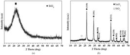
XRD pattern for the nanocomposite is shown in Figure 1b and reveals a small shoulder at 2θ = 21.7° that can be attributed to amorphous SiO2 and other peaks assigned to crystalline SnO2. All values of diffraction peaks are in accordance with JCPDS pattern (card No. 00-041-1485). The main diffraction peaks observed for SnO2 are centered approximately at 2θ values of 27.1°, 34.2° and 52.1°. In addition, the crystallite size based on the major diffraction peak is 27.8 nm for the nanocomposite.
3.3 Morphology of nanocomposite
Surface morphology of the nanocomposite is presented in Figure 2. SEM micrographs reveal formation of a heterogeneous mixture of agglomerates with irregular shapes and sizes.

3.4 FTIR and Raman analyses
FT-IR and Raman spectra for the SnO2/SiO2 nanocomposite are shown in Figure 3a and 3b, respectively. Comparisons between wavenumber obtained in this work with literature values are exhibited in Tables 2 and 3.
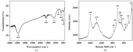
Infrared spectrum shows a broad band at 3455 cm–1 attributed to O-H stretching from hydroxyl groups presents on surface of the material and the band at 1628 cm–1 is associated with bending H-O-H bond groups of adsorbed water molecules3131 Yang S and Gao L. Facile and surfactant-free route to nanocrystalline mesoporous tin oxide. Journal of the American Ceramic Society. 2006; 89(5):1742-1744. http://dx.doi.org/10.1111/j.1551-2916.2006.00947.x.
http://dx.doi.org/10.1111/j.1551-2916.20...
. Bands at 467, 807 and 1097 cm–1 are assigned respectively to vibrational modes of O-Si-O bending, to symmetric stretching of Si-O-Si group and to the asymmetric stretching of Si-O-Si structural bond of siloxane3232 Jung HY, Gupta RK, Oh EO, Kim YH and Whang CM. Vibrational spectroscopic studies of sol–gel derived physical and chemical bonded ORMOSILs. Journal of Non-Crystalline Solids. 2005; 351(5):372-379. http://dx.doi.org/10.1016/j.jnoncrysol.2005.01.004.
http://dx.doi.org/10.1016/j.jnoncrysol.2...
. Vibrations in the range from 500 to 700 cm–1 are assigned to Sn-O-Sn group as result of condensation reactions3333 Li Z, Shen W, Zhang X, Fang L and Zu X. Controllable growth of SnO2 nanoparticles by citric acid assisted hydrothermal process. Colloids and Surfaces. A, Physicochemical and Engineering Aspects. 2008; 327(1-3):17-20. http://dx.doi.org/10.1016/j.colsurfa.2008.05.043.
http://dx.doi.org/10.1016/j.colsurfa.200...
. The bands characterized by the peaks 548 and 664 cm–1 are assigned to the Sn-O stretching vibration and Sn-O-Sn asymmetric vibration, respectively3434 Mohamed SH. SnO2 dendrites–nanowires for optoelectronic and gas sensing applications. Journal of Alloys and Compounds. 2012; 510(1):119-124. http://dx.doi.org/10.1016/j.jallcom.2011.09.006.
http://dx.doi.org/10.1016/j.jallcom.2011...
.
SnO2 has a tetragonal rutile crystalline structure with point group D4h[3535 Katiyar RS, Dawson P, Hargreave MM and Wilkinson GR. Dynamics of the rutile structure. III. Lattice dynamics, infrared and Raman spectra of SnO . 2Journal of Physics. C. Solid State Physics. 1971; 4(15):2421-2431. http://dx.doi.org/10.1088/0022-3719/4/15/027.
http://dx.doi.org/10.1088/0022-3719/4/15...
]. According to Li et al. there are three typical modes to SnO2 in Raman spectrum (474 cm−1 (Eg), 631 cm−1(A1g) and 775 cm−1 (B2g). When the particle size decreases, A1g and B2g modes of SnO2 are shifted to lower wavenumbers and Eg mode is shifted to higher wavenumber3636 Diéguez A, Rodríguez AR, Vilà A and Morante JR. The complete Raman spectrum of nanometric SnO[sub 2] particles. Journal of Applied Physics. 2001; 90(3):1550-1557. http://dx.doi.org/10.1063/1.1385573.
http://dx.doi.org/10.1063/1.1385573...
. Obtained results have shown that mode at 475 cm–1 is assigned to translational mode (Eg) of the oxide. On the other hand, mode at 620 cm–1 is assigned to symmetric O-Sn-O stretching (A1g) and the third mode at 752 cm–1 is assigned to asymmetric O-Sn-O stretching (B2g)3737 Li L. Growth and photoluminescence properties of SnO2 nanobelts. Materials Letters. 2013; 98:146-148. http://dx.doi.org/10.1016/j.matlet.2013.02.038.
http://dx.doi.org/10.1016/j.matlet.2013....
.
The position of SnO2 peak in Raman spectrum is dependent of particle size and a fourth vibrational mode (B1g) peak appears only to nanomaterials. Thus, in the Raman spectrum the presence of an intense peak centered at 125 cm–1 is associated to non degenerated B1g mode of SnO2 and is assigned to rotation of the oxygen atoms, with all oxygen atoms participating in the vibration at tetragonal unit cell of rutile3636 Diéguez A, Rodríguez AR, Vilà A and Morante JR. The complete Raman spectrum of nanometric SnO[sub 2] particles. Journal of Applied Physics. 2001; 90(3):1550-1557. http://dx.doi.org/10.1063/1.1385573.
http://dx.doi.org/10.1063/1.1385573...
. All modes and assignments are presented in Table 3.
The effects of particle size and disorder in the material lead to a relaxation of the Raman selection rule, and some modes that are usually inactive in Raman become actives. Furthermore, the peaks at 242 cm–1, 347 cm–1, 540 cm–1and 681 cm–1were assigned to optical phonon modes of SnO2, Eu (1) TO, Eu (2) LO, A2uTO and A2uLO of SnO2, where LO and TO are longitudinal and transverse optical phonons, respectively3434 Mohamed SH. SnO2 dendrites–nanowires for optoelectronic and gas sensing applications. Journal of Alloys and Compounds. 2012; 510(1):119-124. http://dx.doi.org/10.1016/j.jallcom.2011.09.006.
http://dx.doi.org/10.1016/j.jallcom.2011...
,3838 Mendes PG, Moreira ML, Tebcherani SM, Orlandi MO, Andrés J, Li MS, et al. SnO2 nanocrystals synthesized by microwave-assisted hydrothermal method: towards a relationship between structural and optical properties. Journal of Nanoparticle Research. 2012; 14(3):750-762. http://dx.doi.org/10.1007/s11051-012-0750-7.
http://dx.doi.org/10.1007/s11051-012-075...
39 Zhou JX, Zhang MS, Hong JM and Yin Z. Raman spectroscopic and photoluminescence study of single-crystalline SnO2 nanowires. Solid State Communications. 2006; 138(5):242-246. http://dx.doi.org/10.1016/j.ssc.2006.03.007.
http://dx.doi.org/10.1016/j.ssc.2006.03....
-4040 Sun SH, Meng GW, Zhang GX, Gao T, Geng BY, Zhang LD, et al. Raman scattering study of rutile SnO2 nanobelts synthesized by thermal evaporation of Sn powders. Chemical Physics Letters. 2003; 376(1-2):103-107. http://dx.doi.org/10.1016/S0009-2614(03)00965-5.
http://dx.doi.org/10.1016/S0009-2614(03)...
.
Peaks at 1354 and 1587 cm–1 are termed D and G bands, respectively4141 Faria DLA and Lopes FN. Análise de pinturas rupestres do Abrigo do Janelão (Minas Gerais) por microscopia raman. Quimica Nova. . 2011; 34(8):1358-1364. http://dx.doi.org/10.1590/S0100-40422011000800012.
http://dx.doi.org/10.1590/S0100-40422011...
, and confirm the presence of amorphous carbon that is typical of the temperature and of the sol-gel route used for obtain the nanocomposite.
4 Conclusion
Biogenic SiO2 extracted from rice husk and the SnO2/SiO2nanocomposite were characterized by XRF, XRD, SEM, FTIR and Raman. The method used in SiO2 extraction was efficient to obtain amorphous biogenic SiO2 of high purity in nanometer scale. SnO2/SiO2 nanocomposite behaved as a solid mixture of SiO2 of low crystallinity and crystalline SnO2. Therefore, from rice rusk is possible obtain biogenic SiO2 of high purity that added to SnO2 provide the SnO2/SiO2 nanocomposite.
Acknowledgements
The authors acknowledge financial assistance from FAPEAM (Grant # 2985/2012) and the Laboratory of Advanced Optical Spectroscopy (LMEOA/IQ-UNICAMP/FAPESP Grant # 2009/54066-7) for use of Raman equipment.
References
-
1Umeda J, Kondoh K and Michiura Y. Process parameters optimization in preparing high-purity amorphous silica originated from rice husks. Materials Transactions. 2007; 48(12):3095-3100. http://dx.doi.org/10.2320/matertrans.MK200715.
» http://dx.doi.org/10.2320/matertrans.MK200715 -
2Shen J, Liu X, Zhu S, Zhang H and Tan J. Effects of calcination parameters on the silica phase of original and leached rice husk ash. Materials Letters. 2011; 65(8):1179-1183. http://dx.doi.org/10.1016/j.matlet.2011.01.034.
» http://dx.doi.org/10.1016/j.matlet.2011.01.034 -
3Adam F, Appaturi JN, Khanam Z, Thankappan R and Nawi MAM. Utilization of tin and titanium incorporated rice husk silica nanocomposite as photocatalyst and adsorbent for the removal of methylene blue in aqueous medium. Applied Surface Science. 2013; 264:718-726. http://dx.doi.org/10.1016/j.apsusc.2012.10.106.
» http://dx.doi.org/10.1016/j.apsusc.2012.10.106 -
4Gorthy P and Pudukottah M. Production of silicon carbide from rice husks. Journal of the American Ceramic Society. 1999; 82(6):1393-1400. http://dx.doi.org/10.1111/j.1151-2916.1999.tb01929.x.
» http://dx.doi.org/10.1111/j.1151-2916.1999.tb01929.x -
5Adam F, Appaturi JN and Iqbal A. The utilization of rice husk silica as a catalyst: review and recent progress. Catalysis Today. 2012; 190(1):2-14. http://dx.doi.org/10.1016/j.cattod.2012.04.056.
» http://dx.doi.org/10.1016/j.cattod.2012.04.056 -
6Adam F and Chua JH. The adsorption of palmytic acid on rice husk ash chemically modified with Al(III) ion using the sol-gel technique. Journal of Colloid and Interface Science. 2004; 280(1):55-61. http://dx.doi.org/10.1016/j.jcis.2004.07.006. PMid:15476773.
» http://dx.doi.org/10.1016/j.jcis.2004.07.006 -
7Gaydhankar TR, Joshi PN, Kalita P and Kumar R. Optimal synthesis parameters and application of Sn-MCM-41 as an efficient heterogeneous catalyst in solvent-free Mukaiyama-type aldol condensation. Journal of Molecular Catalysis A Chemical. 2007; 265(1-2):306-315. http://dx.doi.org/10.1016/j.molcata.2006.10.041.
» http://dx.doi.org/10.1016/j.molcata.2006.10.041 -
8Naskar MK and Chatterjee M. A novel process for the synthesis of lithium aluminum silicate powders from rice husk ash and other water-based precursor materials. Materials Letters. 2005; 59(8-9):998-1003. http://dx.doi.org/10.1016/j.matlet.2004.06.075.
» http://dx.doi.org/10.1016/j.matlet.2004.06.075 -
9Ganesan K, Rajagopal K and Thangavel K. Rice husk ash blended cement: Assessment of optimal level of replacement for strength and permeability properties of concrete. Construction & Building Materials. 2008; 22(8):1675-1683. http://dx.doi.org/10.1016/j.conbuildmat.2007.06.011.
» http://dx.doi.org/10.1016/j.conbuildmat.2007.06.011 -
10Lakshmanan R, Okoli C, Boutonnet M, Jaras S and Rajarao GK. Microemulsion prepared magnetic nanoparticles for phosphate removal: Time efficient studies. Journal of Environmental Chemical Engineering. 2014; 2(1):185-189. http://dx.doi.org/10.1016/j.jece.2013.12.008.
» http://dx.doi.org/10.1016/j.jece.2013.12.008 -
11Yang W, Gao Z, Wang J, Wang B and Liu L. Hydrothermal synthesis of reduced graphene sheets/Fe2O3 nanorods composites and their enhanced electrochemical performance for supercapacitors. Solid State Sciences. 2013; 20:46-53. http://dx.doi.org/10.1016/j.solidstatesciences.2013.03.011.
» http://dx.doi.org/10.1016/j.solidstatesciences.2013.03.011 -
12Ebrahimi A, Pirouz A, Abdi Y, Azimi S and Mohajerzadeh S. Selective deposition of CuO/SnO2 sol–gel on porous SiO2 suitable for the fabrication of MEMS-based H2S sensors. Sensors and Actuators. B, Chemical. 2012; 173:802-810. http://dx.doi.org/10.1016/j.snb.2012.07.104.
» http://dx.doi.org/10.1016/j.snb.2012.07.104 -
13Manjula P, Satyanarayana L, Swarnalatha Y and Manorama SV. Raman and MASNMR studies to support the mechanism of low temperature hydrogen sensing by Pd doped mesoporous SnO2. Sensors and Actuators. B, Chemical. 2009; 138(1):28-34. http://dx.doi.org/10.1016/j.snb.2009.02.051.
» http://dx.doi.org/10.1016/j.snb.2009.02.051 -
14Zhao Y, Liu J, Liu Q, Sun Y, Song D, Yang W, et al. One-step synthesis of SnO2 hollow microspheres and its gas sensing properties. Materials Letters. 2014; 136:286-288. http://dx.doi.org/10.1016/j.matlet.2014.08.073.
» http://dx.doi.org/10.1016/j.matlet.2014.08.073 -
15Wang F, Song X, Yao G, Zhao M, Liu R, Xu M, et al. Carbon-coated mesoporous SnO2 nanospheres as anode material for lithium ion batteries. Scripta Materialia. 2012; 66(8):562-565. http://dx.doi.org/10.1016/j.scriptamat.2012.01.003.
» http://dx.doi.org/10.1016/j.scriptamat.2012.01.003 -
16Shimizu K, Katagiri M, Satokawa S and Satsuma A. Sintering-resistant and self-regenerative properties of Ag/SnO2 catalyst for soot oxidation. Applied Catalysis B: Environmental. 2011; 108–109:39-46. http://dx.doi.org/10.1016/j.apcatb.2011.08.003.
» http://dx.doi.org/10.1016/j.apcatb.2011.08.003 -
17Dou X, Sabba D, Mathews N, Wong LH, Lam YM and Mhaisalkar S. Hydrothermal Synthesis of High Electron Mobility Zn-doped SnO 2 Nanoflowers as Photoanode Material for Efficient Dye-Sensitized Solar Cells. Chemistry of Materials. 2011; 23(17):3938-3945. http://dx.doi.org/10.1021/cm201366z.
» http://dx.doi.org/10.1021/cm201366z -
18Feng YS, Zhou SM, Li Y and Zhang LD. Preparation of the SnO2/SiO2 xerogel with a large specific surface area. Materials Letters. 2003; 57(16-17):2409-2412. http://dx.doi.org/10.1016/S0167-577X(02)01245-4.
» http://dx.doi.org/10.1016/S0167-577X(02)01245-4 -
19Granger G, Restoin C, Roy P, Jamier R, Rougier S, Lecomte A, et al. Nanostructured optical fibers in the SiO2/SnO2 system by the sol-gel method. Materials Letters. 2014; 120:292-294. http://dx.doi.org/10.1016/j.matlet.2014.01.104.
» http://dx.doi.org/10.1016/j.matlet.2014.01.104 -
20An GH and Ahn HJ. Fabrication of SnO2 and SiO2 nanoparticle-embedded carbon nanofiber composites via co-electrospinning. Ceramics International. 2012; 38(4):3197-3201. http://dx.doi.org/10.1016/j.ceramint.2011.12.024.
» http://dx.doi.org/10.1016/j.ceramint.2011.12.024 -
21Grieken RV, Martos C, Sánchez MS, Serrano DP, Melero JA, Iglesias J, et al. Synthesis of Sn–silicalite from hydrothermal conversion of SiO2–SnO2 xerogels. Microporous and Mesoporous Materials. 2009; 119:176-185.
-
22Adam F, Appaturi JN, Thankappan R and Nawi MAM. Silica–tin nanotubes prepared from rice husk ash by sol–gel method: Characterization and its photocatalytic activity. Applied Surface Science. 2010; 257(3):811-816. http://dx.doi.org/10.1016/j.apsusc.2010.07.070.
» http://dx.doi.org/10.1016/j.apsusc.2010.07.070 -
23Lorenzi R, Lauria A, Mochenova N, Chiodini N and Paleari A. Study of the absorption edge of SnO2 nanoparticles embedded in silica films. Journal of Non-Crystalline Solids. 2011; 357(8-9):1888-1891. http://dx.doi.org/10.1016/j.jnoncrysol.2010.12.045.
» http://dx.doi.org/10.1016/j.jnoncrysol.2010.12.045 -
24Wei TY, Kuo CY, Hsu YJ, Lu SY and Chang YC. Tin oxide nanocrystals embedded in silica aerogel: Photoluminescence and photocatalysis. Microporous and Mesoporous Materials. 2008; 112(1-3):580-588. http://dx.doi.org/10.1016/j.micromeso.2007.10.040.
» http://dx.doi.org/10.1016/j.micromeso.2007.10.040 -
25Zhu Y, Chen J, Li H, Zhu Y and Xu J. Synthesis of mesoporous SnO2–SiO2 composites and their application as quartz crystal microbalance humidity sensor. Sensors and Actuators. B, Chemical. 2014; 193:320-325. http://dx.doi.org/10.1016/j.snb.2013.11.091.
» http://dx.doi.org/10.1016/j.snb.2013.11.091 -
26Pechini MPMethod of preparing lead and alkaline earth titanates and niobates and coating method using the same to form a capacitorUS33306971967July11
-
27Yalcà N and Sevinç V. Studies on silica obtained from rice husk. Ceramics International. 2001; 27:219-224. http://dx.doi.org/10.1016/S0272-8842(00)00068-7.
» http://dx.doi.org/10.1016/S0272-8842(00)00068-7 -
28Della VP, Hotza D, Junkes JA and Oliveira APN. Estudo comparativo entre sílica obtida por lixívia ácida da casca de arroz e sílica obtida por tratamento térmico da cinza de casca de arroz. Quimica Nova. 2006; 29(6):1175-1179. http://dx.doi.org/10.1590/S0100-40422006000600005.
» http://dx.doi.org/10.1590/S0100-40422006000600005 -
29Yu X, Tian J, Xie H, Shen H and Wang Q. The integrated production of microbial lipids and bio-SiO2 from rice husks by an organic electrolytes pretreatment technology. Bioresource Technology. 2014; 153:403-407. http://dx.doi.org/10.1016/j.biortech.2013.12.039. PMid:24398252.
» http://dx.doi.org/10.1016/j.biortech.2013.12.039 -
30Watari TI, Nakata A, Kiba Y, Torikai T and Yada M. Fabrication of porous SiO2/C composite from rice husks. Journal of the European Ceramic Society. 2006; 26(4-5):797-801. http://dx.doi.org/10.1016/j.jeurceramsoc.2005.06.013.
» http://dx.doi.org/10.1016/j.jeurceramsoc.2005.06.013 -
31Yang S and Gao L. Facile and surfactant-free route to nanocrystalline mesoporous tin oxide. Journal of the American Ceramic Society. 2006; 89(5):1742-1744. http://dx.doi.org/10.1111/j.1551-2916.2006.00947.x.
» http://dx.doi.org/10.1111/j.1551-2916.2006.00947.x -
32Jung HY, Gupta RK, Oh EO, Kim YH and Whang CM. Vibrational spectroscopic studies of sol–gel derived physical and chemical bonded ORMOSILs. Journal of Non-Crystalline Solids. 2005; 351(5):372-379. http://dx.doi.org/10.1016/j.jnoncrysol.2005.01.004.
» http://dx.doi.org/10.1016/j.jnoncrysol.2005.01.004 -
33Li Z, Shen W, Zhang X, Fang L and Zu X. Controllable growth of SnO2 nanoparticles by citric acid assisted hydrothermal process. Colloids and Surfaces. A, Physicochemical and Engineering Aspects. 2008; 327(1-3):17-20. http://dx.doi.org/10.1016/j.colsurfa.2008.05.043.
» http://dx.doi.org/10.1016/j.colsurfa.2008.05.043 -
34Mohamed SH. SnO2 dendrites–nanowires for optoelectronic and gas sensing applications. Journal of Alloys and Compounds. 2012; 510(1):119-124. http://dx.doi.org/10.1016/j.jallcom.2011.09.006.
» http://dx.doi.org/10.1016/j.jallcom.2011.09.006 -
35Katiyar RS, Dawson P, Hargreave MM and Wilkinson GR. Dynamics of the rutile structure. III. Lattice dynamics, infrared and Raman spectra of SnO . 2Journal of Physics. C. Solid State Physics. 1971; 4(15):2421-2431. http://dx.doi.org/10.1088/0022-3719/4/15/027.
» http://dx.doi.org/10.1088/0022-3719/4/15/027 -
36Diéguez A, Rodríguez AR, Vilà A and Morante JR. The complete Raman spectrum of nanometric SnO[sub 2] particles. Journal of Applied Physics. 2001; 90(3):1550-1557. http://dx.doi.org/10.1063/1.1385573.
» http://dx.doi.org/10.1063/1.1385573 -
37Li L. Growth and photoluminescence properties of SnO2 nanobelts. Materials Letters. 2013; 98:146-148. http://dx.doi.org/10.1016/j.matlet.2013.02.038.
» http://dx.doi.org/10.1016/j.matlet.2013.02.038 -
38Mendes PG, Moreira ML, Tebcherani SM, Orlandi MO, Andrés J, Li MS, et al. SnO2 nanocrystals synthesized by microwave-assisted hydrothermal method: towards a relationship between structural and optical properties. Journal of Nanoparticle Research. 2012; 14(3):750-762. http://dx.doi.org/10.1007/s11051-012-0750-7.
» http://dx.doi.org/10.1007/s11051-012-0750-7 -
39Zhou JX, Zhang MS, Hong JM and Yin Z. Raman spectroscopic and photoluminescence study of single-crystalline SnO2 nanowires. Solid State Communications. 2006; 138(5):242-246. http://dx.doi.org/10.1016/j.ssc.2006.03.007.
» http://dx.doi.org/10.1016/j.ssc.2006.03.007 -
40Sun SH, Meng GW, Zhang GX, Gao T, Geng BY, Zhang LD, et al. Raman scattering study of rutile SnO2 nanobelts synthesized by thermal evaporation of Sn powders. Chemical Physics Letters. 2003; 376(1-2):103-107. http://dx.doi.org/10.1016/S0009-2614(03)00965-5.
» http://dx.doi.org/10.1016/S0009-2614(03)00965-5 -
41Faria DLA and Lopes FN. Análise de pinturas rupestres do Abrigo do Janelão (Minas Gerais) por microscopia raman. Quimica Nova. . 2011; 34(8):1358-1364. http://dx.doi.org/10.1590/S0100-40422011000800012.
» http://dx.doi.org/10.1590/S0100-40422011000800012
Publication Dates
-
Publication in this collection
May-Jun 2015
History
-
Received
22 Apr 2015 -
Reviewed
09 June 2015


