ABSTRACT
This study aimed at comparing the growth and mineralization of the femur, tibia, and metatarsus of male and female broiler chicks. On the day of hatch, 100 male and 100 female Ross 308 broiler chicks were transferred stainless cages with 10 birds per cage. On d 7, 14, 21, 28, 35, and 42, five males and five females were sacrificed and their femur, tibia, and metatarsus were collected. Results showed that the tibia was the heaviest and the longest and contained the highest content of ash and calcium (Ca) among the three leg bones. The femur had the greatest diameter. The weight, length, diameter, and ash weight of the femur, tibia, and metatarsus linearly increased with age. The ash, Ca, and phosphorus (P) content in the femur and the tibia quadratically increased with age; by contrast, these parameters in the metatarsus linearly increased with age. The bones grew faster in 1 to 21 d of age. The weight, diameter, and ash weight of the three bones of males were higher than those of females. The Ca to P ratio of the three bones (femur, tibia, and metatarsus) was approximately 2.0:1. These data indicate that there are differences in bone growth and mineralization among the femur, tibia, and metatarsus of male or female broiler chicks.
Keywords:
Bone; mineralization; calcium; phosphorus; broiler chick.
INTRODUCTION
Rapid growth of modern broilers has resulted in skeletal problems, particularly in leg bones, thereby reducing broiler growth performance and increasing mortality. The leg bones of birds include the femur, the tibia, and the metatarsus. Studies have been conducted to determine several characteristics of these leg bones. Skinner & Waldroup (1995Skinner JT, Waldroup PW. Allometric bone development in floor-reared broilers. Journal of Applied Poultry Research 1995;4:265-270.) measured the length, diameter, and ash, calcium (Ca), and phosphorus (P) contents of the tibia of broilers. Barreiro et al.(2011Barreiro FR, Baraldi-Artoni SM, Amaral LA, Barbosa JC, Girardi AM, Pacheco MR, Amoroso L. Determination of broiler femur parameters at different growth phases. International Journal of Poultry Science2011;10:849-853.) determined the growth of the femur of broilers. Applegate & Lilburn (2002Applegate TJ, Lilburn MS. Growth of the femur and tibia of a commercial broiler line. Poultry Science 2002;81:1289-1294. ) characterized the relationship between growth performance and the development of the tibia and femur in broilers. However, the growth of the metatarsus has not yet been examined. The difference in the growth and mineralization among femur, tibia, and metatarsus remains unknown.
Therefore, this study was conducted to compare the growth and mineralization of the femur, the tibia, and the metatarsus of male and female broilers.
MATERIAL AND METHODS
All of the procedures used in this study were approved by the Animal Care Committee of Shangqiu Normal University.
Birds, diets, and management
On the day of hatch, 100 male and 100 female Ross 308 broiler chicks were housed in stainless-steel cages with 10 birds per cage. Between d 1 and d 14, birds were reared in starter cages (70 cm × 70 cm × 30 cm) and on d 15, they were transferred to grower-finisher cages (190 cm × 50 cm × 35 cm). The birds were supplied with a mash diet (Table 1) and water ad libitum. The lighting program consisted of 24 h of light from d 0 to d 14, and 20 h of light from d 15 to d 42. Room temperature was maintained at 33 °C from d 0 to d 3, 30 °C from d 4 to d 7, 27 °C from d 8 to d 14, and 20 °C from d 15 to d 42.
Sample collection
On d 7, 14, 21, 28, 35, and 42, five males and five females were selected and sacrificed. The left femur, tibia, and metatarsus of the birds were excised and frozen at -20 °C for subsequent analysis (weight, length, diameter, ash weight, and ash, Ca, and P contents).
Sample analysis
Following the method of Hall et al. (2003Hall LE, Shirley RB, Bakalli RI, Aggrey SE, Pesti GM, Edwards HMJr. Power of two methods for the estimation of bone ash of broilers. Poultry Science 2003;82:414-418. ), the femur, tibia, and metatarsus were boiled for 5 min to loosen muscle tissues. Muscles, connective tissue, and the fibula bone were completely removed using scissors and forceps. The femur, tibia, and metatarsus were cleaned and then placed in a container with ethanol for 48 h for the removal of water and polar lipids. Non-polar lipids were extracted by immersing the bones in anhydrous ether for 48 h. Bones were then dried at 105 °C for 24 h and weighed. Bone diameter was measured at the medial point. Bones were burnt in a muffle furnace at 600 °C for 48 h to determine bone ash weight. Bone ash content is expressed as a percentage of dry bone weight.
Dietary and bone Ca and P contents were determined using the ethylene diamine tetraacetic acid (EDTA) titration method and the photometric method after reaction with ammonium molybdate and ammonium metavanadate, respectively. Crude protein (CP) content in the diets was determined by the Kjeldahl method (PN-1430, Barcelona, Spain).
Statistical analysis
Data were submitted to two-way analysis of variance using SAS software (SAS Institute, 2002). Orthogonal comparisons were performed to determine the linear and quadratic effects of age on bone growth and mineralization. Means were compared by Tukey's test when probability values were significant (p < 0.05).
RESULTS AND DISCUSSION
Bone weight
The bones weighed from highest to lowest in the following order: tibia > femur > metatarsus (Table 2). Bone weight linearly increased with age. Femur and metatarsus weights were influenced by broiler age and sex (p < 0.05). Males presented heavier femur, tibia, and metatarsus than females (p < 0.05), particularly in broilers between d 28 and d 42.
Bone weight increase resulted from the increasing feed intake and mineral retention as broilers age; however, dietary Ca and P contents decreased from starter to grower-finisher phase. Bones grew faster in the starter phase (1 to 21 d of age). The bone weight of 14-d-old birds increased by 3.6 times compared with that of 7-d-old birds; by contrast, the weight of 42-d-old broilers only increased by 1.3 times compared with that of 35-d-old broilers.
Male broilers had heavier bone weight than females because males consumed more feed (Skinner & Waldroup, 1995Skinner JT, Waldroup PW. Allometric bone development in floor-reared broilers. Journal of Applied Poultry Research 1995;4:265-270.) and presented greater mineral retention than females. Studies have shown that the weight of the tibia of male chicks is higher than that of females (Bond et al., 1991Bond PL, Sullivan TW, Douglas JH, Robeson LG. Influence of age, sex, and method of rearing on tibia length and mineral deposition in broilers. Poultry Science 1991;70:1936-1942. ; Yalcin et al., 2001Yalcin S, Ozkan S, Coskuner E, Bilgen G, Delen Y, Kurtulmus Y, Tanyalcin T. Effects of strain, maternal age and sex on morphological characteristics and composition of tibial bone in broilers. British Poultry Science 2001;42:184-190.).
Effects of age on bone weight: femur (Linear, p < 0.001; Quadratic, p < 0.001); tibia (Linear, p < 0.001; Quadratic, p < 0.001); and metatarsus (Linear, p < 0.001; Quadratic, p < 0.001).
Bone length
The tibia was the longest leg bone (Table 3). The femur was longer than the metatarsus from 7 to 28 d of age (p < 0.05). However, no differences in length were observed between the femur and the metatarsus from 35 to 42 d of age (p > 0.05). Bone length linearly increased with age. Age and sex affected the length of the tibia and the metatarsus (p < 0.01).
The tibia was longer than the femur and the metatarsus in broilers. Goetting-Fuchs et al.(2012Goetting-Fuchs C, Günther R, Liesner VG, Liesner BG, Beyerbach M, Kamphues J. Investigations on skeletal development, bone mineralisation as well as calcium and phosphorus levels in blood of male fattening turkeys. European Poultry Science 2012;76:121-130.) observed similar results in turkeys; in particular, the tibia is the longest bone, followed by the metatarsus and the femur in 2- to 22-wk-old turkeys. In the present study, no differences in the bone length were observed between male and female broilers. However, Bondet al. (1991Bond PL, Sullivan TW, Douglas JH, Robeson LG. Influence of age, sex, and method of rearing on tibia length and mineral deposition in broilers. Poultry Science 1991;70:1936-1942. ) found that males had longer tibia than females.
Bone diameter
Femur had the greatest diameter (Table 4). No differences (p > 0.05) in the diameter of the tibia and the metatarsus were observed (except at 7 d of age). Bone diameter linearly increased with age. Age and sex influenced the diameter of the femur and the metatarsus (p < 0.05). The bone diameter of males was greater than that of females (p < 0.05), particularly from 35 to 42 d of age.
The bone diameter of 12- to 20-wk-old turkeys varied in the following order: tibia > femur > metatarsus (Goetting-Fuchset al., 2012Goetting-Fuchs C, Günther R, Liesner VG, Liesner BG, Beyerbach M, Kamphues J. Investigations on skeletal development, bone mineralisation as well as calcium and phosphorus levels in blood of male fattening turkeys. European Poultry Science 2012;76:121-130.). In the present study, the bone diameter of the males was greater than that of the females. Applegate & Lilburn (2002Applegate TJ, Lilburn MS. Growth of the femur and tibia of a commercial broiler line. Poultry Science 2002;81:1289-1294. ) reported that 28- to 43-d-old male broilers had greater tibia diameter than females of the same age; however, no differences were observed in tibia diameter between males and females from 8 to 21 d of age.
Bone ash weight
Among the three bones, tibia had the heaviest ash weight (Table 5). Bone ash weight linearly increased with age. Age and sex influenced the ash weight of the femur and the metatarsus (p < 0.05); in particular, the ash weight of males was higher than that of females.
Minerals are retained faster in the bones during the starter phase. Ash weight at 14 d of age was 3.9 times higher compared with that obtained at 7 d of age. By contrast, ash weight at 42 d of age was 1.3 times higher compared with that of 35 d of age. The ash weight of the femur and metatarsus of males was higher than that of females in the present study. Bondet al. (1991Bond PL, Sullivan TW, Douglas JH, Robeson LG. Influence of age, sex, and method of rearing on tibia length and mineral deposition in broilers. Poultry Science 1991;70:1936-1942. ) found that the tibia ash weight of male broilers was higher than that of females.
Bone ash content
Tibia presented the highest ash content (Table 6). No differences were observed in the ash content of the femur and the metatarsus from 7 to 21 d of age (p > 0.05). The ash content of the femur was higher than that of the metatarsus at 28 d of age (p < 0.05). However, the ash content of the femur was lower than that of the metatarsus from 35 to 42 d of age (p < 0.05). The bone ash content of the femur and the tibia quadratically increased with age. The highest ash content was observed at 28 d of age. By contrast, the ash content of the metatarsus linearly increased with age. Age and sex influenced the ash content of the metatarsus (p = 0.01).
Yair et al. (2012Yair R, Uni Z, Shahar R. Bone characteristics of late-term embryonic and hatchling broilers: Bone development under extreme growth rate. Poultry Science 2012;91:2614-2620.) observed different ash content among leg bones; in particular, the ash content of the tibia was lower than that of the femur in embryos between 14 d and 19 d of incubation, whereas the tibia presented higher ash content than the femur in broilers post-hatch, between 3 and 7 d of age.
The rapid formation and mineralization of the tibia of broiler chicks occur from d 4 to d 18 and from d 4 to d 11, respectively (Williams et al., 2000Williams B, Solomon S, Waddington D, Thorp B, Farquharson C. Skeletal development in the meat-type chicken. British Poultry Science 2000;41:141-149. ). The ash content of the tibia increases from 8 to 22 d of age; however, this value decreased at 42 d of age (Barreiro et al., 2011Barreiro FR, Baraldi-Artoni SM, Amaral LA, Barbosa JC, Girardi AM, Pacheco MR, Amoroso L. Determination of broiler femur parameters at different growth phases. International Journal of Poultry Science2011;10:849-853.), indicated that bones grow faster during the starter phase compared with the finisher phase. In the present study, the ash content of the femur and tibia quadratically increased with age; by contrast, the ash content of the metatarsus linearly increased with age. The metatarsus exhibited the lowest breaking-strength among leg bones (femur, tibia, and metatarsus); however, the highest body weight (g) endured by per breaking-strength (N) was observed in the metatarsus (g/N) (Gu et al., 2010Gu R, Tang XJ, Lu JX, Ge QL, Gao YS. Comparison on bone strength of four breeds of commercial generation broilers. China Poultry 2010;32:28-30.). Therefore, the metatarsus should constantly retain minerals to sustain the body weight of broiler chicks.
Sex did not affect the ash content of leg bones in the present study and no differences were observed in the ash content of the tibia of male and female broilers in previous studies (Bond et al., 1991Bond PL, Sullivan TW, Douglas JH, Robeson LG. Influence of age, sex, and method of rearing on tibia length and mineral deposition in broilers. Poultry Science 1991;70:1936-1942. ; Yalcinet al., 2001Yalcin S, Ozkan S, Coskuner E, Bilgen G, Delen Y, Kurtulmus Y, Tanyalcin T. Effects of strain, maternal age and sex on morphological characteristics and composition of tibial bone in broilers. British Poultry Science 2001;42:184-190.).
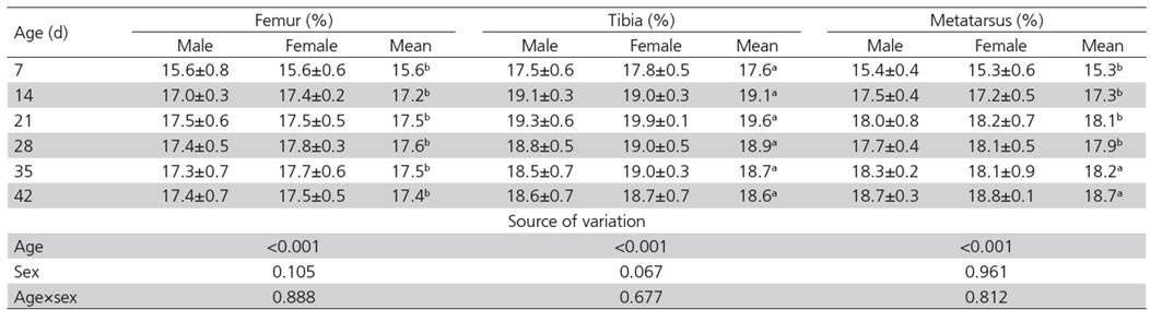
Bone Ca content
The highest Ca content was observed in the tibia (Table 7). No differences in Ca content were observed between the femur and the metatarsus from 7 to 28 d of age (p > 0.05). However, the femur exhibited a lower Ca content than the metatarsus from 35 to 42 d of age (p < 0.05). The Ca content of the femur and the tibia quadratically increased with age. The highest Ca content was observed on 28 and 21 d of age in the femur and the tibia, respectively. By contrast, the Ca content of the metatarsus linearly increased with age. No differences in Ca content were observed between male and female broilers (p > 0.05).
- Comparison of the calcium (Ca) content of the femur, tibia, and metatarsus of male and female broilers1.
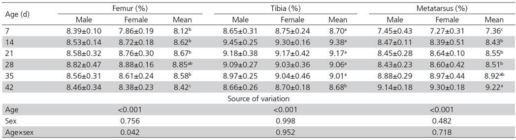
- Comparison of the phosphorus (P) content of the femur, tibia, and metatarsus of male and female broilers1.
Differences in the Ca content of leg bones were observed in turkeys. The femur contained higher Ca than the tibia in 2- to 22-wk-old turkeys (Goetting-Fuchs et al., 2012Goetting-Fuchs C, Günther R, Liesner VG, Liesner BG, Beyerbach M, Kamphues J. Investigations on skeletal development, bone mineralisation as well as calcium and phosphorus levels in blood of male fattening turkeys. European Poultry Science 2012;76:121-130.). These data showed the different ability of leg bones to retain minerals.
In our study, the Ca content of the tibia and femur quadratically increased with age. This result is consistent with that reported by Barreiro et al. (2009Barreiro FR, Sagula AL, Junqueira OM, Pereira GT, Baraldi-Artoni, SM. Densitometric and biochemical values of broiler tibias at different ages. Poultry Science2009;88:2644-2648.). Talaty et al. (2009Talaty PN, Katanbaf MN, Hester PY. Life cycle changes in bone mineralization and bone size traits of commercial broilers. Poultry Science 2009;88:1070-1077. ) also found that chicks exhibited the greatest tibia mineral density at 28 d of age. Similarly to ash content, metatarsus Ca content linearly increased with age.
No differences in bone Ca content were observed between males and females in the present study. A previous research showed that male broilers present lower tibia Ca than females from 42 to 56 d of age (Skinner & Waldroup, 1995Skinner JT, Waldroup PW. Allometric bone development in floor-reared broilers. Journal of Applied Poultry Research 1995;4:265-270.).
Bone P content
Tibia had the highest P content from 7 to 35 d of age. No differences were observed in the P content of the femur and the metatarsus from 14 to 35 d of age (p > 0.05). The metatarsus had the largest P content, followed by tibia and femur at 42 d of age (p < 0.05). The P content of the femur and the tibia quadratically increased with age and reached the highest value at 28 and 14 d of age, respectively. By contrast, the P content of the metatarsus linearly increased with age. No differences in bone P content were observed between males and females (p > 0.05).
Goetting-Fuchs et al.(2012Goetting-Fuchs C, Günther R, Liesner VG, Liesner BG, Beyerbach M, Kamphues J. Investigations on skeletal development, bone mineralisation as well as calcium and phosphorus levels in blood of male fattening turkeys. European Poultry Science 2012;76:121-130.) reported that femur P content was higher than that of the tibia in 2- to 22-wk-old turkeys.
The lowest P content of the tibia was observed at 8 d of age and then increased at 22 d of age; however, P content did not increase at 42 d of age (Barreiro et al., 2009Barreiro FR, Sagula AL, Junqueira OM, Pereira GT, Baraldi-Artoni, SM. Densitometric and biochemical values of broiler tibias at different ages. Poultry Science2009;88:2644-2648.). The percentage of Ca and P of the whole body decreased in broilers from 3 to 6 wk of age (Nieß et al., 2005Nieß E, Hovenjürgen M, Pfeffer E. Whole body concentrations of major minerals and of some trace elements in 3, 5 and 6 weeks old broiler chicks. European Poultry Science 2005;69:16-22.). These data suggested that the P is retained faster in the tibia and the whole body during starter phase.
Skinner & Waldroup (1995Skinner JT, Waldroup PW. Allometric bone development in floor-reared broilers. Journal of Applied Poultry Research 1995;4:265-270.) and Barreiro et al. (2009Barreiro FR, Sagula AL, Junqueira OM, Pereira GT, Baraldi-Artoni, SM. Densitometric and biochemical values of broiler tibias at different ages. Poultry Science2009;88:2644-2648.) found that the Ca to P ratio in the tibia of broilers was 2.0:1. Similar results were observed in the present study; in particular, the Ca to P ratio in the femur, tibia, and metatarsus was also approximately 2.0:1.
In conclusion, the leg bones of broiler chicks grew faster at 1 to 21 d of age compared with 22 to 42 d of age. The retention of Ca and P was related with the ash content in the three leg bones. These data indicate that there are differences in femur, tibia, and metatarsus growth and mineralization of male and female broilers.
ACKNOWLEDGMENTS
This study was supported by the National Natural Science Foundation of China (31101732), Innovation Scientists and Technicians Troop Construction Projects of Henan Province, and Shangqiu Normal University Foundation (2013GGJS10).
REFERENCES
- Applegate TJ, Lilburn MS. Growth of the femur and tibia of a commercial broiler line. Poultry Science 2002;81:1289-1294.
- Barreiro FR, Baraldi-Artoni SM, Amaral LA, Barbosa JC, Girardi AM, Pacheco MR, Amoroso L. Determination of broiler femur parameters at different growth phases. International Journal of Poultry Science2011;10:849-853.
- Barreiro FR, Sagula AL, Junqueira OM, Pereira GT, Baraldi-Artoni, SM. Densitometric and biochemical values of broiler tibias at different ages. Poultry Science2009;88:2644-2648.
- Bond PL, Sullivan TW, Douglas JH, Robeson LG. Influence of age, sex, and method of rearing on tibia length and mineral deposition in broilers. Poultry Science 1991;70:1936-1942.
- Goetting-Fuchs C, Günther R, Liesner VG, Liesner BG, Beyerbach M, Kamphues J. Investigations on skeletal development, bone mineralisation as well as calcium and phosphorus levels in blood of male fattening turkeys. European Poultry Science 2012;76:121-130.
- Gu R, Tang XJ, Lu JX, Ge QL, Gao YS. Comparison on bone strength of four breeds of commercial generation broilers. China Poultry 2010;32:28-30.
- Hall LE, Shirley RB, Bakalli RI, Aggrey SE, Pesti GM, Edwards HMJr. Power of two methods for the estimation of bone ash of broilers. Poultry Science 2003;82:414-418.
- Nieß E, Hovenjürgen M, Pfeffer E. Whole body concentrations of major minerals and of some trace elements in 3, 5 and 6 weeks old broiler chicks. European Poultry Science 2005;69:16-22.
- SAS Institute. SAS user's guide. Version 9 ed. Cary; 2002.
- Skinner JT, Waldroup PW. Allometric bone development in floor-reared broilers. Journal of Applied Poultry Research 1995;4:265-270.
- Talaty PN, Katanbaf MN, Hester PY. Life cycle changes in bone mineralization and bone size traits of commercial broilers. Poultry Science 2009;88:1070-1077.
- Williams B, Solomon S, Waddington D, Thorp B, Farquharson C. Skeletal development in the meat-type chicken. British Poultry Science 2000;41:141-149.
- Yair R, Uni Z, Shahar R. Bone characteristics of late-term embryonic and hatchling broilers: Bone development under extreme growth rate. Poultry Science 2012;91:2614-2620.
- Yalcin S, Ozkan S, Coskuner E, Bilgen G, Delen Y, Kurtulmus Y, Tanyalcin T. Effects of strain, maternal age and sex on morphological characteristics and composition of tibial bone in broilers. British Poultry Science 2001;42:184-190.
Publication Dates
-
Publication in this collection
Sept 2015
History
-
Received
Apr 2014 -
Accepted
Aug 2014