
Abstract
Studies evaluating the influence of nutrients on plant anatomy are very important because nutritional deficiencies can alter the thickness and shape of certain tissues, compromising their functionality what can explain the reduction of productivity. The aim of this study was to characterize the anatomical changes in cherry tomato plants subjected to calcium (Ca) and magnesium (Mg) deficiencies. The experiment was conducted in nutrient solution and the plants subjected to three treatments: complete solution (Control), Ca restriction (1 mmol L-1 of Ca) and Mg omission (without Mg). The experimental design was completely randomized, with three repetitions. Sixty days after seedling transplanting leaves and stem were collected and submitted to anatomical evaluations. Ca or Mg deficiency promotes most evident anatomical changes in chlorophyllous and vascular tissues of the leaves, rather than in the stems. Leaves of ‘Sindy’ tomato plants with a concentration of 1.7 g kg-1 of Mg and visual symptoms of Mg deficiency present hyperplasia of both tissues, phloem and xylem. This deficiency also promotes increases in the thickness of mesophyll, spongy parenchyma and palisade parenchyma, and consequently of leaf thickness. The midrib of the leaves with a concentration of 10 g kg-1 of Ca, without visual symptoms of deficiency presented phloem hypertrophy and hyperplasia.
Keywords:
Solanum lycopersicum Mill; xylem; phloem; mineral nutrition; nutrient solution
INTRODUCTION
It is known that macronutrients and micronutrients can directly or indirectly influence the anatomical structure of plants. An insufficient or excess supply of these minerals may cause alterations in plant anatomy. Anatomical studies that aim to evaluate the influence of nutrients on plant anatomy are of fundamental importance because, aside from causing damage primarily to metabolic pathways [11 Fontes P.C.R. Diagnóstico do estado nutricional das plantas. Editora UFV; 2001. pp.122], alterations in mineral nutrition can compromise the structure and function of tissues and hence the development of the plant [22 Marschner H. Marschner’s Mineral Nutrition of Higher Plants, 3rd ed. London: Academic Press; 2012. pp.684].
Proper mineral nutrition is important because lack or excess quantity of nutrients affects firstly plant metabolism, triggering adjustment mechanisms. If the lack or excess persist these adjustment mechanisms fail to maintain the plant homeostasis reflecting in low growth, anatomical disarrangement and visual symptoms [33 Cantarutti, R.B.; Barros, N.F.; Martinez, H.E.P.; Novais, R.F. Avaliação da fertilidade do solo e recomendação de fertilizantes. In: Novais, R.F.; Alvarez, V.H.; Barros, N.F.; Fontes, R.L.F.; Cantarutti, R.B.; Neves, J.C. Fertilidade do solo. Viçosa: Sociedade Brasileira de Ciência do Solo, 2007; pp. 769-850.].
Magnesium (Mg), one of the essential nutrients, is involved in numerous physiological processes during plant growth and development [44 Cakmak, I.; Yazici, A.M. Magnesium: a forgotten element in crop production. Better Crops 2010, 94, 23-25., 55 Gransee, A.; Fuhrs, H. Magnesium mobility in soils as a challenge for soil and plant analysis, magnesium fertilization and root uptake under adverse growth conditions. Plant Soil 2013, 368, 5-21.]. We can highlight its function as central atom of chlorophyll and as metal component that activates more than 300 enzymes [22 Marschner H. Marschner’s Mineral Nutrition of Higher Plants, 3rd ed. London: Academic Press; 2012. pp.684]. Calcium (Ca), by the other hand, is responsible for growth, cell wall thickness and remodeling, as well as for plant tissue development [66 Hepler, P.K.; Winship, L.J. Calcium at the cell wall-cytoplast interface. J. Integr. Plant Biol. 2010, 52, 147-160., 77 Kudla, J.; Batistic, O.; Hashimoto, K. Calcium signals: the lead currency of plant information processing. Plant Cell 2010, 22, 541-563.].
The effect of Ca and Mg on the anatomy of the leaves is justified by their functions and compartmentation in plant cells. High Ca concentrations are found in the middle lamella of the cell wall, at the exterior surface of the plasma membrane, in the endoplasmic reticulum, and in the vacuole. Thus, Ca is involved in the cell wall stabilization, cell extension and secretory processes, membrane stabilization, cation-anion balance and osmoregulation, and acts as second messenger [22 Marschner H. Marschner’s Mineral Nutrition of Higher Plants, 3rd ed. London: Academic Press; 2012. pp.684, 88 White, P.J.; Broadley, M.R. Calcium in plants. Ann. Bot. 2003, 92, 487-511.]. The Mg ion, besides the presence in the chlorophyll molecule is firmly bound to pectin in cell walls, precipitate as sparingly soluble salts in the vacuole, and act as bridging element for the aggregation of ribosome subunits [22 Marschner H. Marschner’s Mineral Nutrition of Higher Plants, 3rd ed. London: Academic Press; 2012. pp.684].
Although the mechanisms by which plants respond to nutritional stress are similar, small differences are observed among species and/or cultivars. Thus, knowing these differences is very import in plant production because production costs are greater when plants are experiencing nutritional stress [99 Cambraia, J. Aspectos bioquímicos, celulares e fisiológicos dos estresses nutricionais em plantas, In: Estresses ambientais danos e benefícios em plantas. Nogueira, R.J.M.C.; Araújo, E.L.; Willadin, O.L.G.; Cavalcante, U.M.T. eds., UFRPE, Imprensa Universitária, Recife, 2005. pp. 127-137.]. Many studies have explored different aspects of tomato mineral nutrition, including nutrient accumulation, growth and production responses, effects of nutrients on quality, deficiency symptoms and foliar diagnostic standards [1010 Fernandes, P.D.; Churata-Masca, M.G.C.; Oliveira, G.D.; Haag, H.P. Nutrição mineral de hortaliças: XXVII - Absorção de nutrientes pelo tomateiro (Lycopersicon esculentum, Mill.), em cultivo rasteiro. An. Esc. Super. Agric. Luiz de Queiroz 1976, 32, 595-608.
11 Dechen, A.R.; Haag, H.P.; Sarruge, J.R.; Oliveira, G.D. Nutrição mineral de hortaliças: XXXV Efeitos de doses de cálcio na solução nutritiva, no desenvolvimento e nos teores de nitrogênio, fósforo, potássio, cálcio, magnésio e enxofre, em plantas de tomateiro (Lycopersicon esculentum Mill.). An. Esc. Super. Agric. Luiz de Queiroz 1980, 37, 1009-1057.
12 Fontes, P.C.R.; Sampaio, R.A.; Mantovani, E.C. Tomato yield and potassium concentrations in soil and in plant petioles as affected by potassium fertirrigation. Pesq. Agropec. Bras. 2000, 35, 575-580.-1313 Oliveira, R.H.; Lima, M.J.S.; Pereira-Junior, H.A.; Rebouças, T.N.H.; Morais, O.M.; Guimarães, B.V.C., et al. Caracterização de sintomas visuais de deficiência de micronutrientes em tomateiro do grupo salada. Semina: Ciênc. Agrar. 2009, 30, 1093-1100.]. There are also studies related to the physiological responses to Mg deficiency showing decline in transpiration, accumulation of sugars and starch, increased oxidative stress, metabolite alterations, interactions between Mg and decline in photosynthetic activity [1414 Tanoi, K.; Kobayashi, N.I. Leaf Senescence by Magnesium Deficiency. Plants. 2015; 4: 756-772.
15 Guo, W.; Nazimc, H.; Lianga, Z.; Yang, D. Magnesium deficiency in plants: An urgent problem. The Crop Journal. 2016, 4, 83 - 91.-1616 Farhat, N.; Elkhouni, A.; Zorrig, W.; Smaoui, A.; Abdelly, C.; Rabhi, M. Effects of magnesium deficiency on photosynthesis and carbohydrate partitioning. Acta Physiol Plant. 2016, 38, 145.]. However, in general there are few studies addressing the anatomical changes associated with nutrient visual or foliar diagnosis, and none was found in cherry tomato plant. Thus, the aim of this study was to characterize the anatomical changes in cherry tomato plants subjected to Ca and Mg deficiencies.
MATERIAL AND METHODS
The experiment was conducted in a greenhouse of the Laboratory of Mineral Nutrition of Plants, Department of Fitotecnia, Universidade Federal de Viçosa, Brazil. The cherry tomato seedlings (Solanum lycopersicum Mill. Hybrid ‘Sindy’) were obtained by germinating seeds in trays containing commercial substrate. After the plants had developed two definitive leaves, they were selected based on uniformity and transferred to plastic pots (8 L capacity). Two plants were placed in a vessel containing a specific nutrient solution, and each vessel represented an experimental plot. The treatment solutions were complete (Control), Ca restriction and Mg omission.
The complete nutrient solution had the following composition: N = 8 mmol L-1; P = 2 mmol L-1; K = 4 mmol L-1; Ca = 2 mmol L-1; Mg = 1 mmol L-1; S = 1 mmol L-1; Na = 2 mmol L-1; B = 21 μmol L-1; Cu = 0.9 μmol L-1; Fe = 35 μmol L-1; Mn = 15 μmol L-1; Mo = 0.7 μmol L-1; Zn = 0.4 μmol L-1. The treatment minus Mg have received “zero” mmol L-1 of Mg and the minus Ca received a low dose of this nutrient (1 mmol L-1 of Ca). We have decided supplying such a dose to avoid high concentration of Na, as companion ion of the anionic macronutrients, in case “zero” Ca would be supplied. These omission treatments have received the same concentrations of the other macro and micronutrients. The electrical conductivities (EC) of the nutrient solutions were of 1.22; 1.23 and 1.27 dS m-1 for the treatments complete, minus Ca and minus Mg respectively. The nutrient solutions were maintained under constant aeration. The volume of water lost by evapotranspiration was supplemented with deionized water and the pH was monitored and adjusted to 5.5 ± 0.5 with 1 mol L-1 HCl or 1 mol L-1 NaOH on a daily basis. The nutrient solutions were completely renewed whenever electrical conductivity was reduced by 30%.
The experimental design was completely randomized, with three treatments: complete solution, Ca restriction and Mg omission, and three repetitions were performed. Sixty days after the start of the experiment we picked out some leaves to the anatomical evaluations. It was used the median third of the leaves in each evaluation. Considering the nutrient mobility in the phloem it was taken leaves at different positions: For Mg, we sampled the third fully expanded leaf from the apex; for Ca, we sampled older leaves from the middle third of the plants that were not showing visible senescence. Sections of the stem (approximately 5 cm long) at the internode just below the third fully expanded leaf from the apex were also sampled from each plant. The samples were fixed in a solution of FAA50 (formaldehyde, acetic acid and 50% ethyl alcohol) for 72 hours and stored in 70% ethanol [1717 Johansen, D.A. Plant Microtechnique. McGraw-Hill Book Company, New York; 1940; pp. 523].
To make the slides, samples of approximately 0.25 cm2 were dehydrated in an ascending ethanol series and embedded in methacrylate (Historesin-Leica) according to the manufacturer's recommendations. The material was transversely sectioned (5 μm thickness) in a rotary microtome, and the sections were stained with toluidine blue for structural characterization and micromorphometric analysis. The slides were mounted in synthetic resin (Permount). For each experimental repetition, digital images of stem and leaf cross sections were obtained using a photomicroscope (Olympus AX70) with a U-Photo system.
In each leaf sample, it was measured the total thickness of the lamina and the thickness of the following components: adaxial epidermis, abaxial epidermis, palisade parenchyma, spongy parenchyma, and mesophyll (palisade plus spongy parenchyma). These data were transformed into percentage of the total thickness of the lamina. Length and width of palisade parenchyma cells were also measured. In the midrib, the number of sieve tube elements and the number, diameter and total area of vessel elements of the main vascular bundle were also measured.
In the stems, it was measured the total thickness of the sampled region and the thickness of epidermis, chlorenchyma, collenchyma, ground parenchyma (cortical and medullar parenchyma), phloem, xylem, sclereids and sclereids cell wall, and the number of cambium zone cells. Except the last two, all other measurements were transformed into percentage in relation to the total thickness.
The measurements were taken with Image Pro-Plus software (Media Cybernetics, Silver Spring, MD, USA). For all measurements at least ten replicates (measures) for each repetition were used. The data were subjected to analysis of variance. The mean values obtained to the plants Ca and Mg deprived were compared with the obtained in control plants (Complete treatment) by means of Dunnett’s test at 5% probability.
RESULTS
A Ca dose as low as 1 mmol L-1 in the nutrient solution did not promote the development of visual symptoms of deficiency (Figure 1B) even with a reduction of 53% in Ca leaf concentration, that fell from 21.3 to 10 g kg-1. As a result of Ca starvation the dry matter accumulation suffered a reduction of 19%. The plants submitted to the treatment minus Mg presented interveinal chlorosis of older and intermediate leaves that evolved from the edges toward the midrib (Figure 1 C). In such conditions the dry matter accumulation by the plants was depressed by 55% and the leaf Mg concentration fell from 5.4 to 1.7 g kg-1 [1818 Maia, J.T.L.S.; Martinez, H.E.P.; Clemente, J.M.; Ventrella, M.C.; Milagres, C.C. Growth, nutrient concentration, nutrient accumulation and visual symptoms of nutrient deficiencies in cherry tomato plants. Semina: Ciênc. Agrar. 2019, 40, 585-598.]. These results are in accordance to the fact that when visual symptoms are evident the plant growth is severely depressed. These results also stud out that probably, the tomato cultivar Sindy has low Ca requirement.
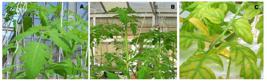
Visual aspect of ‘Sindy’ tomato plants subjected to 60 days of calcium and magnesium deprivation: (A) Complete nutrient solution (Control); (B) Treatment minus Ca; (C) Treatment minus Mg.
Regarding the anatomic features the cherry tomato leaf has a uniseriate epidermis with glandular and non-glandular trichomes, and stomata on both sides; thus, it is classified as amphistomatic. The mesophyll is dorsiventral, with the palisade facing the adaxial side and the spongy parenchyma facing the abaxial side. In the midrib, the chlorenchyma extends across the adaxial side, where it is interrupted by collenchyma cells. The vascular bundle of the midrib is bicollateral with phloem facing both sides of the leaf and is surrounded by ground parenchyma. The other vascular bundles are collateral, with bundle sheaths discretely arranged in the mesophyll.
The stem of the cherry tomato plant also presents a uniseriate epidermis with glandular and non-glandular trichomes and stomata. The cortex shows subepidermal chlorenchyma, followed by collenchyma and ground parenchyma. The vascular tissue is formed by primary (internal and external) and secondary phloem, bounded externally by sclereids and primary and secondary xylem Phloem and xylem are joined by vascular cambium and the ground parenchyma fills the medulla of the stem.
The general anatomical features of the leaf and stem of cherry tomato, described above, were not qualitatively affected by the treatments; however, quantitative differences were very evident.
In Ca-deficient plants, although the proportion of spongy parenchyma was 25% higher than that observed in the complete treatment, the final thickness of the leaf was not affected as the length of palisade parenchyma cells was lower in this condition (Table 1). Phloem hypertrophy and hyperplasy in midrib can be observed as the main anatomical alteration of Ca-deficient plants and confirmed by the increase of 235% in the number of sieve tube elements (Table 1). In relation to the xylem, there was no change in the number and diameter of vessel elements in Ca-deficient plants, but the total area occupied by vessel elements was 147% higher than in the control.
Comparatively to the control, in the stem, only the percentage of chlorenchyma and the thickness of the cell wall of the sclereids were reduced in the Ca-deficient plants. The decrease in the wall thickness of the sclereids reached 74% (Figure 2, Table 1). In the stem of Mg deficient plants, only the chlorenchyma and the thickness of the sclereid cell wall decreased 30% and 64%, respectively (Figure 2, Table 1).

Stems of cherry tomato plants grown in complete solution (A, D, G), in Ca-deficient medium (B, E, H) or in Mg-deficient medium (C, F, I). (Photomicrographs; cross sections). A-C, general view of the stems. D-F, detail of the cortex, vascular tissue and pith region. G-I, detail of the vascular cambium region. bs, bundle sheath; cc, companion cell; co, collenchyma; ch, chlorenchyma; ct, cortex; ep, epidermis; fi, fiber; pa, parenchyma; pc, parenchyma cell; ph, phloem; pi, pith; sc, sclereids; se, sieve tube element; st, stomata; vc, vascular cambium; v, vessel element; vt, vascular tissue; xy, xylem;. Bar = 500 µm (A-C); 200 µm (D-F); 50 µm (G-I).
When compared to the control there was a variation of a large number of anatomical features in Mg-deficient plants. There was a 133% increase in leaf thickness of Mg-deficient plants accompanied by the increase of mesophyll 107%, spongy parenchyma 123%, palisade parenchyma cell length and width. Phloem tissue hyperplasia was also one of the main characteristics of Mg-deficient plants, and results mainly from 234% increase in number of sieve tube elements and their associated companion cells (Figure 3, Table 1). In the xylem there was a significant increase in the number (hyperplasia) and in the total area of vessel elements in Mg-deficient plants that reached 167% of the control.

Leaves of cherry tomato plants grown in complete solution (A, D, G), in Ca-deficient medium (B, E, H) or in Mg-deficient medium (C, F, I). (Photomicrographs; cross sections). A-C, general view of midrib region. D-F, detail of the midrib. G-I, detail of the lamina. ab, abaxial epidermis; ad, adaxial epidermis; bs, bundle sheath; co, collenchyma; ch, chlorenchyma; pa, parenchyma; ph, phloem; pp, palisade parenchyma; sp, spongy parenchyma; st, stomata; vc, vascular cambium; vt, vascular tissue; xy, xylem. Bar = 300 µm (A-C); 100 µm (D-I).
DISCUSSION
The anatomy of leaves and stems of cherry tomato were just affected quantitatively by Ca or Mg deficiency. It must be considered that anatomical symptoms can vary with respect to the time of appearance and the sequence in which the plant organs are affected, the age and species of plant studied, the environment, and the severity of the deficiency [3333 Dickison, W.C. Integrative plant anatomy. Academic Press, San Diego; 2000; pp. 533]. The most evident anatomical changes in the plants occurred in the leaves, in chlorophyllous and in vascular tissues. Therefore, Ca or Mg deficiency may interfere with plant photosynthetic activity and substance transport by phloem and xylem.
Ca-deficient plants produced leaves with slight changes in the proportion of chlorophyllous tissues, but the percentage of mesophyll remained unchanged. Regarding the vascular tissues, the total area occupied by the xylem vessel elements was increased by 1.47 times, while the number of sieve tube elements of the phloem was increased by 2.35 times. This suggests that leaf anatomical changes would not directly affect the photosynthetic sites, but rather the supply of water and salts by the xylem and the transport of organic material by the phloem in Ca-deficient plants. Hyperplasia of phloem may be understood as an adaptive strategy of the plant under conditions of Ca or Mg deficiency to maximize the sap translocation in this vascular tissue. Changes in the activity of meristems as procambium and vascular cambium may be related to hyperplasy of phloem and to the increase of the area occupied by xylem vessel elements in cherry tomato plants.
The Mg deficiency imposed greater anatomical leaf changes compared to Ca deficiency in cherry tomato plants. However, it should be considered that the nutrient solution minus Mg had total absence of the element, while the solution minus Ca had 1 mmol L-1 of Ca.
Under Mg deficiency, the thickness of the leaf was greater, with larger chlorenchyma cells (hypertrophy). The hypertrophy of chlorenchyma cells in leaves of cherry tomato, especially under Mg deficiency, could be related to changes in osmotic equilibrium of vacuoles and in the cell wall extensibility [22 Marschner H. Marschner’s Mineral Nutrition of Higher Plants, 3rd ed. London: Academic Press; 2012. pp.684]. The chlorenchyma with larger and vacuolated cells and larger intercellular spaces had a lower number of chloroplasts per unit area. Farther on, in addition of the involvement of Mg in chlorophyll molecule [22 Marschner H. Marschner’s Mineral Nutrition of Higher Plants, 3rd ed. London: Academic Press; 2012. pp.684] we can conclude that this anatomical configuration by itself can cause yellowing of leaves under Mg deficiency. Mg is a structural component of the chlorophyll molecule and its deficiency compromises its function [1616 Farhat, N.; Elkhouni, A.; Zorrig, W.; Smaoui, A.; Abdelly, C.; Rabhi, M. Effects of magnesium deficiency on photosynthesis and carbohydrate partitioning. Acta Physiol Plant. 2016, 38, 145., 2222 Suutarinen, J.; Heiska, K.; Autio, K. The effects of calcium chloride and sucrose prefreezing treatments on the structure of strawberry tissues. LWT-FoodSci. Technol. 2000, 33, 89-102., 2323 Alquini, Y.; Bona, C.; Boerger, M.R.T.; Costa, C.G.; Barros, C.F. Epiderme. In: Anatomia Vegetal. Apezzato, G.B.; Carmello-Guerreiro, S.M. eds., Universidade Federal de Viçosa, Viçosa; 2006; pp. 87-108.]. Therefore, Mg is associated with leaf yellowing in the form of interveinal chlorosis in leaves under Mg deficiency [44 Cakmak, I.; Yazici, A.M. Magnesium: a forgotten element in crop production. Better Crops 2010, 94, 23-25.]. This Mg deficiency may involve a programmed cell damage related to oxidative damage in chloroplasts [2424 Foyer, C.H.; Noctor, G. Redox homeostasis and antioxidant signalling: a metabolic interface between stress perception and physiological responses. Plant Cell. 2005, 17, 1866-1875.]. In Arabidopsis thaliana leaves show a disorganization of thylakoid membranes due to Mg-deficient [2525 Hermans, C.; Jhonson, G.N.; Strasser, R.J.; Verbruggen, N. Physiological characterization of magnesium deficiency in sugar beet: acclimation to low magnesium differentially affects photosystems I and II. Planta. 2004, 220, 344-355.]. Transverse sections of the interveinal region of the leaf revealed malformed parenchyma cells, the presence of which resulted in an increased area of intercellular space that increased the total thickness of the leaf. As a result of the anatomical changes observed in these tissues, plants experiencing Mg deficiency exhibited interveinal chlorosis in mature leaves [1414 Tanoi, K.; Kobayashi, N.I. Leaf Senescence by Magnesium Deficiency. Plants. 2015; 4: 756-772., 1919 Taiz, L.; Zeiger, E. Fisiologia Vegetal. 5ª ed. Artmed, Porto Alegre; 2013; pp.918].
Interestingly, the Mg deficiency in the tomato caused an hyperplasia of booth phloem and xylem, which affects the transport of organic material and water and salts, respectively. Under Mg deficit, the vascular tissues especially the phloem [2626 Zhong, W.; Schobert, C.; Komor, E. Transport of magnesium ions in the phloem of Ricinus communis L. seedlings. Planta. 1993, 190, 114-119.] may be affected, compromising the translocation of carbohydrates [2727 Puech, L.; Mehne-Jakobs, B. Histology of magnesium-deficient Norway spruce needles influenced by nitrogen source. Tree Phys. 1997, 17, 301-310., 2828 Cakmak, I.; Hengeler, C.; Marschner, H. Changes in phloem export of sucrose in leaves in response to phosphorus, potassium and magnesium deficiency in bean plants. J. Exp. Bot. 1994, 45, 1251-1257.]. Additionally, Mg is essential for the activation of ATPases involved in phloem loading [2929 Fink, S. Strutural changes in conifer needles due to Mg and K deficiency. Fert. Res. 1991, 27, 23-27., 3030 Cakmak, I.; Kirkby, E.A. Role of magnesium in carbon partitioning and alleviated photo oxidative damage. Physiol. Plant. 2008, 133, 692-704.], under Mg-deficient, phloem loading inhibition into is explained by an impairment of the membrane integral protein, H+/ATPase. Mg lack reduces the level of Mg-ATP complexes required by plasma membrane-bound ATPases. Therefore, there is inhibition of photosynthate export via phloem [3131 Hanstein, S.; Wang, X.Z.; Qian, X.Q.; Friedhoff, P.; Fatima, A.; Shan, Y.H., et al. Changes in cytosolic Mg2+ levels can regulate the activity of the plasma membrane H+-ATPase in maize. Biochem J. 2011, 435, 93-101., 3232 White PJ. Ion uptake mechanisms of individual cells and roots: short-distance transport. In Marschner’s mineral nutrition of higher plants. Marschner, P. ed., Academic Press, London, 2012; p. 7-47.]. Needles of damaged spruce (Picea abies L. Karst.) growing at a Mg-deficient and ozone polluted mountain site, produced rapid needle yellowing, hypertrophy and anomalous divisions of cambium cells, phloem collapse, and production of atypical xylem tracheids [3434 Boxler-Baldoma, C.; Lütz, C.; Heumann, H.G.; Siefermann-Harms, D. Structural changes in the vascular bundles of light-exposed and shaded spruce needles suffering from Mg deficiency and ozone pollution. J. Plant Physiol. 2006, 163, 195-205.].
In the stem, the chlorenchyma tissue decreased 16.08% in plants deficient in Ca, but what is more evident is the reduction of the wall thickness of the sclereides external to the phloem. Similar response is observed in the stem of Mg-deficient plants, which indicates that both Ca and Mg deficiency would be related to the problems in secondary cell wall synthesis. Phloem fiber formation may be compromised in Ca-deficient plants, indicating that Ca deficiency has a greater effect on the formation of phloem compared with xylem [2022 Algan, G.; Tilekliogly, B. The effects of N, P, Ca and Fe deficiency in Linum usitatissimum L. in cambial activity and differentiation. C. Fac. Sci. Univ of Ankara Series C. 1992, 10, 1-10.].
As observed in this study, the cell walls of phloem sclereids were thinner under Ca deficiency compared with the control treatment. The cell wall, the middle lamella and the outer surface of the plasma membrane are all areas with high concentrations of Ca, which has essential structural functions; therefore, any change in the supply of this element can undermine the formation of these structures [2121 Trevisam, R. Análises histológicas e bioquímicas em calos de Eucalyptus uronphylla S. T. Blake cultivados in vitro sob interação nutricional de boro e cálcio. Tese de Doutorado, Escola Superior de Agricultura “Luiz de Queiroz”, Universidade de São Paulo, Piracicaba 2005; pp.167.]. When calcium chloride (CaCl2) was applied to pre-frozen strawberry fruits, their cell wall structure remained unchanged compared with to those receiving no treatment, becoming less firm and resistant [2222 Suutarinen, J.; Heiska, K.; Autio, K. The effects of calcium chloride and sucrose prefreezing treatments on the structure of strawberry tissues. LWT-FoodSci. Technol. 2000, 33, 89-102.]. The most obvious symptom of both Ca or Mg deficiency in cherry tomato stems is the marked decrease in the cell wall thickness of support cells (sclereids) located outside the phloem.
The decrease in cell wall thickness of sclereids may be related to the contribution of these ions through the process of cell wall deposition, since Ca and Mg are strongly associated with cell wall pectins [22 Marschner H. Marschner’s Mineral Nutrition of Higher Plants, 3rd ed. London: Academic Press; 2012. pp.684]. In Ca-deficient tissue polygalacturonase activity is increased, and a typical symptom of Ca deficiency is the disintegration of cell walls and the collapse of the affected tissues [22 Marschner H. Marschner’s Mineral Nutrition of Higher Plants, 3rd ed. London: Academic Press; 2012. pp.684]. The decrease in cell wall thickening of supporting cells in cherry tomato plants may decrease the mechanical strength of the body as a whole.
CONCLUSION
Ca or Mg deficiency promotes most evident anatomical changes in chlorophyllous and vascular tissues of the leaves, rather than in the stem of ‘Sindy’ tomato plants.
Leaves of ‘Sindy’ tomato plants with a concentration of 1.7 g kg-1 of Mg and visual symptoms of Mg deficiency present hyperplasia of both tissues, phloem and xylem. This deficiency also promotes increases in the thickness of mesophyll, spongy parenchyma and palisade parenchyma, and consequently of leaf thickness.
The midrib of the leaves of ‘Sindy’ tomato plants with a concentration of 10 g kg-1 of Ca, without visual symptoms of deficiency presented phloem hypertrophy and hyperplasia.
Acknowledgments:
The authors thank the Conselho Nacional de Desenvolvimento Científico e Tecnológico (CNPq) and Coordenação de Aperfeiçoamento de Pessoal de Nível Superior (CAPES - Finance Code 001) for financial support.
REFERENCES
-
1Fontes P.C.R. Diagnóstico do estado nutricional das plantas. Editora UFV; 2001. pp.122
-
2Marschner H. Marschner’s Mineral Nutrition of Higher Plants, 3rd ed. London: Academic Press; 2012. pp.684
-
3Cantarutti, R.B.; Barros, N.F.; Martinez, H.E.P.; Novais, R.F. Avaliação da fertilidade do solo e recomendação de fertilizantes. In: Novais, R.F.; Alvarez, V.H.; Barros, N.F.; Fontes, R.L.F.; Cantarutti, R.B.; Neves, J.C. Fertilidade do solo. Viçosa: Sociedade Brasileira de Ciência do Solo, 2007; pp. 769-850.
-
4Cakmak, I.; Yazici, A.M. Magnesium: a forgotten element in crop production. Better Crops 2010, 94, 23-25.
-
5Gransee, A.; Fuhrs, H. Magnesium mobility in soils as a challenge for soil and plant analysis, magnesium fertilization and root uptake under adverse growth conditions. Plant Soil 2013, 368, 5-21.
-
6Hepler, P.K.; Winship, L.J. Calcium at the cell wall-cytoplast interface. J. Integr. Plant Biol. 2010, 52, 147-160.
-
7Kudla, J.; Batistic, O.; Hashimoto, K. Calcium signals: the lead currency of plant information processing. Plant Cell 2010, 22, 541-563.
-
8White, P.J.; Broadley, M.R. Calcium in plants. Ann. Bot. 2003, 92, 487-511.
-
9Cambraia, J. Aspectos bioquímicos, celulares e fisiológicos dos estresses nutricionais em plantas, In: Estresses ambientais danos e benefícios em plantas. Nogueira, R.J.M.C.; Araújo, E.L.; Willadin, O.L.G.; Cavalcante, U.M.T. eds., UFRPE, Imprensa Universitária, Recife, 2005. pp. 127-137.
-
10Fernandes, P.D.; Churata-Masca, M.G.C.; Oliveira, G.D.; Haag, H.P. Nutrição mineral de hortaliças: XXVII - Absorção de nutrientes pelo tomateiro (Lycopersicon esculentum, Mill.), em cultivo rasteiro. An. Esc. Super. Agric. Luiz de Queiroz 1976, 32, 595-608.
-
11Dechen, A.R.; Haag, H.P.; Sarruge, J.R.; Oliveira, G.D. Nutrição mineral de hortaliças: XXXV Efeitos de doses de cálcio na solução nutritiva, no desenvolvimento e nos teores de nitrogênio, fósforo, potássio, cálcio, magnésio e enxofre, em plantas de tomateiro (Lycopersicon esculentum Mill.). An. Esc. Super. Agric. Luiz de Queiroz 1980, 37, 1009-1057.
-
12Fontes, P.C.R.; Sampaio, R.A.; Mantovani, E.C. Tomato yield and potassium concentrations in soil and in plant petioles as affected by potassium fertirrigation. Pesq. Agropec. Bras. 2000, 35, 575-580.
-
13Oliveira, R.H.; Lima, M.J.S.; Pereira-Junior, H.A.; Rebouças, T.N.H.; Morais, O.M.; Guimarães, B.V.C., et al. Caracterização de sintomas visuais de deficiência de micronutrientes em tomateiro do grupo salada. Semina: Ciênc. Agrar. 2009, 30, 1093-1100.
-
14Tanoi, K.; Kobayashi, N.I. Leaf Senescence by Magnesium Deficiency. Plants. 2015; 4: 756-772.
-
15Guo, W.; Nazimc, H.; Lianga, Z.; Yang, D. Magnesium deficiency in plants: An urgent problem. The Crop Journal. 2016, 4, 83 - 91.
-
16Farhat, N.; Elkhouni, A.; Zorrig, W.; Smaoui, A.; Abdelly, C.; Rabhi, M. Effects of magnesium deficiency on photosynthesis and carbohydrate partitioning. Acta Physiol Plant. 2016, 38, 145.
-
17Johansen, D.A. Plant Microtechnique. McGraw-Hill Book Company, New York; 1940; pp. 523
-
18Maia, J.T.L.S.; Martinez, H.E.P.; Clemente, J.M.; Ventrella, M.C.; Milagres, C.C. Growth, nutrient concentration, nutrient accumulation and visual symptoms of nutrient deficiencies in cherry tomato plants. Semina: Ciênc. Agrar. 2019, 40, 585-598.
-
19Taiz, L.; Zeiger, E. Fisiologia Vegetal. 5ª ed. Artmed, Porto Alegre; 2013; pp.918
-
22Algan, G.; Tilekliogly, B. The effects of N, P, Ca and Fe deficiency in Linum usitatissimum L. in cambial activity and differentiation. C. Fac. Sci. Univ of Ankara Series C. 1992, 10, 1-10.
-
21Trevisam, R. Análises histológicas e bioquímicas em calos de Eucalyptus uronphylla S. T. Blake cultivados in vitro sob interação nutricional de boro e cálcio. Tese de Doutorado, Escola Superior de Agricultura “Luiz de Queiroz”, Universidade de São Paulo, Piracicaba 2005; pp.167.
-
22Suutarinen, J.; Heiska, K.; Autio, K. The effects of calcium chloride and sucrose prefreezing treatments on the structure of strawberry tissues. LWT-FoodSci. Technol. 2000, 33, 89-102.
-
23Alquini, Y.; Bona, C.; Boerger, M.R.T.; Costa, C.G.; Barros, C.F. Epiderme. In: Anatomia Vegetal. Apezzato, G.B.; Carmello-Guerreiro, S.M. eds., Universidade Federal de Viçosa, Viçosa; 2006; pp. 87-108.
-
24Foyer, C.H.; Noctor, G. Redox homeostasis and antioxidant signalling: a metabolic interface between stress perception and physiological responses. Plant Cell. 2005, 17, 1866-1875.
-
25Hermans, C.; Jhonson, G.N.; Strasser, R.J.; Verbruggen, N. Physiological characterization of magnesium deficiency in sugar beet: acclimation to low magnesium differentially affects photosystems I and II. Planta. 2004, 220, 344-355.
-
26Zhong, W.; Schobert, C.; Komor, E. Transport of magnesium ions in the phloem of Ricinus communis L. seedlings. Planta. 1993, 190, 114-119.
-
27Puech, L.; Mehne-Jakobs, B. Histology of magnesium-deficient Norway spruce needles influenced by nitrogen source. Tree Phys. 1997, 17, 301-310.
-
28Cakmak, I.; Hengeler, C.; Marschner, H. Changes in phloem export of sucrose in leaves in response to phosphorus, potassium and magnesium deficiency in bean plants. J. Exp. Bot. 1994, 45, 1251-1257.
-
29Fink, S. Strutural changes in conifer needles due to Mg and K deficiency. Fert. Res. 1991, 27, 23-27.
-
30Cakmak, I.; Kirkby, E.A. Role of magnesium in carbon partitioning and alleviated photo oxidative damage. Physiol. Plant. 2008, 133, 692-704.
-
31Hanstein, S.; Wang, X.Z.; Qian, X.Q.; Friedhoff, P.; Fatima, A.; Shan, Y.H., et al. Changes in cytosolic Mg2+ levels can regulate the activity of the plasma membrane H+-ATPase in maize. Biochem J. 2011, 435, 93-101.
-
32White PJ. Ion uptake mechanisms of individual cells and roots: short-distance transport. In Marschner’s mineral nutrition of higher plants. Marschner, P. ed., Academic Press, London, 2012; p. 7-47.
-
33Dickison, W.C. Integrative plant anatomy. Academic Press, San Diego; 2000; pp. 533
-
34Boxler-Baldoma, C.; Lütz, C.; Heumann, H.G.; Siefermann-Harms, D. Structural changes in the vascular bundles of light-exposed and shaded spruce needles suffering from Mg deficiency and ozone pollution. J. Plant Physiol. 2006, 163, 195-205.
HIGHLIGHTS
-
1
In cherry tomato leaves the deficiency of Ca and Mg affects the elements of vessels.
-
2
Ca-deficient plants produced leaves with changes in the of chlorophyll tissues.
-
3
The Mg deficiency in the cherry tomato caused an hyperplasia of phloem and xylem.
-
4
Hyperplasia of phloem may be understood as an adaptive strategy of the plant.
Publication Dates
-
Publication in this collection
08 May 2020 -
Date of issue
2020
History
-
Received
19 Nov 2018 -
Accepted
25 Nov 2019


