Abstracts
OBJECTIVE: To compare the myoelectric activity before and after ground contact between single leg (SL) and double leg (DL) landings in male athletes. PARTICIPANTS: Fifteen male volleyball athletes without signs and symptoms of lesions in the lower extremities, with a minimum of three years experience in the sport (13 ± 1 years, 1.70 ± 0.12 m, 60 ± 12 kg). MEASUREMENTS: Participants performed two vertical jumps, landing unilaterally and bilaterally. The myoelectric activity of the rectus femoris (RF), biceps femoris (BF), hip adductors (HA) and the BF/RF ratio were compared between the two landings and between the phases characterized by 100ms before (PRE) and after 100 ms (POST) ground contact using ANOVA two-way test with post hoc test of Bonferroni (α = 5%). RESULTS: In both landings activation of RF was higher in the POST in relation to the PRE (p <0.0001). Comparing the landings in the same phase statistical differences (p = 0.2212) were not found. Although the BF did not present significant differences between the PRE and POST in each landing (p = 0.2321), its activation was higher in SL (p = 0.0051). The HA showed greater activation in the POST during the SL (p = 0.0013), however there were no differences when comparing the two landings (p = 0.9233). The BF/RF ratio was higher in both landings during PRE (p = 0.0012). Nevertheless, no differences between the landings (p = 0.7037) were found. CONCLUSION: The results suggest that each muscle has a different role during landing tasks in men. While RF has the main function to decelerate the knee and the downward movement, characterized by increased activation in the POST, BF seems to attenuate the loads on the knee in activities of higher impact, staying more active throughout the cycle in the SL. The increased activation of HA after ground contact in the SL highlights the importance of core region in stabilizing the pelvis in situations of great instability. Further studies are needed to determine the effects of muscle activation at the imposition of mechanical load on the knee that are potentially harmful to male athletes.
biomechanics; EMG; risk factor; males; anterior cruciate ligament
OBJETIVO: Comparar a atividade muscular antes e após o contato com o solo entre as aterrissagens unilateral (AU) e bilateral (AB) em atletas do sexo masculino. PARTICIPANTES: Quinze atletas masculinos de voleibol sem sinais e sintomas de lesões nas extremidades inferiores (13 ± 1 ano, 1,70 ± 0,12m, 60 ± 12kg). MENSURAÇÕES:Os participantes realizaram dois saltos verticais, aterrissando unilateralmente e bilateralmente. A atividade mioelétrica do reto femoral (RF), bíceps femoral (BF), adutores de quadril (AQ) e a relação BF/RF foram comparados entre as duas aterrissagens e entre as fases caracterizadas por 100ms antes (PRE) e 100ms após (POS) o contato com o solo. RESULTADOS: Em ambas as aterrissagens, a ativação do RF foi maior na fase POS em relação à PRE. Na comparação entre as aterrissagens dentro da mesma fase não encontramos diferenças estatísticas. Apesar de o BF não ter apresentado diferenças entre as fases PRE e POS em cada aterrissagem, sua ativação foi maior na AU. Os AQ apresentaram maior ativação na fase POS durante a AU, no entanto não houve diferenças quando comparadas as duas aterrissagens. A relação BF/RF apresentou valores maiores em ambas as aterrissagens na fase PRE. No entanto, não encontramos diferenças entre as aterrissagens. CONCLUSÃO: Os resultados sugerem que cada músculo apresenta um papel diferente durante a fase de aterrissagem em homens. Enquanto que o RF possui como principal função a frenagem da articulação do joelho e do movimento descendente, caracterizada pelo aumento da ativação na fase pós-contato, o BF parece atenuar a tensão articular do joelho em atividades de maior impacto, mantendo-se mais ativo durante todo o ciclo da AU. Já a maior ativação dos AQ após o contato com solo na AU evidencia a importância da região lombo-pélvica na estabilização pélvica em situações de grande instabilidade. Estudos futuros são necessários para determinar os efeitos da ativação muscular apresentada na imposição de cargas mecânicas potencialmente lesivas no joelho em atletas do sexo masculino.
biomecânica; EMG; fator de risco; homens; ligamento cruzado anterior
ORIGINAL ARTICLE
LOCOMOTOR APPARATUS IN EXERCISE AND SPORTS
ILaboratory of Biomechanics and Motor Behavior (LaBiCoM) State University of Rio de Janeiro, Brazil
IIInstitute Brazil of Health Technologies Rio de Janeiro, Brazil
IIIProgram of Biomedical Engineering PEB/COPPE Federal University of Rio de Janeiro, Brazil
IVPost-graduation Program in Mechanical Engineering UNESP/FEG São Paulo, Brazil
VPost-graduation Program in Medical Sciences State University of Rio de Janeiro, Brazil
Mailing address
ABSTRACT
OBJECTIVE: To compare the myoelectric activity before and after ground contact between single-leg (SL) and double-leg (DL) landings in male athletes.
PARTICIPANTS:Fifteen male volleyball athletes without signs and symptoms of lesions in the lower extremities, with a minimum of three years experience in the sport (13 ± 1 years, 1.70 ± 0.12 m, 60 ± 12 kg).
MEASUREMENTS: Participants performed two vertical jumps, landing unilaterally and bilaterally. The myoelectric activity of the rectus femoris (RF), biceps femoris (BF), hip adductors (HA) and the BF/RF ratio were compared between the two landings and between the phases characterized by 100ms before (PRE) and after 100 ms (POST) ground contactusing ANOVA two-way test with post hoc test of Bonferroni (α = 5%).
RESULTS: In both landings activation of RF was higher in the POST in relation to the PRE (p <0.0001). Comparing the landings in the same phase statistical differences (p = 0.2212) were not found. Although the BF did not present significant differences between the PRE and POST in each landing (p = 0.2321), its activation was higher in SL (p = 0.0051). The HA showed greater activation in the POST during the SL (p = 0.0013), however there were no differences when comparing the two landings (p = 0.9233). The BF/RF ratio was higher in both landings during PRE (p = 0.0012). Nevertheless, no differences between the landings (p = 0.7037) were found.
CONCLUSION: The results suggest that each muscle has a different role during landing tasks in men. While RF has the main function to decelerate the knee and the downward movement, characterized by increased activation in the POST, BF seems to attenuate the loads on the knee in activities of higher impact, staying more active throughout the cycle in the SL. The increased activation of HA after ground contact in the SL highlights the importance of core region in stabilizing the pelvis in situations of great instability. Further studies are needed to determine the effects of muscle activation at the imposition of mechanical load on the knee that are potentially harmful to male athletes.
Keywords: biomechanics, EMG, risk factor, males, anterior cruciate ligament.
INTRODUCTION
Tillman et al.(1) reported that 90% of the offensive and defensive jumps in volleyball are performed bilaterally and 40% of the landings are performed with only a lower limb. Consequently, great part of the mechanical loads is absorbed by only one lower limb. Kovacs et al.(2) proposed that the most common mechanism of knee injury in volleyball is the asymmetrical landing , when the lower limbs are in contact with the ground at different moments.
Thus, the study of the motor demands in landings with different constraints is essential to the assessment of the potential risks for injuries in the lower extremities and develop intervention programs to reduce the incidence of these injuries(3).
Concerning the myoelectric activity, few studies have compared the motor demands between single-leg and double-leg landings. The anterior and posterior muscle coactivation with the ground plays a crucial role in the control of the articular stiffness as well as in the maintenance of the dynamic articular stability(4). The articular stiffness determines the resistance of a segment to movements, being responsible for the maintenance of postures considered of low risk(5).
To our knowledge, only two studies have compared the myoelectric activity between single-leg and double-leg landings (6,7). Tillman et al.(1) compared single-leg landings with the dominant limb and the non-dominant limb and double-leg landings to verify the differences in the myoelectric activity and vertical forces of the ground reaction and to examine the possible influence of the dominance of one of the limbs in these parameters in different phases of the jump. However, the sample used in this study was composed only of women. It is known that women and men are different about the myoelectric activation in landings, making it impossible hence to generalize the results(8,9). Pappas et al.(7) examined the effects of the type of jump, namely single-leg or double-leg, and the sex in kinetic and kinematic variables, besides assessing the muscular activity of the rectus femoris, hamstrings and lateral gastrocnemius when the knee was at 40º of flexion.
Nevertheless, none of these studies has examined the difference in the electrical activity in muscles which act over the anterior and posterior knee in contact with the ground in single-leg and double-leg landings in men, neither examined the difference between the activation rate between the biceps femoris and the rectus femoris. This step is considered important in the evaluation of the risk factors for injuries in the lower extremities. According to Zebis et al.(10), the excessive activation of the quadriceps over the hamstrings muscles is a possible risk factor for injuries of the anterior cruciate ligament (ACL) in women. Despite the population used in this study, we believe that these mechanisms are true for men, since in vitro studies have demonstrated that the excessive activation of the quadriceps in relation to the hamstrings muscles increases the anterior shear forces of the tibia in relation to the femur and the tension in the referred ligament(11) as well as evidence suggests that this is the main injury mechanism in men(12).
Another aspect little investigated is the difference in the electrical activity in muscles of the lumbopelvic region between different landings in the male population(13). This region is crucial in the control of the positioning of the lower limbs as well as in the absorption of mechanical loads(14), with increase in the risk of injury in individuals with strength and proprioception attenuation in this region(13,15).
Based on the findings in the literature, we believe that a first step in identifying the risk factors for injuries in the ACL in men is to characterize the myoelectric activity presented in motor conduct, normally associated with the injury mechanism in the referred ligament. Thus, the aim of this study was to compare the muscular activity of the rectus femoris, biceps femoris, hip adductors and the BF/RF ratio before and after contact with the ground between single-leg and double-leg landings in male athletes.
METHODS
The sample was composed of 15 male athletes from a regional volleyball team (13 ± 1 year, 1.70 ± 0.12m, 60 ± 12kg), with a minimum of three years of experience. All parents and legal tutors of the participants signed the Free and Clarified Consent Form allowing them to participate in the study. This study was approved by the Ethics Committee of the State University of Rio de Janeiro.
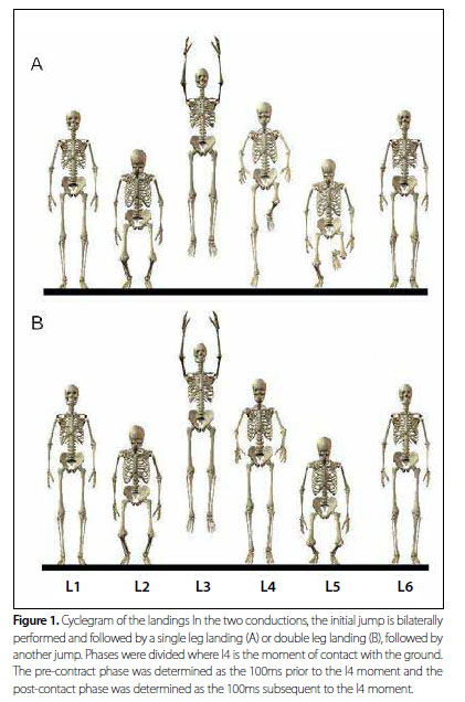
Each individual performed two types of vertical jumps. For each one, the athletes performed the propulsion phase with both lower limbs and landed with one lower limb (single-leg landing) or with the two lower limbs (double-leg landing). Immediately after landing, the athletes performed another jump, in the same condition of the first landing (Figure 1). These jumps were selected, since usually in volleyball the impulsion phase for attack and defense jumps is performed with the two lower limbs, and the landings are performed both with the two and only with one lower limb(6).
Initially, the athletes performed familiarization with the motor conduct used. Subsequently, each athlete performed three landings with one lower limb and three landings with the two lower limbs for the myoelectric activity (EMG). Data collection was performed in the dominant lower limb(7,9). The jumps were randomly performed to minimize the possible fatigue or learning process effects. Randomization was performed through an algorithm programmed in MatLab (The MathWorks, USA). One-minute interval was given between trials.
In order to have the myoelectric activity taken, Ag/AgCl electrodes (KOBME, Bio Protection Corp., Korea) were placed on the rectus femoris, biceps femoris and adductor longus/gracilis, which Will be referred in this work as hip adductors, according to the terminology by Cram et al.(16) for surface bipolar electrodes. The electrodes on the rectus femoris were placed on the Center of the anterior portion of the thigh, approximately on the half of the distance between the knee and the antero superior iliac spine. On the biceps femoris, the electrodes were placed on the lateral region of the thigh, two thirds of distance between the trochanter major and the knee posterior region, and the electrodes of the hip adductors were placed on the medial region of the thigh, in an oblique direction, four centimeters below the pubis(16). The electrodes were placed parallel to the muscle fibers and the interelectrode distance was of two centimeters for all muscles (Figure 2).
Before the application of the electrodes, the skin was prepared by shaving the area and cleansing with alcohol to reduce surface impedance. To prevent movement artifacts in the signals, the electrode cables were fixed to the skin using adhesive tape (3M, Brazil).
The electrical signals of all muscles were collected at acquisition frequency of 2 kHz (EMG 100B, BIOPAC Systems Inc., Santa Barbara, CA, USA), amplified (Differential Bipolar Amplification, input impedance = 2MΩ, common mode rejection ratio > 110 dB, gain = 1,000), converted analog to digital (12bit, MP100WSW BIOPAC Systems Inc.) and stored in a personal computer to be analyzed in the Acknowledge 3.5 software (BIOPAC Systems Inc., Holliston, MA, USA).
The signals were filtered using a fourth-order Butterworth filter, in the direct and reverse direction to avoid phase distortions, with cutoff frequencies of 20Hz and 500Hz. Subsequently, root mean square values RMS were obtained from the filtered signal at every 5ms.
In order to normalize the amplitude of the myoelectric signal, a RMS value of one maximum isometric voluntary contraction (MVIC) with six seconds of duration was used as reference. These six seconds were divided in six windows of one second each and the highest value obtained in these windows was used to normalize data. The MVIC processing was equal to the other signals previously described.
Concerning the rectus femoris, the MVICT was performed through a resisted isometric contraction with knee at 60º of flexion, in a trial to perform one extension, while for the biceps femoris, a resisted isometric contraction with knee at 45º of flexion, was performed in a trial to perform one flexion and for the hip adductors, a resisted isometric contraction with hip at 0º of abduction was performed in a trial to perform one adduction.
To determine the initial ground contact, an electrical circuit was structured so that a terminal located in the sole in the first meta-tarsus region of the subject's shoe emitted a digital electrical signal upon contact with a metal platform fixed the ground that was then captured by the UMI 100B module (BIOPAC Systems, USA).
STATISTICAL ANALYSIS
The myoelectric activity was analyzed with the determination of two phases, through the generated signal in relation to the contact with the ground. The first, named pre-contact phase, was determined as the 100ms prior to the contact with the ground, while the second one, named post-contact phase, was determined by the 100ms after the contact with the ground. The values used in the statistics analysis were the arithmetic means of the normalized RMS values, obtained in the three trials in each phase for each landing. This procedure was adopted as a strategy to reduce the variability of the myoelectric signal.
In order to compare the myoelectric activity between the landings and the pre and post-contact with the ground periods, the two-way ANOVA test with the TYPE OF JUMP (single and double-leg) x JUMP PHASE (pre and post-contact) factors with the Bonferroni post hoc test were used. Significant level of 5% was used in this study. The statistics analyses used were performed with the GraphPad Prism software (Version 5.0).
RESULTS
Statistics analysis of the contraction of the rectus femoris muscle revealed significant differences for the jump phase factor (F1,.8 = 121.6, p < 0.0001), but not for the type of jump factor (F1,.8 = 1.56, p = 0.2212). The Bonferroni post hoc test confirmed the differences, demonstrating higher values for the post-contact phase compared to the pre-contact phase both for the single-leg landing (difference = 28.89, p < 0.001) and the double-leg landing (difference= 29.29, p < 0.001). The biceps femoris presented differences only in the type of jump factor (F1,28 = 9,23, p = 0,0051), but not for the jump phase factor (F1.28 = 1.49, p = 0.2321). The Bonferroni post hoc test confirmed the differences, presenting higher values for the single-leg landing than in the double-leg landing in the pre-contact phase (difference = 11.35, p < 0.05) and in the post-contact phase (difference = 9.33, p < 0.05).
The hip adductors demonstrated significant differences for the jump phase factor (F1.28 = 13.55, p = 0.0013), but not for the type of jump factor (F1.28 = 0.01, p = 0.9233). The Bonferroni post hoc test revealed differences between phases in the single-leg landing (difference = 8.67, p < 0.01), but not for the double-leg landing (difference = 3.47, p > 0.05).
Likewise for the rectus femoris and hip adductors, the ANOVA test revealed significant differences in the jump phase factor (F1.28 = 13.01, p = 0.0012) for the BF/RF ratio. There were not differences in the type of jump factor (F1.28 = 0.15, p = 0.7037). The Bonferroni test identified differences between the two phases both in the single-leg (difference = 0.69, p < 0.05) and double-leg landing (difference = 0.74, p < 0.05).
The means, standard errors and variation coefficients of the EMG data are described in table 1.
DISCUSSION
In this study, the electric activation of hip and knee muscles in two different landing types before and after contact with the ground was compared. Our results suggest that each muscle assessed presents a specific role in the landing control.
The rectus femoris presented increase in its activation in the pre-contact phase for the post-contact phase, as reported in other studies for other quadriceps muscles(17,18) and for the rectus femoris itself (9,18). These findings seem to be related to the knee joint function, more specifically through the eccentric contraction of the quadriceps in absorbing the energy generated by the forces ground reaction(19) and, consequently, stop the movement. Thus, after contact with the ground, increase in the myoelectric activity was expected.
However, an intriguing finding of out study was the lack of differences between the activity of this muscle in the two types of landing. Studies have demonstrated that the ground reaction forces and the energy generated during the landing with one lower limb are higher than with two lower limbs(1). Thus, it is expected that the quadriceps activity is higher in the former than in the latter type of landing. The lack of statistical differences in this study may be related to different heights reached by the sample used in this study in the vertical jump in each landing situation presented by Leporace et al.(20). Our population reached higher vertical dislocation in the jump with subsequent double-leg landing. Therefore, the potential energy generated in the jump in the single-leg landing was lower, which means lower activity of the rectus femoris to stop the movement.
The biceps femoris presented activation strategies different from those of the rectus femoris. Although the statistics analysis has revealed differences between the two landing types in each phase, the activation of this musculature remained with no statistical differences during the pre and post-contact phases in the two conducts. This maintenance of the activation leads us to believe in pre-set strategies based on previous experiences(21), especially under the perspective that the activation of this musculature is able to reduce stress on the ACL(22,23) through increase of stiffness on the knee(24,25), reduction of anterior tibial shear force on the femur (26,27) and of the knee internal rotation(28). These findings corroborate to some studies in the literature which presented tendency of maintenance of the activity of the biceps femoris in men before and after contact with the ground,(18).
However, data analysis of the female population in the study by Hanson et al.(18) and Tillman et al.(6) suggest that the gender may be an important variable concerning the activation of this musculature, constituting in a potential factor for injury in the ACL in women(10).
Besides gender, other factors which can have influenced on these differences between studies, such as training status(29), the sport practiced by the tested individuals(30) and the motor conducts used in the test, since to some studies used running with change of direction(18,31) and other different jump and landing types(1,7,17,29). However, even if all variables were controlled, there could have been discrepancy in the results of the studies in the literature, since the study by Fagenbaum and Darling(17) demonstrated high variability of the activation of the biceps femoris between trials, corroborated in the present study through the high variation coefficient (table 1), which generates the hypothesis of different strategies of motor coordination based on the absence of global strategies, making the individual strategies necessary (32).
The results of the BF/RF ratio corroborate the discussed findings. Excessive quadriceps activation with no sufficient activation of the hamstrings muscles increases the tension in the ACL(11), constituting in a possible risk factor for injuries in this ligament(23). Decrease in the values after landing demonstrates increase of activation of the rectus femoris with more magnitude than presented by the biceps femoris, which is according to the literature(9,18). Nevertheless, an important factor was the absence of differences between the two types of landing. Concerning gender comparison, the studies demonstrated that women present lower BF/RF ratio after contact with the ground(9,17,18), which is usually called quadriceps dominance. However, in the present study, differences between the two types of landing have not been found, which suggests that in order to attenuate the articular stress men present different strategies of muscular activation to keep the BF/RF ratio constant even between motor conducts with different mechanical stress. These findings should be carefully interpreted, though, since the maximum vertical dislocation reached was not controlled, which may interfere in the results.
The results of the hip adductors demonstrated absence of statistical differences between the two types of landing within each phase, despite the fact that in the single-leg landing after contact with the ground there is greater activity compared to the activity in the pre-contact phase. The increase in the activity of this group after contact with the ground in this type of landing seems to be related to the function of the adductors in aiding in the maintenance of the suitable positioning as well as of the pelvis stability during motor conducts which involve some specific instability levels(33). From this point of view, the absence of statistical differences in the double-leg landing may be explained by higher stability of this conduct, since contact with the ground with the two lower limbs leads to more suitable symmetry and greater balance condition than in the single-leg landing.
Hewett et al.(34) suggested that decrease in the knee adduction and abduction torques after plyometric training would be related to alterations in the muscular behavior of the lower extremities on the frontal plane. Concerning the importance of the muscles which act in this hip joint, Olmstead et al.(35) found that the tensor fasciae latae (hip abdutor) synergically acts with the quadriceps during the knee extension,while the gracillis (hip adductor) synergically acts with the semitendinosus and semimembranosus during the knee flexion, indicating that the hip adductor and abductor musculatures may have direct influence on the maintenance of the dynamic stability of the knee.
Our study presents some limitations. Firstly, the tests were performed in laboratory environment. Although this situation contributes to the control of intervenient variables during the tests, it does not necessarily represent which occurs during the sports practice, due to several variables which are present in the game situations, which are controlled in laboratory situations, such as the ball, opponents, fatigue, attention to the movement, among others. It seems that these variables may influence on the injury mechanism in sports(3,36). Another aspect not controlled was the lack of standardization between the peak of vertical dislocation performed in the jumps. This aspect undoubtedly influences on the muscle activation(37). However, the aim of this study was to compare motor techniques present during the sports practice, in the case of this study, volleyball(6). Leporace et al.(20) showed that the vertical dislocation between the two conducts was different for the population in the present study, which possibly justifies some findings.
CONCLUSION
The results suggest that each muscle tested presents a different role during the lading phase in men. While the rectus femoris presents as main role the deceleration of the knee articulation and the descending movement, characterized by the increase in the activation in the post-contact phase, the biceps femoris seems to attenuate the articular tension of the knee in activities of greater impact, remaining more active during the entire single-leg landing cycle. Higher activation of the hip adductors after contact with the ground in the single-leg landing justifies the importance of the lumbopelvic region in the pelvic stabilization in situations of great instability. Further investigation is necessary to determine the effects of the muscle activation presented in the potentially harmful mechanical loads imposed to the knee in male athletes.
REFERENCES
- 1. Tillman MD, Hass CJ, Brunt D, Bennett GR. Jumping and landing techniques in elite women's volleyball. J Sports Sci Med 2004;3:30-6.
- 2. Kovacs I, Tihanyi J, DeVita P, Rácz L, Barrier J, Hortobágyi T. Foot placement modifies kinematics and kinetics during drop jumping. Med Sci Sport Exerc 1999;31:708-71.
- 3. Brito J, Soares J, Rebelo AN. Prevenção de lesões do Ligamento Cruzado Anterior em Futebolistas. Rev Bras Med Esp 2009;15:62-9.
- 4. Solomonow M, Krogsgaard M. Sensory-motor control of knee stability. Scand J Med Sci Sport 2001;11:64-80.
- 5. Butler RJ, Crowell HP, Davis IM. Lower extremity stiffness: implications for performance and injury. Clin Biomech 2003;18:511-7.
- 6. Tillman MD, Criss RM, Brunt D, Hass CJ. Landing constraints influence ground reaction forces and lower extremity EMG in female volleyball players. J Appl Biomech 2004;20:36-50.
- 7. Pappas E, Hagins E, Sheikhzadeh A, Nordin M, Rose D. Biomechanical differences between unilateral and bilateral landings from a jump: gender differences. Clin J Sport Med 2007;17:263-8.
- 8. Hewett TE, Zazulak BT, Myer GD, Ford KR. A review of electromyographic activation levels, timing differences and increased anterior cruciate ligament injury incidence in female athletes. Br J Sports Med 2005;39:347-50.
- 9. Nagano Y, Ida H, Akai M, Fukubayashi T. Gender differences in knee kinematics and muscle activity during single limb drop landing. Knee 2007;14:218-23.
- 10. Zebis MK, Andersen LL, Bencke J, Kjaer M, Aagaard P. Identification of Athletes at Future Risk of Anterior Cruciate Ligament Ruptures by Neuromuscular Screening. Am J Sports Med 2009;37:1967-73.
- 11. Markolf K, O'Neil G, Jackson S. Effects of applied quadriceps and hamstrings muscle loads on forces in the anterior and posterior cruciate ligaments. Am J Sports Med 2004;32:1144-9.
- 12. Quatman CE, Hewett TE. The anterior cruciate ligament injury controversy: is "valgus collapse" a sex-specific mechanism? Br J Sports Med 2009;43:328-35.
- 13. Zazulak BT, Hewett TE, Reeves NP, Goldberg B, Cholewicki J. Deficits in neuromuscular control of the trunk predict knee injury risk: a prospective biomechanical-epidemiologic study. Am J Sports Med 2007;35:1123-30.
- 14. Myer GD, Chu DA, Brent JL, Hewett TE. Trunk and hip control neuromuscular training for the prevention of knee joint injury. Clin Sports Med 2008;27:425-48.
- 15. Leetun DT, Ireland ML, Willson JD, Ballantyne BT, Davis IM. Core stability measures as risk factors for lower extremity injury in athletes. Med Sci Sports Exerc 2004;36:926-34.
- 16. Cram J, Kasman G, Holtz J. Introduction to surface electromyography. Gaithersburg: Aspen Publishers, 1998.
- 17. Fagenbaum R, Darling WG. Jump landing strategies in male and female college athletes and the implications of such strategies for Anterior Cruciate Ligament Injury. Am J Sports Med 2003;31:233-40.
- 18. Hanson AM, Padua DA, Blackburn JT, Prentice WE, Hirth CJ. Muscle activation during side-step cutting maneuvers in male and female soccer athletes. J Athl Train. 2008;43:133-43.
- 19. Elmer SJ, Madigan ML, LaStayo PC, Martin JC. Joint-specific power absorption during eccentric cycling. Clin Biomech. 2010;25:154-8.
- 20. Leporace G, Praxedes J, Fonseca R, Chagas D, Brandão Junior JD, Rodrigues C, Pereira GR, Batista LA. Diferenças na cinemática entre dois tipos de aterrissagens em atletas de voleibol masculinos. Rev Bras Cineantropom Desempenho Hum 2010;12:464-70.
- 21. Bastian AJ. Learning to predict the future: the cerebellum adapts feedfoward movement control. Curr Opin Neurobiol 2006;16:645-9.
- 22. Pandy MG, Shelbourne KB. Dependence of cruciate-ligament loading on muscles forces and external load. J Biomech 1997;30:1015-24.
- 23. Renstrom P, Arms SW, Stanwyck TS, Johnson RJ, Pope MH. Strain within the anterior cruciate ligament during hamstrings and quadriceps activity. Am J Sports Med. 1986;14:83-7.
- 24. Louie JK, Mote CD Jr. Contribution of the musculature to rotator laxity and torsional stiffness at the knee. J Biomech 1987;20:281-300.
- 25. Markolf KL, Graff-Radford A, Amstutz HC. In vivo knee stability. A quantitative assessment using an instrumented clinical testing apparatus. J Bone J Surg Am 1978;60:664-74.
- 26. Imran A, O'Conner J. Control of knee stability after ACL injury or repair: interaction between hamstrings contraction and tibial translation. Clin Biomech 1998;13:153-62.
- 27. McNair P, Marshall R. Loading characteristics in subjects with normal and anterior cruciate ligament deficient knee joints. Arch Phys Med Rehabil 1994;75:584-9.
- 28. MacWilliams B, Wilson D, DesJardins J, Romero J, Chao EY. Hamstrings co-contraction reduces internal rotation, anterior translation and anterior cruciate ligament loading in weightbearing flexion. J Orthop Res 1999;17:817-22.
- 29. Medina JM, McLeod TCV, Howell SK, Kingma JJ. Timing of neuromuscular activation of the quadriceps and hamstrings prior to landing in high school male athletes, female athletes and female non-athletes. J Electromyo Kinesiol 2008;18:591-7.
- 30. Herrington L. Knee valgus angle during landing tasks in female volleyball and basketball players. J Strength Cond Res. 2009. Published Ahead of Print, 4 Dez. doi: 10.1519/JSC.0b013e3181b62c77.
- 31. Beaulieu ML, Lamontagne M, Xu L. Gender differences in Time-Frequency EMG analysis of unanticipated cutting maneuvers. Med Sci Sports Exerc 2008;40:1795-804.
- 32. Latash, M. Synergy. Oxford University Press, New York. 2008.
- 33. Chimera NJ, Swanik NJ, Swanik KA, Straub SJ. Effects of plyometric training on muscle-activation strategies and performance in females athletes. J Athl Train 2004;39:24-31.
- 34. Hewett TE, Stroupe AL, Nance TA, Noyes FR. Plyometric training in female athletes. Am J Sports Med 1996;24:765-73.
- 35. Olmstead TG, Wevers HW, Bryant JT, Gouw GJ. Effect of muscular activity on valgus/varus laxity and stiffness of the knee. J Biomech 1986;19:565-77.
- 36. Bahr R, Krosshaug T. Understanding injury mechanisms: a key component of preventing injuries in sport. Br J Sports Med 2005;39:324-9.
- 37. Arampatzis A, Morey-Klapsing G, Brüggemann GP. The effect of falling height on muscle activity and foot motion during landings. J Electromyogr Kinesiol 2003;13:533-44.
Activation of hip and knee muscles during two landing tasks performed by male volleyball athletes
Publication Dates
-
Publication in this collection
20 Jan 2012 -
Date of issue
Oct 2011