Abstract
Introduction:
sepsis is a serious public health problem, affecting millions of people in the world each year, with a high mortality rate (one out of four patients) and an increasing incidence. Sepsis is one of the main causes of maternal mortality and an important cause of admission to obstetric intensive care units.
Case description:
In this study, the authors report the case of a woman having been submitted to cesarean section three days before presenting clinical signs of sepsis and septic shock caused by a liver abscess. The patient had a set of complications secondary to shock, such as thrombocytopenia, coagulopathy, toe ischemia and acute kidney failure. The patient had cholelithiasis and recurrent pain in the right hypochondrium during pregnancy. During hospitalization, the mechanism involved in the development of hepatic abscess was infection of the biliary tract. The patient was treated in an obstetric intensive care unit with antibiotics and drainage of the liver abscess. Progress was favorable and the patient was discharged in good health.
Discussion:
pyogenic liver abscess during pregnancy and puerperium is a serious condition which represents a diagnostic and therapeutic challenge, with few cases reported. The normally nonspecific clinical and laboratory findings can lead to a late diagnosis, which increases the risk of maternal morbidity and mortality.
Key words
Liver abscess; Sepsis; Puerperium; Cholecystitis; Percutaneous drainage
Resumo
Introdução:
a sepse é um problema grave de saúde pública, afetando milhões de pessoas no mundo a cada ano. Apresenta uma alta mortalidade, um em quatro doentes, e vem aumentando sua incidência. É uma das principais causas de mortalidade materna, sendo uma causa importante de admissões emunidades de terapia intensiva obstétrica.
Descrição do caso:
puérpera, no terceiro dia pós-cesariana, apresentou quadro clinico de sepse e choque séptico secundário a um abscesso hepático piogênico.Evoluiu com complicações secundárias ao choque: plaquetopenia, coagulopatia, isquemia de pododáctilos e insuficiência renal aguda. A paciente era portadora de colelitíase e apresentou repetidas crises álgicas em hipocôndrio direitodurante a gestação.Durante o internamento, ficou definido que a causa do abscesso hepático foi a infecção das vias biliares.Realizou-se tratamento em unidade de terapia intensiva obstétrica, tendo a paciente sido submetida à drenagem do abscesso hepático e antibioticoterapia. A evolução foi satisfatória e a paciente obteve alta em boas condições.
Discussão:
o abscesso hepático piogênico durante a gravidez e o puerpério é uma condição extremamente grave, com poucos casos relatados, representando um desafio diagnóstico e terapêutico. Como os achados clínicos e laboratoriais são usualmente inespecíficos, um diagnóstico retardado é possível, aumentando os riscos de morbidade e mortalidade materna em casos não tratados precocemente.
Palavras-chave
Abscesso hepático; Sepse; Puerpério; Colecistite; Drenagem percutânea
Introduction
Sepsis is a serious public health problem affecting millions of individuals throughout the world each year. The mortality rate is high (one out of every four patients) and the incidence has been increasing.11 Rhodes A, Evans L, Alhazzani W, Levy MM, Antonelli M, Ferrer R, et al. Surviving sepsis campaign: international-guidelines for management of severe sepsisand septic shock, 2016. Crit Care Med. 2017; 45 (3): 486-552. Estimates indicate approximately 600 thousand new cases of sepsis each year in Brazil, with an approximate cost of US$ 9.6 thousand per patient.22 Brasil. Instituto Latino-americano de Sepse. Sepse: um problema de saúdepública / Instituto Latino-americano de Sepse. Brasília, DF; 2015. p. 1-90. This is a life-threatening organic dysfunction caused by a harmful systemic response secondary to infection. Septic shock is characterized by the occurrence of metabolic, cellular and circulator dysfunction and is associated with a high risk of mortality.11 Rhodes A, Evans L, Alhazzani W, Levy MM, Antonelli M, Ferrer R, et al. Surviving sepsis campaign: international-guidelines for management of severe sepsisand septic shock, 2016. Crit Care Med. 2017; 45 (3): 486-552.
Sepsis is one of the main causes of mortality among pregnant women and a major cause of admission to obstetric intensive care unit.33 Creanga A, Berg C, Syverson C, Seed K, Bruce F, Callaghan W. Pregnancy-related mortality in the United States, 2006-2010. Obstet Gynecol. 2015; 125 (1): 5-12. The prevalence ranges from 0.001% to 0.02% of all child births and is related to infections of an obstetric origin, such as infected abortion, chorioamnionitis and puerperal infection, or a non-obstetric origin, such as the result of primary infections affecting other sites.44 Louis J, Menard M, Gee R.Racial and ethnic disparities in maternal morbidity and mortality.Obstet Gynecol. 2015;125(3):690-4. doi: 10.1097/A0G.0000000000000704.
https://doi.org/10.1097/A0G.000000000000...
The mortality rate among women with septic shock in the pregnancy-puerperium phase can reach 28%.55 Barton J, Sibai B. Severe sepsis and septic shock in pregnancy. Obstet Gynecol 2012; 120: 689. However, some factors contribute to a better prognosis in obstetrics, such as the fact that pelvis is the most common infection site, which is a region in which surgical intervention can be performed and there is greater sensitivity among the main microorganisms to broad-spectrum antibiotic therapy. Moreover, pregnant women are usually younger and have fewer comorbidities, which improves the prognosis of such patients.55 Barton J, Sibai B. Severe sepsis and septic shock in pregnancy. Obstet Gynecol 2012; 120: 689.
Pyogenic liver abscesses are the most common types of visceral abscesses. The annual incidence is estimated at around two cases per 100,000 patients, with a more frequent occurrence in men than women. Risk factors include diabetes, underlying pancreatic or hepatobiliary disease and liver transplant.66 Yüksel B, Seven A, Kucur S, Gozükara I, Keskin N. Presentation and management of pyogenic liver abscess in a 23-week pregnant woman.Case Rep Obstet Gynecol. 2013; 2013: 845215. Such abscesses commonly occur due to direct dissemination caused by peritonitis or infection of the bile ducts or, indirectly, secondary to hematogenic dissemination. An underlying bile duct condition, such as gallstones or malignant obstruction, is found in 40 to 60% of cases of liver abscesses.66 Yüksel B, Seven A, Kucur S, Gozükara I, Keskin N. Presentation and management of pyogenic liver abscess in a 23-week pregnant woman.Case Rep Obstet Gynecol. 2013; 2013: 845215.
Many pathogens have been reported and this variability reflects different etiologies, types of medical interventions and geographic differences. Typical clinical manifestations are fever (approximately 90% of patients) and abdominal pain (50 to 75% of patients).77 Rahimian J, Wilson T, Oram V, Holzman R. Pyogenic liver abscess: recent trends in etiology and mortality.Clin Infect Dis. 2004; 39 (11): 1654-9. Signs and symptoms are usually located in the upper right quadrant and may include pain upon palpation and signs of peritoneal irritation. Around half of patients have hepatomegaly.77 Rahimian J, Wilson T, Oram V, Holzman R. Pyogenic liver abscess: recent trends in etiology and mortality.Clin Infect Dis. 2004; 39 (11): 1654-9.
Pyogenic liver abscess in pregnancy and puerperium is an extremely serious and rare complication that should be considered in the occurrence of fever accompanied by upper abdominal pain and hepatomegaly.
The case report described herein received approval from the human research ethics committee of Instituto de Medicina Integral Prof. Fernando Figueira (IMIP) (certificate number: 48398715.6.0000.5201) and was described only after the patient signed a statement of informed consent.
Case Report
A 26-year-old white woman with a stable partner and an incomplete high school education, resident of the municipality of Glória do Goitá in the state of Pernambuco (PE), Brazil, was in her third pregnancy, having had two previous cesarean sections. She underwent prenatal care at a private healthcare service in the city of Vitória de Santo Antão, PE. She had chronic hypertension and took α-methyldopa (1g/day). During the pregnancy, the patient experienced recurrent upper abdominal pains stemming from cholelithiasis, which had been diagnosed by ultrasound prior to the pregnancy.
The patient was submitted to elective cesarean section at a maternity in the municipality of Vitória de Santo Antão, PE in the 39th week of pregnancy due to repeated episodes of pain in the upper abdomen secondary to the cholelithiasis and refractory to analgesic medications. The newborn was a healthy male weighing 2,830 g, with Apgar scores of 8 and 9 in the first and fifth minutes, respectively. The mother, who continued to experience pain even with the use of analgesic medication, and the infant received discharge from hospital 48 hours after cesarean section.
On the third day of puerperium, the patient presented important dyspnea and was treated in another hospital located in the city of Jaboatão dos Guararapes, PE. The patient presented a worsening of the dyspnea as well as hypotension and oliguria, requiring intubation for mechanical ventilation and the use of vasoactive drugs. Investigating the possibility of pulmonary thromboembolism, computed tomography of the thorax was performed, which suggested thrombosis at the base of the right lung.
The patient was transferred to the obstetric intensive care unit of the Instituto de Medicina Integral Prof. Fernando Figueira (IMIP) in the city of Recife, PE, still on the third day after the cesarean section, presenting a poor general state, anasarca, hypotension, tachycardia, fever spikes and toe ischemia perceptible during the physical examination. The diagnostic hypotheses were pulmonary thromboembolism, disseminated intravascular coagulation, HELLP syndrome and toe ischemia, which were the reasons for referral to the reference unit. At IMIP, the diagnostic hypotheses of sepsis and acute kidney failure were added. The following blood findings were recorded upon admission to IMIP: blood type = A; Rh factor = negative; rapid anti-HIV test = non-reactive; VDRL = non-reactive; urea = 238 mg/dL; creatinine = 5.2 mg/dL; hemoglobin = 9.2 g/dL; hematocrit = 26.9%; leukocytes = 20,200/≠L (band cells = 1%, segmented cells = 86%); platelets = 17,000/≠L; INR = 2.09; fibrinogen = 349.7 mg/dL; sodium = 135 mmol/L; potassium = 4.9 mmol/L; lactic lactate dehydrogenase= 544 u/L; aspartate aminotransferase = 183 u/L; alanine aminotransferase = 156 u/L; alkaline phosphatase= 247 u/L; gamma-glutamyltransferase = 65 u/L; total bilirubin = 3.6 mg/dL; direct bilirubin = 3.2 mg/dL; and indirect bilirubin = 0.4 mg/dL.
Antibiotic therapy was initiated with piperacillin/tazobactam 4.5 g every six hours. Fresh plasma was prescribed. A nephrology exam and ultrasound of the abdomen were requested. On the same day, the nephrology team determined that dialysis was not necessary.
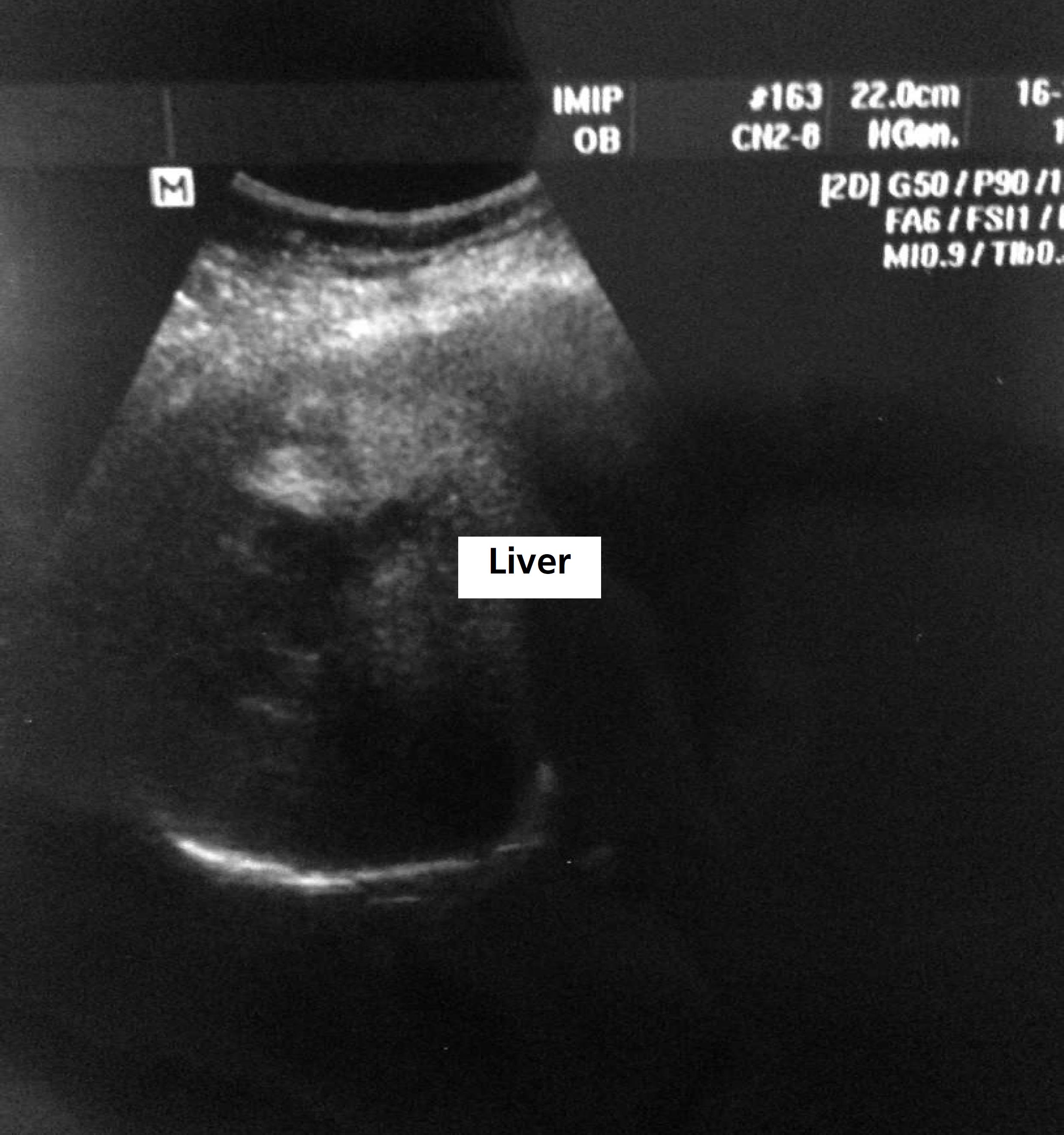
On the sixth day of puerperium, the patient remained on mechanical ventilation with a fever, tachycardia and a worsening of kidney function (urea = 505 mg/dL; creatinine = 6.0 mg/dL) and daily dialysis was indicated. The results of the blood cultures were positive in two samples for a Gram negative bacterium (Citrobacter freundii) sensitive to piperacillin and tazobactam and the decision was made to maintain the antibiotic therapy. As the platelet count began to drop again (7000/≠l), 10 units of platelet concentrate were prescribed. The following day, the vascular surgeon visited the patient and advised maintaining heparin, which had been suspended, and scheduling the placement of an inferior vena cava filter. Abdomen ultrasound revealed an image of low echogenicity with irregular contours and relatively imprecise limits, measuring approximately 5.8 x 2.6 cm, with no evidence of flow, located in the projection of the right lobe of the liver (Figure 1).
Computed tomography (CT) of the abdomen confirmed a liver abscess measuring 9.4 x 7.9 x 7.3 cm, occupying segments VII and VIII. The CT image also revealed that the main right branch of the portal vein and its peripheral segments were not opacified in post-contrast sequences and had hypoechoic content in the interior, suggesting the possibility of thrombosis as a secondary complication.
The general surgeon suggested puncturing the abscess, but this procedure was contraindicated at the time due to the patient's coagulation disorder. After ten days of puerperium, an improvement occurred in the clinical status, but the patient began to have blood pressure spikes and anti-hypertensive therapy was optimized. On the 11th day of puerperium, sedation was suspended for the daily arousal and the patient was extubated. As improvements occurred in the clinical and laboratory variables, the decision made was to use a Venturi mask at 50%, which was well adapted. Improvements occurred in the laboratory findings, thrombocytopenia (151,000/≠l) and kidney function (urea = 86 mg/dL and creatinine = 1.3 mg/dL). Dialysis was suspended on the 12th day of puerperium.
The liver abscess was drained on the 18th day of puerperium, with no complications. The patient was discharged from obstetric intensive care on the 21st day after the cesarean section. On the fifth day after drainage of the liver abscess, with the resolution of sepsis and acute kidney failure, bed sores on the back and intergluteal region, toe ischemia and chronic arterial hypertension, the patient was in good clinical condition and was sent for rooming-in taking piperacillin/tazobactam (4.5 g every six hours), clonidine (0.3 mg/day), nifedipine (30 mg/day) and propranolol (80 mg/day).
During rooming-in, debridement was performed on the bed sore on the back and Dersani bandages were placed. The ischemic toes were monitored and amputation was not required. The CT of the abdomen on the 31st day of puerperium for the assessment of the regression of the liver abscess revealed a large area of plegmon in the right lobe (Figure 2). As the patient was stable, with normalized white blood cell counts and a reduction in drainage output, the drain was removed and the patient was scheduled to undergo another CT of the abdomen after 30 days as an outpatient.
The patient was discharged from hospital on the 34th day of puerperium in a good general state after having undergone 31 days of piperacillin/tazobactam and was oriented to undergo outpatient follow up, during which she reported episodes of pain in the upper abdomen and occasional fever. CT of the abdomen revealed that dimensions of the liver were preserved, with an area of attenuation with calcic points in the periphery of segment VII, which could be a consequence of the drained abscess. Ultrasound of the abdomen revealed a scleroatrophic gallbladder filled with calculus. The patient was submitted to cholecystectomy at the municipal hospital in Vitória, PE, and is currently fine and without complaints.
Discussion
In the present case, the clinical condition of a woman in the puerperium deteriorated and the diagnosis of liver abscess was delayed, which led to significant morbidity. However, treatment led to a successful outcome. The delay in the diagnosis of such a case may be explained by the extremely rare condition. Although fever and pain in the upper right abdomen are common symptoms, the clinical presentation is nonspecific in many cases and diagnosis becomes a challenge. Thus, liver abscess is generally only investigated when patients have a clinical case of severe sepsis.66 Yüksel B, Seven A, Kucur S, Gozükara I, Keskin N. Presentation and management of pyogenic liver abscess in a 23-week pregnant woman.Case Rep Obstet Gynecol. 2013; 2013: 845215.
Liver abscesses commonly occur due to direct dissemination caused by peritonitis or bile duct infection or, indirectly, secondary to hematogenic dissemination.77 Rahimian J, Wilson T, Oram V, Holzman R. Pyogenic liver abscess: recent trends in etiology and mortality.Clin Infect Dis. 2004; 39 (11): 1654-9. In the present case, the mechanism involved in the development of liver abscess was a bile duct infection, as the patient had cholelithiasis, with recurring episodes of pain in the gallbladder during pregnancy.
Different microorganisms are associated with liver abscess, such as amoebiasis, Listeria monocytogenes, Brucellosis, Staphylococcus aureus, Escherichia coli and polymicrobial infections.88 Sherer D, Dalloul M, Shah T, Walsh A, Sokolovski M, Zinn H, Serur E, Abulafia O. Sonography and magnetic resonance imaging of a maternal methicillin-resistant Staphylococcus aureus liver abscess at 33 weeks' gestation. J Ultrasound Med. 2010;29(6):989-92. The bacterium found in two blood culture samples was Citrobacter freundii, which is sensitive to piperacillin and tazobactam. No reference to this Gram-negative agent was found in the literature on liver abscess.
Like the clinical symptoms, laboratory tests are also nonspecific for the diagnosis of liver abscess. The most frequent findings are leukocytosis, increased alkaline phosphatase and hyperfibrinogenemia, which are nonspecific during pregnancy and puerperium.66 Yüksel B, Seven A, Kucur S, Gozükara I, Keskin N. Presentation and management of pyogenic liver abscess in a 23-week pregnant woman.Case Rep Obstet Gynecol. 2013; 2013: 845215. Leukocytosis only merits attention during pregnancy and puerperium when above 25,000/≠L or in the occurrence of left shift.99 Souza A, Filho M, Ferreira L. Hematological changes and pregnancy. Rev Bras Hematol Hemoter. 2002; 24 (1): 2936.
The present case demonstrates the importance of abdominal imaging exams in postpartum women who have unusual symptoms, such as pain in the upper right quadrant associated with known conditions, such as cholelithiasis and acute cholecystitis. Ultrasound, in particular, is a low-cost exam with high sensitivity for the diagnosis of pyogenic liver abscess (approximately 85%).66 Yüksel B, Seven A, Kucur S, Gozükara I, Keskin N. Presentation and management of pyogenic liver abscess in a 23-week pregnant woman.Case Rep Obstet Gynecol. 2013; 2013: 845215.
Treatment for pyogenic liver abscess consists of percutaneous drainage and the use of antibiotics for two to six weeks.88 Sherer D, Dalloul M, Shah T, Walsh A, Sokolovski M, Zinn H, Serur E, Abulafia O. Sonography and magnetic resonance imaging of a maternal methicillin-resistant Staphylococcus aureus liver abscess at 33 weeks' gestation. J Ultrasound Med. 2010;29(6):989-92. Rapid diagnosis and treatment are vital due to the high mortality rate described in the literature, which can reach 100% in untreated cases. Mortality rates of 28% are reported for patients treated solely with antimicrobial agents versus 4% among patients retreated with drainage and antimicrobial agents.88 Sherer D, Dalloul M, Shah T, Walsh A, Sokolovski M, Zinn H, Serur E, Abulafia O. Sonography and magnetic resonance imaging of a maternal methicillin-resistant Staphylococcus aureus liver abscess at 33 weeks' gestation. J Ultrasound Med. 2010;29(6):989-92. In the present case, drainage was delayed due to a coagulation disorder, but was performed at the earliest possible time.
The diagnosis of HELLP syndrome raised initially was based on the findings of thrombocytopenia and increased transaminases in a patient in the immediate postpartum period with chronic hypertension. However, this diagnosis was discarded due to the increase in total bilirubin in detriment to direct bilirubin, which did not characterize hemolysis. Moreover, septic shock explained thrombocytopenia and the increase in transaminases.
The patient exhibited portal vein thrombosis. The diagnosis of this condition was incidental and thrombosis was likely the result of the hepatobiliary infectious process, although the possibility of septic thrombosis cannot be discarded. Indeed, the patient had risk factors for portal vein thrombosis, such as cholecystitis, liver abscess and puerperal period itself, in which the risk of thromboembolism increases more than 20 fold compared to women who are not pregnant or who are not in postpartum period.1010 Jackson E, Curtis K, Gaffield M. Risk of venous thromboembolism during the postpartum period: a systematic review. Obstet Gynecol. 2011; 117 (3): 691-703.
The favorable outcome in the present study is believed to be the result of the timely diagnosis of sepsis/septic shock and prompt care in the intensive care unit, with treatment initiated within the golden hours.
References
-
1Rhodes A, Evans L, Alhazzani W, Levy MM, Antonelli M, Ferrer R, et al. Surviving sepsis campaign: international-guidelines for management of severe sepsisand septic shock, 2016. Crit Care Med. 2017; 45 (3): 486-552.
-
2Brasil. Instituto Latino-americano de Sepse. Sepse: um problema de saúdepública / Instituto Latino-americano de Sepse. Brasília, DF; 2015. p. 1-90.
-
3Creanga A, Berg C, Syverson C, Seed K, Bruce F, Callaghan W. Pregnancy-related mortality in the United States, 2006-2010. Obstet Gynecol. 2015; 125 (1): 5-12.
-
4Louis J, Menard M, Gee R.Racial and ethnic disparities in maternal morbidity and mortality.Obstet Gynecol. 2015;125(3):690-4. doi: 10.1097/A0G.0000000000000704.
» https://doi.org/10.1097/A0G.0000000000000704. -
5Barton J, Sibai B. Severe sepsis and septic shock in pregnancy. Obstet Gynecol 2012; 120: 689.
-
6Yüksel B, Seven A, Kucur S, Gozükara I, Keskin N. Presentation and management of pyogenic liver abscess in a 23-week pregnant woman.Case Rep Obstet Gynecol. 2013; 2013: 845215.
-
7Rahimian J, Wilson T, Oram V, Holzman R. Pyogenic liver abscess: recent trends in etiology and mortality.Clin Infect Dis. 2004; 39 (11): 1654-9.
-
8Sherer D, Dalloul M, Shah T, Walsh A, Sokolovski M, Zinn H, Serur E, Abulafia O. Sonography and magnetic resonance imaging of a maternal methicillin-resistant Staphylococcus aureus liver abscess at 33 weeks' gestation. J Ultrasound Med. 2010;29(6):989-92.
-
9Souza A, Filho M, Ferreira L. Hematological changes and pregnancy. Rev Bras Hematol Hemoter. 2002; 24 (1): 2936.
-
10Jackson E, Curtis K, Gaffield M. Risk of venous thromboembolism during the postpartum period: a systematic review. Obstet Gynecol. 2011; 117 (3): 691-703.
Publication Dates
-
Publication in this collection
Oct-Dec 2017
History
-
Received
03 Apr 2017 -
Reviewed
23 Sept 2017 -
Accepted
05 Oct 2017