Abstracts
The present work studied the prevalence and histopathology of Neoechinorhynchus curemai Noronha, 1973 (Acanthocephala: Neoechinorhynchidae) from curimbatá, Prochilodus lineatus Valenciennes, 1836. Eighteen fishes with averages of 46.7 + 1.1 cm length and 1,674.8 + 75.6 g weight were collected with net, bimonthly from December 1995 thru December 1996 in the hydroelectric power station of Volta Grande Reservoir (Cemig), Minas Gerais, Brazil. From analysed fishes, 15 were infected with acanthocephalans in the intestine (prevalence 83.3%). The greatest mean intensity occurred in August 1996 with 66.5 (16 to 208) parasites. Histopathological analysis showed complete desquamation of the intestinal epithelium with severe hyperplasia and hypertrofia of the goblet cells. Severe inflammatory reaction at the submucosa, displacement of their sheaf, associated with oedema and mononuclear and eosinophilic infiltration were observed.
Acanthocephala; Neoechinorhynchus curemai; histopathology; Prochilodus lineatus; Minas Gerais; Brazil
O presente trabalho estudou a prevalência e a histopatologia de Neoechinorhynchus curemai Noronha, 1973 (Acanthocephala: Neoechinorhynchidae) em curimbatá, Prochilodus lineatus Valenciennes, 1836. Dezoito peixes com comprimento total médio de 46,7 + 1,1 cm e peso médio de 1.674,8 + 75,6 g foram coletados com rede, bimestralmente, de dezembro de 1995 a dezembro de 1996 na usina hidrelétrica do Reservatório de Volta Grande (Cemig), Minas Gerais, Brasil. Dos peixes analisados, 15 estavam infectados com acantocéfalos no intestino (prevalência de 83,3%). A maior intensidade média ocorreu em agosto de 1996, com 66,5 (16 a 208) parasitos. A análise histopatológica revelou completa descamação do epitélio intestinal com severa hiperplasia e hipertrofia das células caliciformes. Observou-se, ainda, forte reação inflamatória na submucosa, deslocamento de feixes, associado a edemas, bem como infiltração mononuclear e eosinofílica.
Acanthocephala; Neoechinorhynchus curemai; histopatologia; Prochilodus lineatus; Minas Gerais; Brasil
PREVALENCE AND HISTOPATHOLOGY OF Neoechinorhynchus curemai NORONHA, 1973 (ACANTHOCEPHALA: NEOECHINORHYNCHIDAE) IN Prochilodus lineatus VALENCIENNES, 1836 FROM VOLTA GRANDE RESERVOIR, MG, BRAZIL
MARTINS, M. L.,1 MORAES, F. R. de,2 FUJIMOTO, R. Y.,1 ONAKA, E. M.1 and QUINTANA, C. I. F.1
1Centro de Aqüicultura, Unesp, Via Prof. Paulo Donato Castellane, s/n, CEP 14870-000, Jaboticabal, SP, Brazil
2Departamento de Patologia Veterinária, FCAV, Unesp, Rod. Carlos Tonanni, km 5, CEP 14870-000, Jaboticabal, SP, Brazil
Correspondence to: Maurício L. Martins, Centro de Aqüicultura, Unesp, Via Prof. Paulo Donato Castellane, s/n, CEP 14870-000, Jaboticabal, SP, Brazil, e-mail: mlaterca@caunesp.unesp.br
Received June 2, 2000 ¾ Accepted August 8, 2000 ¾ Distributed August 31, 2001
(With 5 figures)
ABSTRACT
The present work studied the prevalence and histopathology of Neoechinorhynchus curemai Noronha, 1973 (Acanthocephala: Neoechinorhynchidae) from curimbatá, Prochilodus lineatus Valenciennes, 1836. Eighteen fishes with averages of 46.7 + 1.1 cm length and 1,674.8 + 75.6 g weight were collected with net, bimonthly from December 1995 thru December 1996 in the hydroelectric power station of Volta Grande Reservoir (Cemig), Minas Gerais, Brazil. From analysed fishes, 15 were infected with acanthocephalans in the intestine (prevalence 83.3%). The greatest mean intensity occurred in August 1996 with 66.5 (16 to 208) parasites. Histopathological analysis showed complete desquamation of the intestinal epithelium with severe hyperplasia and hypertrofia of the goblet cells. Severe inflammatory reaction at the submucosa, displacement of their sheaf, associated with oedema and mononuclear and eosinophilic infiltration were observed.
Key words: Acanthocephala, Neoechinorhynchus curemai, histopathology, Prochilodus lineatus, Minas Gerais, Brazil.
RESUMO
Prevalência e histopatologia de Neoechinorhynchus curemai Noronha, 1973 (Acanthocephala: Neoechinorhynchidae) em Prochilodus lineatus Valenciennes, 1836 do Reservatório de Volta Grande, MG, Brasil
O presente trabalho estudou a prevalência e a histopatologia de Neoechinorhynchus curemai Noronha, 1973 (Acanthocephala: Neoechinorhynchidae) em curimbatá, Prochilodus lineatus Valenciennes, 1836. Dezoito peixes com comprimento total médio de 46,7 + 1,1 cm e peso médio de 1.674,8 + 75,6 g foram coletados com rede, bimestralmente, de dezembro de 1995 a dezembro de 1996 na usina hidrelétrica do Reservatório de Volta Grande (Cemig), Minas Gerais, Brasil. Dos peixes analisados, 15 estavam infectados com acantocéfalos no intestino (prevalência de 83,3%). A maior intensidade média ocorreu em agosto de 1996, com 66,5 (16 a 208) parasitos. A análise histopatológica revelou completa descamação do epitélio intestinal com severa hiperplasia e hipertrofia das células caliciformes. Observou-se, ainda, forte reação inflamatória na submucosa, deslocamento de feixes, associado a edemas, bem como infiltração mononuclear e eosinofílica.
Palavras-chave: Acanthocephala, Neoechinorhynchus curemai, histopatologia, Prochilodus lineatus, Minas Gerais, Brasil.
INTRODUCTION
Observations about taxonomy on Neoechinorhynchus curemai Noronha, 1973 were carried out by Noronha (1973, 1984), Kohn et al. (1985) and Martins et al. (2000). Nothing is known relationed to seasonality, prevalence or histopathology of such species. Prevalence or seasonality studies in brazilian freshwater fishes are scarce. Muzzal (1980), Lassiere & Crompton (1988), Amin & Vignieri (1986) and Amin (1986) related prevalence and seasonality of acanthocephalan. Recently, in Brazil, Brasil-Sato & Pavanelli (1999) studied an ecological and reproductive aspects of N. pimelodi from Pimelodus maculatus of the São Francisco River, Minas Gerais. From histopathology, Esch & Huffines (1973) related reduced villus size by mucosal erosion, fibrosis and eosinophilic infiltration in smallmouth bass, Micropterus dolomieui infected with the acanthocephalan Leptorhynchoides thecatus. Nevertheless, Amin & Heckmann (1992) observed obstruction, compression of villi, including some inflammatory response and haemorrhages in the intestinal epithelium of Catostomus columbianus. Necrosis, macrophages, collagenous fiber production were related by the authors at the point of proboscis attachment. The unique histopathological investigation performed in Brazil was from pacu, Piaractus mesopotamicus in Pirassununga, SP (Ferraz de Lima et al., 1990), parasitized with Metechinorhynchus jucundus that included an inflammatory reaction with mononuclear cells and haemorrhages in the intestinal epithelium.
The present work describes for the first time prevalence and histopathology of N. curemai from curimbatá, Prochilodus lineatus collected in the Volta Grande Reservoir, MG, Brazil.
MATERIAL AND METHODS
The present work is part of an accord between Aquaculture Center of Unesp and hydroelectric power station of Volta Grande Reservoir (Cemig), MG, Brazil that relates parasites fauna of their fish. The reservoir has an inundated area of 195 km2.Eighteen specimens of curimbatá, Prochilodus lineatus Valenciennes, 1836 (Osteichthyes: Prochilodontidae) were collected bimonthly with the aid of a net from December/1995 through December/1996. Acanthocephalans were carefully collected on Petri dishes with distilled water, refrigerated and fixed in AFA for 24 hours to posterior storage in alcohol 70% for count. For histopathological analysis parasitized tissue were fixed in 10% buffered formalin and embedded in paraffin-block, sectioned (6 mm) and stained with haematoxilin-eosin. Prevalence (number of infected host/number of examined host) and mean intensity (total number of parasite/number of infected host) were calculated according to Bush et al. (1997).
RESULTS
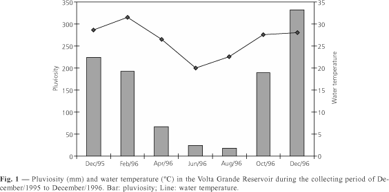
From 18 collected specimens of P. lineatus, 15 presented acanthocephalans in the intestine showing a prevalence of 83.3%. Fishes were present only in August/96, October/96 and December/96 samples. Prevalence of acanthocephalans in those three months was 83.3% (Table 1). However, the higher mean intensity was observed in August (66.5) varying from 16 to 208 helminths. Relation between number of parasites and fish size was not related. Although the acanthocephalans were present in three collecting months the higher pluviosity was observed in December/96 (Fig. 1).
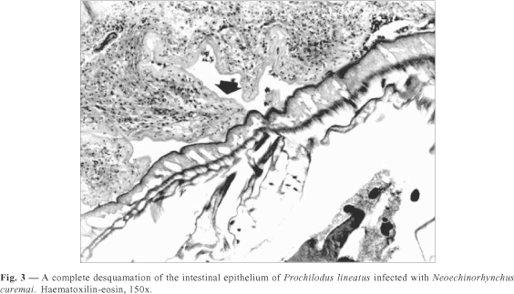
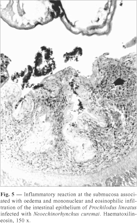
Histopathological analysis showed complete desquamation of the epithelium of intestinal mucosa. In the intestinal lumen it was present cell debris and parasite structures (Figs. 2 and 3). Severe hyperplasia and hypertrofia of the goblet cells were observed (Fig. 4). Nevertheless, a severe inflammatory reaction at the submucosa, displacement of their sheaf, associated with oedema and mononuclear and eosinophilic infiltration were the host responses (Fig. 5).
DISCUSSION
In Brazil, the endoparasitic infections of cultivated fish has a little importance while ectoparasites may cause significant mortality as related by Martins (1998) and Pavanelli et al. (1998). The present work, is one of a sequence of papers reporting parasites of fishes collected from Volta Grande Reservoir. In Plagioscion squamosissimus from this Reservoir, Martins et al. (1999) observed the highest prevalences of larvae of Diplostomum sp. (Diplostomatidae) from the eyes, in April, June and August/96. Nevertheless, Martins et al. (2000) related the highest prevalences of larvae of Thynnascaris sp. (Anisakidae) from the mesentery of fish, in December/95, February, October and December/96.
During a period of 1 year, 15 out of 18 specimens of P. lineatus showed acanthocephalans in the intestine with a prevalence of 83.3%. Nevertheless, only in August, October and December fish were present in nets.
Similar prevalence of 75% was reported by Muzzal (1980) from C. commersoni infected with N. cristatus. By the other hand, an amplitude of variation about 1% to 100% prevalence of N. cylindratus in a several fish species in the Silver Lake and Tichigan Lake at South-eastern Wisconsin was observed (Amin, 1986).
Nevertheless, Lasee (1989) related 47% prevalence of N. pungitius from brook stickleback collected in the Sioux Creek, Wisconsin. In Brazil, Brasil-Sato & Pavanelli (1999) observed 39.3% prevalence of N. pimelodi from P. maculatus during the studied period in the São Francisco River, Minas Gerais State. In the present work, it was difficult to verify seasonality of N. curemai due to the presence of fish in only three months (August, October and December).
This is the first record about histopathology of the P. lineatus infected with N. curemai.
In Brazil, a brief description of the histopathological aspects caused by M. jucundus in pacu (P. mesopotamicus) was performed by Ferraz de Lima et al. (1990). Those authors observed an inflammatory foci of mononuclear cells and haemorrhages in the intestine. In addition, the present study showed complete desquamation of the epithelium of the intestinal mucosa. Severe hyperplasia and hypertrofia of the goblet cells were also observed. Moreover, a severe inflammatory reaction at the submucosa, displacement of their sheaf, associated with oedema and mononuclear and eosinophilic infiltration were the host responses.
When Esch & Huffines (1973) studied the tissue reaction of M. dolomieui infected with L. thecatus the deep penetration of the acanthocephalan proboscis showed fibrosis and eosinophilic granulocytosis as observed in the present work. Moreover, the authors commented that heavy parasitism with other parasites larvae (plerocercoids of Proteocephalus ambloplitis) may contribute to the reduction in reproductive potential. The presence of a great number of helminths may cause inapettence and susceptibility to secondary infection by fungus and bacteria. This is specially true when the analysis of cultured fish is performed (Hedrick, 1998; Martins, 1998).
Intestinal obstruction, compression of villi, haemorrhages, presence of macrophages and fibroblasts were tissue changes related by Amin & Heckmann (1992) from C. columbianus infected with N. idahoensis. This confirmed the present data in which the proboscis attachment provoked the host tissue response. In spite of the presence of a great number (208) of N. curemai in the intestine of P. lineatus it was not observed cell necrosis as caused by N. idahoensis infection.
However, future studies must be done with acanthocephalan infection in the native and cultured Brazilian fish to evaluate other possible tissue changes, population dynamics and the consequences of the parasite to the host reproduction.
Acknowledgments We are gratefull to Companhia Energética de Minas Gerais ¾ Cemig, MG, by the finnancial support.
- AMIN, O. M., 1986, Acanthocephala from lake fishes in Wisconsin: host and seasonal distribution of species of the genus Neoechinorhynchus Hamann, 1892. J. Parasitol., 72(1): 111-118.
- AMIN, O. M. & VIGNIERI, J. C., 1986, Acanthocephala from lake fishes in Wisconsin: Numerical and structural-functional relationships of the giant nuclei in Neoechinorhynchus cylindratus (Neoechinorhynchidae). J. Parasitol, 72(1): 88-94.
- AMIN, O. M. & HECKMANN, R. A., 1992, Description and pathology of Neoechinorhynchus idahoensis n. sp. (Acanthocephala: Neoechinorhynchidae) in Catostomus columbianus from Idaho. J. Parasitol, 78(1): 34-39.
- BRASIL-SATO, M. C. & PAVANELLI, G. C., 1999, Ecological and reproductive aspects of N. pimelodi Brasil-Sato & Pavanelli (Eoacanthocephala, Neoechinorhynchidae) of Pimelodus maculatus Lacépčde (Siluroidei, Pimelodidae) from the basin of the Săo Francisco River, Brazil. Rev. Brasil. Zool, 16(1): 73-82.
- BUSH, A. O., LAFFERTY, K. D., LOTZ, J. M. & SHOSTAK, W., 1997, Parasitology meets ecology on its own terms: Margolis et al revisited. J. Parasitol, 83(4):575-583.
- ESCH, G. W. & HUFFINES, W. J., 1973, Histopathology associated with endoparasitic helminths in bass. J. Parasitol, 59(2): 306-313.
- FERRAZ DE LIMA, C. L. B., FERRAZ DE LIMA, J. Á. & CECCARELLI, P. S., 1990, Ocorręncia de acantocéfalos parasitando pacu, Piaractus mesopotamicus Holmberg, 1887 (Pisces: Serrasalmidae) em piscicultura. Bol. Téc. CEPTA, 2: 43-51.
- HEDRICK, R. P., 1998, Relationships of the host, pathogen, and environment: Implications for diseases of cultured and wild fish populations. J. Aquat. Anim. Health, 10: 107-111.
- KOHN, A., FERNANDES, B. M. M., MACEDO, B. & ABRAMSON, B., 1985, Helminths parasites of freshwater fishes from Pirassununga, SP, Brazil. Mem. Inst. Oswaldo Cruz, RJ, 80(3): 327-336.
- LASEE, B. A., 1989, Seasonal population dynamics and maturation of Neoechinorhynchus pungitius (Acanthocephala: Neoechinorhynchidae) infecting brook stickleback, Culaea inconstans, from Sioux Creek, Wisconsin, U. S. A. Can. J. Zool, 67: 590-595.
- LASSIERE, O. L. & CROMPTON, D. W. T., 1988, Evidence for post-cyclic transmission in the life-history of Neoechinirhynchus rutili (Acanthocephala). Parasitology, 97: 339-343.
- MARTINS, M. L., 1998, Doenças Infecciosas e Parasitárias de Peixes Boletim Técnico Caunesp n. 3, 2. ed. (Ed. Funesp), Jaboticabal, SP, Brasil, 66p.
- MARTINS, M. L., FUJIMOTO, R. Y., NASCIMENTO, A. A. & MORAES, F. R., 1999, Ocorręncia de Diplostomum sp. Nordmann, 1832 (Digenea: Diplostomatidae) em Plagioscion squamosissimus Heckel, 1840, Proveniente do Reservatório de Volta Grande, MG, Brasil. Acta Scientiarum, 21(2): 263-266.
- MARTINS, M. L., FUJIMOTO, R. Y., MORAES, F. R., ANDRADE, P. M., NASCIMENTO, A. A. & MALHEIROS, E. B., 2000, Description and prevalence of Thynnascaris sp. larvae Dollfus, 1933 (Nematoda: Anisakidae) in Plagioscion squamosissimus Heckel, 1840 from Volta Grande Reservoir, State of Minas Gerais, Brazil. Rev. Brasil. Biol., 60(3): 519-526.
- MARTINS, M. L., FUJIMOTO, R. Y., ANDRADE, P. M. & TAVARES DIAS, M., 2000, Recent studies on Neoechinorhynchus curemai Noronha, 1973 (Acanthocephala: Neoechinorhynchidae) in Prochilodus lineatus, Valenciennes, 1836 from Volta Grande Reservoir, MG, Brazil. Rev. Brasil. Biol., 60(4): 673-682.
- MUZZAL, P. M., 1980, Ecology and seasonal abundance of three acanthocephalan species infecting white suckers in SE New Hampshire. J. Parasitol, 66(1): 127-133.
- NORONHA, D., 1973, Sobre Neoechinorhynchus curemai sp.n. (Acanthocephala, Neoechinorhynchidae). Atas Soc. Biol. RJ, 17(1): 19-21.
- NORONHA, D., 1984, Remarks on Neoechinorhynchus curemai Noronha, 1973 (Eoacanthocephala, Neoechinorhynchidae). Mem. Inst. Oswaldo Cruz, RJ, 79(2): 271.
- PAVANELLI, G. C., EIRAS, J. C. & TAKEMOTO, R. M., 1998, Doenças de Peixes. Profilaxia, Diagnóstico e Tratamento. Ed. Universidade Estadual de Maringá, PR, Brasil, 264p.
Publication Dates
-
Publication in this collection
28 Jan 2002 -
Date of issue
Aug 2001
History
-
Accepted
08 Aug 2000 -
Received
02 June 2000